A rare case of ovarian papillary adenocarcinoma in a bitch
Ashwani Kumar Singh, Mrigank Honparkhe, Jasmer Dalal, Rajat Kumar, Kuldip Gupta, Vinod Kumar Singla
1Department of Veterinary Gynaecology and Obstetrics, Guru Angad Dev Veterinary and Animal Sciences University, Ludhiana, 141 004, Punjab, India
2Department of Veterinary Pathology, Guru Angad Dev Veterinary and Animal Sciences University, Ludhiana, 141 004, Punjab, India
A rare case of ovarian papillary adenocarcinoma in a bitch
Ashwani Kumar Singh1*, Mrigank Honparkhe1, Jasmer Dalal1, Rajat Kumar1, Kuldip Gupta2, Vinod Kumar Singla1
1Department of Veterinary Gynaecology and Obstetrics, Guru Angad Dev Veterinary and Animal Sciences University, Ludhiana, 141 004, Punjab, India
2Department of Veterinary Pathology, Guru Angad Dev Veterinary and Animal Sciences University, Ludhiana, 141 004, Punjab, India
ARTICLE INFO
Article history:
Received
Received in revised form
Accepted
Available online
Bitch Ovary
In the present case report, bilateral ovarian tumour (papillary adenocarcinoma) in a 4.5-year-old Saint Bernard bitch and its surgical management is described. Ovariohysterectomy was done and the surgically removed ovarian masses were prepared for histopathological studies. The tumour was associated with pyometra. Macroscopic and histopathological examination confirmed ovarian tumour. Grossly, the tumour consisted of pedunculated processes. The endometrium showed multifocal squamous metaplasia. The findings are discussed as possible consequences of the functioning ovarian tumour and pyometra.
1. Introduction
Ovarian tumours are common neoplasms of dogs that cause various disorders viz. anoestrus, nymphomania, masculinisation, hyperadrenocorticism and alopecia[1]. Canine ovarian tumours are divided into three groups depending on their origin; germ cell tumours, sex-cord stromal tumours and epithelial cell tumours [2]. In dogs, the reported incidence of epithelial neoplasms ranges from 40% to 50% and papillary adenocarcinomas or cystic adenocarcinomas represent nearly 64% of them[3]. These adenomas can affect one or both ovaries and are composed of multiple thin walled cysts filled with transparent squamous fluid or mucin[4]. The present report records a unique case of bilateral ovarian tumour in a bitch.
2. Case report
A 4.5-year-old Saint Bernard bitch was presented to the University Veterinary Clinic with the history of pussy reddish discharge coming out of vagina. Reportedly, the animal was not mated and/ or suffered any injury to the genitalia and had been anorectic since past 10 d. Physical examination presented a poor general state with vulval swelling and alopecia. Abdominal palpation exhibited a large mass caudal to the left kidney and an enlarged uterus. Clinical examination of genitalia revealed foul smelling discharge from vagina. All the clinical parameters (temperature, pulse rate and heart rate) were within the physiological range. Ultrasonographical examination revealed the presence of a non-homogeneous large hypoechoic mass in the left ovary suggestive of ovarian neoplasm, along with segmental enlargements of the uterine horns containing hypoechoic fluid and several hyperechoic projections indicating pyometra. In order to obtain fast recovery ovariohysterectomy was the only option for this case.
Under necessary pre-operative preparation and general anaesthesia, ovariohysterectomy in bitch was performed with standard surgical procedures. The ovarian tumours along with pus filled uterus were removed. Post-operative treatment involved parental administration of antibiotics, anti-inflammatory, analgesics and liver tonics for 5 d and antiseptic dressing of surgical wound with betadine solution on alternate day till sutures were removed (12 d post operative). The bitch showed an uneventful recovery. The general health status of the bitch promptly improved and the alopecia and vulval swelling progressively decreased.
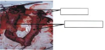
Grossly, the left ovarian tumour (4.5–6.5 cm) was large, encapsulated, firm, irregular and cream to white in colour. Cross-section of the mass revealed an irregular surface that was predominantly covered by cystic areas. These cysts were irregular protruding nodules, variably sized (0.5–1.5 cm) and contained straw-coloured fluid. The right ovarian tumour was comparatively small (0.5–1.5 cm). The uterine horns were enlarged (2–3 cm) and
filled with abundant reddish-brown purulent exudates (Figure 1). The endometrial surface was roughened by the presence of irregular and thickened transverse folds, greyish in colour. Histologically, the tumour was composed of a mixture of various embryonic tissues and cystic structures embedded in a muscular background. The outer margin of the tumour was lined by a squamous epithelium that demonstrated degenerative alterations and all constituents of a normal cutaneous membrane (Figure 2A). Most of the large cystic areas were irregular in shape, filled with keratin and demarcated by a stratified squamous keratinised epithelium (Figure 2B). The solid part of the tumour consisted of a large, dense mixture of irregularly orientated smooth and skeletal muscle fibres and adipose tissue. Within the matrix of connective tissue there were islands of cartilaginous tissue, blood vessels and nerve fibres that were well differentiated but arranged in an ill-defined pattern. A large, extensive band of ovarian parenchyma with prominent neurons, oligodendrocytes and astrocytes was also observed embedded within the tumorous tissue (Figure 2C).

Figure 1. Bilateral ovarian tumour and enlarged pyometric horns.
3. Discussion
The ovarian tumours are often associated with clinical signs like vaginal discharge, alopecia, enlarged vulva, pyometra, cystic endometrial hyperplasia and irregular estrus[5]. In the present case, most clinical signs were exhibited by the bitch. The gross findings displayed large, firm solid and non-homogenous mass of tumours. Similar studies[6] showed that ovarian tumours are mainly suggestive of a solid with inner cystic pattern. The histopathological alterations observed in the present case are in accordance with the findings of Sforna et al. that adenomas consist of pluripotent germ cells that undergo somatic differentiation into two or more germinal cell layers[3]. This explains why these tumours are predominantly intragonadal. Furthermore, canine papillary adenocarcinomas produce both oestrogen and progesterone which play a role in the development of cystic endometrial hyperplasia and subsequent pyometra and are capable of stimulating endometrial and myometrial proliferative changes[7]. Depending on hormonal production, adenomas often induce persistent oestrus, vulval swelling with serosanguineous discharge and alopecia[8]. In the present case, hormonal imbalance together with local growth factor production due to stromal invasion seemed to be the most probable cause for the development of the tumour.
In conclusion, the present report records that papillary adenocarcinoma in the bitch could affect both ovaries and ovariohysterectomy is the most effective treatment option for them.

Figure 2. (A) External surface of the mature ovarian tumour with squamous metaplasia of epithelium (M) and extensive adenoid pattern (P) reflecting adenocarcinoma (100× ), (B) Irregular cystic structure (C) demarcated by stratified squamous keratinised epithelium and filled with keratin (K, 40× ), (C) Hyperplastic changed with marked thickening in ovarian parenchyma (T) containing astrocytes (A) and oligodendrocytes (O, 40× ).
Conflict of interest statement
We declare that we have no conflict of interest.
[1] Lakhani SR, Manek S, Penault-Llorca F, Flanagan F. Pathology of ovarian cancers in BRCA1 and BRCA2 carriers. Clin Cancer Res 2004; 10(7): 2473-2481.
[2] Hofle U, Vicente J, Gortazar C. Bilateral ovarian teratoma in a free-living Iberian red deer (Cervus elaphus hispanicus). New Znd Vet J 2004; 52: 44-45.
[3] Sforna M, Brachelente C, Lepri E, Mechelli L. Canine ovarian tumours: a retrospective study of 49 cases. Vet Res Comm 2003; 27: 359-361.
[4] Schlaffer DH, Miller RB. Female genital system. In: Pathology of domestic animals. San Diego: Academic Press; 2007, p. 431-563.
[5] Zanghi A, Catone G, Marino G, Quartuccio M, Nicotina PA. Endometrial polypoid adenomyomatosis in a bitch with ovarian granulosa cell tumour and pyometra. J Comp Path 2007; 136: 83-86.
[6] Yamaguchi Y, Sato T, Shibuya H, Tsumagari S, Suzuki T. Ovarian teratoma with a formed lens and nonsuppurative inflammation in an old dog. J Vet Med Sci 2004; 66: 861-864.
[7] Niskanen M, Thrusfeld MV. Associations between age, parity, hormonal therapy and breed, and pyometra in Finnish dogs. Vet Rec 1998; 143: 493-498.
[8] Johnston SD, Root Kustritz MV, Olson PNS. Disorders of the canine ovary. In: Canine and feline theriogenology. Philadelphia: WB Saunders Co; 2001, p. 193-205.
ment heading
10.1016/j.apjr.2016.06.016
*Corresponding author: Ashwani Kumar Singh, Assistant Professor, Department of Veterinary Gynaecology and Obstetrics, College of Veterinary Science, Guru Angad Dev Veterinary and Animal Sciences University, Ludhiana, India.
Tel: +91-161-2414003
E-mail: assengar2001@yahoo.co.in
Papillary adenocarcinoma
 Asian Pacific Journal of Reproduction2016年4期
- Asian Pacific Journal of Reproduction的其它文章
- Natural honey as a cryoprotectant to improve Arab stallion post-thawing sperm parameters
- Evaluation of the academic achievement of rural versus urban undergraduate medical students in pharmacology examinations
- The relationship between trace mineral concentrations of amniotic fluid with placenta traits in the pregnancy toxemia Ghezel ewes
- Effects of pomegranate juice in Tris-based extender on cattle semen quality after chilling and cryopreservation
- Evaluation of polymorphonuclear (PMN) cells in cervical sample as a diagnostic technique for detection of subclinical endometritis in dairy cattle
- Spontaneous pregnancy after vaginoplasty in a patient presenting a congenital vaginal aplasia
