A rare cause of infertility: A late complication of female genital mutilation
Ferjaoui Mohamed Aimen, François Monneins, Gharrad Majed, Belaiba Amine
Le service de gynécologie et obstétrique, Le centre hospitalier de Gonesse, 95500 GONESSE, France
A rare cause of infertility: A late complication of female genital mutilation
Ferjaoui Mohamed Aimen*, François Monneins, Gharrad Majed, Belaiba Amine
Le service de gynécologie et obstétrique, Le centre hospitalier de Gonesse, 95500 GONESSE, France
ARTICLE INFO
Article history:
Received
Received in revised form
Accepted
Available online
Female genital mutilation Epidermal inclusion cyst Clitoral reconstruction
Female genital mutilation is a cultural practice in many African and Asian societies based usually on religious beliefs. This practice made by a non-medical and traditional practitioner with non-sterile instruments is a source of many complications such as infection, acute and chronic pain, life-threatening hemorrhage, sexual dysfunction, and rarely epidermal inclusion cysts. We report a case of a large epidermal inclusion cyst in a 36-year-old patient, 30 years after a female genital mutilation. The patient complains of a 2-year secondary infertility with a self-imaging alteration and a sexual dysfunction. The management of this complication was based on surgery with a psychological support and sexual therapies.
1. Introduction
Female genital mutilation (FGM) or excision is an old cultural practice in many African and Asian societies. The most common reason to practice genital mutilation is religious beliefs. The consequences of this practice on the women health are well known, especially psychologic impact, self-imaging alteration, sexual dysfunction and organic or physical impact as obstetrical complications, dyspareunia and chronic pelvic pain. Complications may occur immediately or in several years later. We report a case of rare long-term complications of FGM and how to manage it.
2. Case report
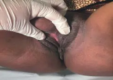
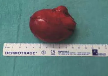
A 36 years-old Malian women, gravida 1 para 1, presents a 2-year secondary infertility. Her medical history is marked by a genital excision at the age of 5 years. She has no memory about this incident and she avoided to talk about. She had a vaginal delivery 2 years ago without complications. Since 2 years, the patient feels embarrassed to do sexual intercourse because of a vulvar tumor which increases in size gradually. She had neither pelvic pain nor dyspareunia, but she describes recently a hypoactive sexual desire. The clinical examination showed a vulvar tumor covered by a regular skin depending on the clitoral area measuring 7 cm. This tumor had a soft consistence (Figure 1) without adhesion to the deep planes. The urinary meatus is individualized and uncompressed. All vulva elements appeared normal (labia majora, labia minora, vestibule and vaginal orifi ce) except clitoral area marked by the vulvar tumor located in the scar of the mutilation (Figure 2). Talking about the vulvar lesion, the patient recognized psychological discomfort, especially during sexual intercourse. An ultrasound imaging was performed, and it showed a multi-locular cystic tumor measuring 4 x 7 cm with heterogeneously dense contained (Figure 3).

Figure 1. A large vulvar tumor, with a soft consistence.

Figure 2. The vulvar elements are normal except the clitoral area. The urinary meatus is individualized and uncompressed.
The decision was to perform a cystectomy and a vulvar plastic procedure under spinal anesthesia. Through a vertical incision we proceeded to a dissection of the cyst. Cystectomy was performed after ligation of the cyst pedicle. The root of the glans clitoris wasindividualized. Finally a clitoral reconstruction and vulvoplasty were performed (Figure 4). The histologic examination showed an epidermic cyst containing abundant laminated keratin.

Figure 3. The cyst measuring 7 cm.

Figure 4. After the cystectomy, we proceeded on a resection of the excess skin (A and B) and observed the immediate post operative aspect (C) and 3-day post operative aspect (D).
3. Discussion
FGM is a cultural practice characterizing a lot of African and Asian societies. This clinical entity can be seen in occidental countries among immigrant communities[1]. According to the World Health Organization (WHO) guidelines, the classifi cation of FGM is based on four types: (i) type I (clitoridectomy) a partial or total removal of the clitoris and, rarely, the prepuce as well; (ii) type II (excision) is the partial or total removal of the clitoris and the labia minora, with or without excision of the labia majora; (iii) type III (infibulation) is the most severe form and involves partial or total removal of the labia minora and majora with or without clitoridectomy; (iv) type IV FGM refers to all other harmful procedures to the female genitalia for nonmedical purposes, e.g., pricking, piercing, incising, scraping, and cauterizing the genital area. Type I and II constitute about 80% of FGM. Our patient is from an African country (Mali) and she had a type I FGM (circumcision).
This procedure is made by a non-medical traditional practitioner commonly an old woman. Non sterile instruments are used without any hemostasis. That’s why FGM is a common source of complications such as life-threatening hemorrhage, infection, acute and chronic pelvic pain, dysmenorrhoea, sexual dysfunction[2], obstetrics complications, psychological complication and infertility[3].
The epidermal inclusion cyst is a frequent complication of FGM[3], and it arises from the development of epidermal cells in the FGM scar within a circumscribed space, and the sebaceous gland secretion and epidermic cells peeling allow the cyst to grow up[4]. The delay from circumcision to development of the inclusion cyst is variable. In the case of our patient the cyst appeared after pregnancy, probably because of the hormonal environment during pregnancy stimulating secretion of sebaceous glands and epidermal cells of the embedded tissue in the FGM scar. This hypothesis was discussed in the medical literature[5].
Clinically, the epidermal inclusion cyst is painless and can be associated with other urogenital symptoms, such as dyspareunia,micturition disturbances, and vulval pain[6,7]. Usually, epidermal inclusion cysts are small sized but huge cysts are reported in a few cases[8]. In the present case, the cyst measured 7 cm. The management of this FGM complication is surgery: enucleation of the cyst in case of small cysts[9] and a clitoral reconstruction with vulvoplasty in larger cysts[10]. No post-operative recurrence is reported[9].
In France, the UNICEF estimated that 53 000 adult women were victims of FGM[11]. In the study of Ndiaye et al.[12] the management of FGM and their complications is not based only on surgery, which can be badly lived, but requires a medical, psychological and sexological management.
Declare of interest statement
We declare that we have no confl ict of interest.
[1] World Health Organization (WHO). Female genital mutilation. Fact sheet no. 241. Geneva, Switzerland: World Health Organization (WHO) Media Center, 2008.
[2] Alsibiani SA, Rouzi AA. Sexual function in women with female genital mutilation. Fertil Steril 2010; 93: 722-724.
[3] Abdulrahim AR. Epidermal clitoral inclusion cyste: not a rare complication of female genital mutilation. Hum Reprod 2010; 25(7): 1672-1674.
[4] Larsen U, Okonofua FE. Female circumcision and obstetric complications. Int J Gynaecol Obstet 2002; 77: 255-265.
[5] Rizk DE. A large clitoral epidermoid inclusion cyst first presenting in adulthood following childhood circumcision. J Obstet Gynaecol 2007; 27: 445-448.
[6] Rouzi AA, Sindi O, Radhan B, Ba’aqeel H. Epidermal clitoral inclusion cyst after type I female genital mutilation. Am J Obstet Gynecol 2001; 185: 569-571.
[7] World Health Organization (WHO). Female genital mutilation: prevalence and distribution. Fact sheet no. 153. Geneva, Switzerland: World Health Organization (WHO) Media Center, 1997.
[8] Ofodile FA, Oluwasanmi JO. Post-circumcision epidermoid inclusion cysts of the clitoris. Plast Reconstr Surg 1979; 63: 485-486.
[9] Asante A, Omurtag K, Roberts C. Epidermal inclusion cyst of the clitoris 30 years after female genital mutilation. Fertil Steril 2010; 94(3): 1097. e1-3.
[10] Abdulcadir J, Rodriguez MI, Say L. A systematic review of the evidence on clitoral reconstruction after female genital mutilation/cutting. Int J Gynaecol Obstet 2015; 129(2): 93-97.
[11] UNICEF. Female genital mutilation/cutting: a statistical overview and exploration of the dynamics of change. Library J 2013; 21(42): 184-190.
[12] Antonetti Ndiaye E, Fall S, Beltran L. Benefi ts of multidisciplinary care for excised women. Gynecol Obstet Biol Reprod 2015; 44: 862-869.
ent heading
10.1016/j.apjr.2016.03.001
*Corresponding author: Ferjaoui Mohamed Aimen, Le service de gynécologie et obstétrique, Le centre hospitalier de Gonesse, 95500 GONESSE, France.
E-mail: ferjaoui16@yahoo.fr
Vulvoplasty
 Asian Pacific Journal of Reproduction2016年3期
- Asian Pacific Journal of Reproduction的其它文章
- Corpus callosum agenesis: Role of fetal magnetic resonance imaging
- Chilled and post-thawed semen characteristics of buffalo semen diluted in tris extender enriched with date palm pollen grains (TPG)
- Establishment, characterization and cryopreservation of Fars native goat fetal fibroblast cell lines
- Somatic embryogenesis and in vitro flowering in Hybanthus enneaspermus (L.) F. Muell.-a rare multipotent herb
- Pollutant exposure in Manila Bay: Effects on the allometry and histological structures of Perna viridis (Linn.)
- Changes in sperm characteristics of the three main breeds of sheep in Algeria after dietary supplementation
