
Primary oral and nasal transmissible venereal tumor in a mix-breed dog
2016-07-02 06:58MahdiehRezaeiShahrzadAziziShimaShahheidaripourSaraRostami1DepartmentofClinicalScienceFacultyofVeterinaryMedicineShahidBahonarUniversityKermanIranDepartmentofPathobiologyFacultyofVeterinaryMedicineShahidBahonarUniversityKerman
Mahdieh Rezaei, Shahrzad Azizi, Shima Shahheidaripour, Sara Rostami1Department of Clinical Science,Faculty of Veterinary Medicine,Shahid Bahonar University,Kerman,IranDepartment of Pathobiology,Faculty of Veterinary Medicine,Shahid Bahonar University,Kerman,IranFaculty of Veterinary Medicine,Shahid Bahonar University,Kerman,Iran
Primary oral and nasal transmissible venereal tumor in a mix-breed dog
Mahdieh Rezaei1*, Shahrzad Azizi2, Shima Shahheidaripour3, Sara Rostami31Department of Clinical Science,Faculty of Veterinary Medicine,Shahid Bahonar University,Kerman,Iran2Department of Pathobiology,Faculty of Veterinary Medicine,Shahid Bahonar University,Kerman,Iran
3Faculty of Veterinary Medicine,Shahid Bahonar University,Kerman,Iran
ARTICLE INFO
Article history:
Received 19 Oct 2015
Received in revised form 9 Nov, 2nd
revised form 24 Nov 2015
Accepted 22 Dec 2015
Available online 18 Mar 2016
Keywords:
Transmissible venereal tumor
Primary
Oral
Nasal
Dog
ABSTRACT
Transmissible venereal tumor(TVT)is a coitally transmitted tumor of dogs with widespread distribution. The present study describes the occurrence of the primary oral and nasal TVT in a 10-year-old, female, mix-breed dog. The case was presented with a history of anorexia, inability to swallow and dyspnea. Clinical examinations revealed the emaciation, muzzle deformity due to the presence of a friable,fleshy, cauliflower-like mass in the oral cavity and submandibular lymphadenopathy. TVT was diagnosed based on histopathological findings. The dog was discharged with therapeutic intervention with vincristine. Unfortunately, the case died before readmission because of the progressive worsening of the general condition. Our findings highlight the need for considering TVT for the differential diagnosis of the extragenital masses in dogs.
Case report http://dx.doi.org/10.1016/j.apjtb.2016.03.006
Tel: +98 9133431675
E-mail: mahdiehrrezaei@gmail.com
Foundation Project: Supported by a grant from Shahid Bahonar University of Kerman, Research Council, Kerman, Iran(No. 92-GR-VS-01).
Peer review under responsibility of Hainan Medical University. The journal implements double-blind peer review practiced by specially invited international editorial board members.
1. Introduction
Canine transmissible venereal tumor(TVT)is a round cell neoplasm with widespread distribution, particularly in region with tropical and subtropical climates[1,2]. This canine tumor transmits coitally with no breed or sex predilection[3,4]. TVT is more commonly observed in the free-roaming, stray, sexually active dogs[5,6]. The most affected site for this tumor is the external genitalia of both sexes[7,8], appearing as a cauliflowerlike growth[6,9]. Extragenital involvement is observed following social behavior including sniffing, licking or scratching[7]. Diagnosis of TVT is based on the history, clinical signs, cytology and histopathology[9]. Chemotherapy is the choice of treatment[2,9]. Although, there are reports of secondary occurrence of oral and nasal TVT in dogs, primary occurrence of these sites is almost rare. The present study describes history, clinical signs, radiographic and histopathological findings of the primary oral and nasal TVT in a dog.
2. Case report
A 10-year-old, female, mix-breed dog was referred to the veterinary animal hospital with a history of anorexia, inability to swallow and dyspnea for several weeks. Clinical examinations revealedtheemaciation,muzzledeformityduetothepresenceofa friable,fleshy, cauliflower-like mass in the oral cavity and submandibular lymphadenopathy. Halitosis and blood-tinged saliva were observed in the oral examination as well. It seemed that the mass extended toward the nose and eye at the same side and resulted in the purulent nasal discharge, breathing problem, hyperemia and conjunctivitis(Figure 1). No abnormalities were found on the examination of other parts of the body such as external genitalia. Deformity and lysis of the facial bones and the presence of tumor-like lesions in bothnasal and oral cavities were detected by radiology(Figure 2). Imaging studies including radiography and ultrasonography did not show any visceral metastasis. No regional lymph node involvement was found. Then, for incisional biopsy, tissue was taken from the oral and nasalmassesandfixedin10%bufferedformalin.Histopathologic investigation showed a large number of round to oval cells withbasophilic cytoplasm containing distinct intracytoplasmic vacuoles and prominent nucleoli. Fibrous stroma was present between basophilic round cells, and frequent mitotic figures were also observed(Figure3).Thedog'sownerdeclaredthattheanimalhad notanycontactwithotherdogswithsimilarlesionsintheexternal genitalia or other sites. Based on histopathological findings, primary oral and nasal TVT was diagnosed. The dogwas discharged with therapeutic intervention with vincristine(0.025–0.050 mg/ kg, i.v.)once weekly for three weeks and broad-spectrum antibiotic administration. Ophthalmic nasal drops, including ciprofloxacin and dexamethasone were also prescribed. Unfortunately, the case died before readmission because of the progressive worsening of the general condition.
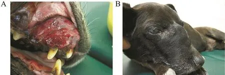
Figure 1. Photograph showing the presence of a friable,fleshy, cauliflower-like mass in the oral cavity(A), muzzle deformity due to the extension of the mass toward the nose and eye at the same side(B).

Figure 2. Deformity and lysis of the facial bones and the presence of the tumorous lesions in both nasal and oral cavities.

Figure 3. Histopathological findings.
3. Discussion
TVT is a common canine reproductive disease throughout the world[8,10]. TVT occurs between 1 and 7 years old(mean: 3 years old)[9,11]with the lowest incidence above 10 years old [12]. Some researchers reported higher prevalence in males [9,12], although others noted that more females were affected than the males[11]. No sex predilection was detected in other reports[4,13]. Higher incidence of TVT has been found in crossbred/nondescript breed by Srivastava et al.[12]. In the present study, we reported the primary oral and nasal TVT in a 10-year-old, female, mix-breed dog.
According to the literature, TVT occurs predominantly in the stray dogs of the regions with tropical and sub-tropical climates[3,11]. In contrary, we reported it in an indoor dog from an arid area. The present case is similar to the other studies reporting the muzzle deformity, existence of friable fleshy cauliflowerlike mass, halitosis, purulent nasal discharge and submandibular lymphadenopathy as the most clinical symptoms of primary or secondary oral and nasal TVT[14]. According to our findings, affected dogs may have signs of respiratory problem due to thickening of the soft tissues in the nares that results in upper airway obstruction. Dyspnea was a striking feature of this dog's history. Inability to swallow, anorexia and blood-tinged saliva are all attributed to the oral neoplasms. Progressive extension of the tumorous mass affected ocular tissue as hyperemia and conjunctivitis of the same side. As we describe here, this benign tumor mostly appears as a friable, hemorrhagic, cauliflower-like mass[6,11,13], but that in the mouth appears more diffuse[4]. In comparison, atypical appearance as an ulcerated lesion with exuberant granulation tissue was also described[15].
Vestibule and vagina of the female dogs, and penis of the male dogs have been documented as the most affected sites of TVT[3]. Secondary involvement at other sites of the body has also been reported. Primary extragenital involvement is almost rare. Compared to our results, Chikweto et al. reported 19.2% of dogs with TVT in extragenital sites in the nasal cavity, eye orbit, spleen, liver, skin, ribs, subcutaneous, submandibular, cervical and inguinal lymph nodes[3]. He also found evidence of metastasis to the ovaries in two dogs. Interestingly, the presence of extragenital lesions without primary genital involvement was determined in 5.1% of cases. In the mentioned study, one dog had primary nasal TVT while primary TVT of subcutaneous tissue was diagnosed in three dogs. In another study performed in Grenada, extragenital TVT lesions were detected in 9(23%)cases with two lesions in the nose, two masses in the mouth and three on the skin[9]. In accordance with our findings, primary intranasal TVT was reported in six dogs by Papazoglou et al.[14]. None of these dogs had TVT-compatible lesions on the external genitalia. Moreover, oral primary TVT was reported in dogs by Filgueira [16]. In comparison with our report of TVT in oral and nasal cavities, extragenital lesion(right hind limb)of TVT was also reported by Lima et al.[15]. Moreover, primary extragenital cutaneous TVT associated with Leishmania infantum infection was described in another study[7]. Diffuse cutaneous canine TVT with primary prepuce mass was also found by other researchers[5].
In the current case, diagnosis of TVT was confirmed by histopathology that was similar to previous reports[3,9,11]. We observed a large number of the round cells with a prominent nucleolus and basophilic cytoplasm on cytology. TVTs should be differentiated from other round cell tumors of the skin including histiocytoma, lymphoma, and mast cell tumor. The site of the tumor plays an important role in diagnosis. TVTs show immunoreactivity with lysozyme, alpha-1-antitrypsin, and vimentin. They are negative with keratins, S100 protein, lambda light-chain immunoglobulins, immunoglobulin G, immunoglobulin M, and CD3 antigen[17]. As we described here, TVT is a neoplastic growth with a low metastatic rate. In this case, no signs of metastasis were detected by radiology and ultrasonography. No primary tumor was also found in the external genitalia making metastatic dissemination unlikely. Treatment should be directed at tumor remission and elimination of secondary infections and other complications.Medical treatment with chemotherapeutic agents such as vincristine has been shown to be effective[2,9,14]. We started treatment by intravenous injection of vincristine sulfate at the dose of 0.025 mg/kg body weight weekly. Broad-spectrum antibiotic and local drops were also administered to resolve secondary complications. Unfortunately, the case died due to the progressive worsening of the general condition.
Our findings highlight the need for considering TVT for the differential diagnosis of extragenital masses in dogs. Neutering of affected dogs can be beneficial to eliminate this transplantable tumor.
Conflict of interest statement
We declare that we have no conflict of interest.
References
[1]Ganguly B, Das U, Das AK. Canine transmissible venereal tumour: a review. Vet Comp Oncol 2016;14: 1-12.
[2]Lapa FAS, Andrade SF, Gervazoni ER, Kaneko VM, Sanches OC, Gabriel Filho LRA. Histopathological and cytological analysis of transmissible venereal tumor in dogs after two treatment protocolos. Colloq Agrar 2012;8(1): 36-45.
[3]Chikweto A, Kumthekar S, Larkin H, Deallie C, Tiwari KP, Sharma RN, et al. Genital and extragenital canine transmissible venereal tumor in dogs in Grenada, West Indies. Open J Vet Med 2013;3: 111-4.
[4]Das U, Das AK. Review of canine transmissible venereal sarcoma. Vet Res Commun 2000;24: 545-56.
[5]Champour M, Ojrati N, Nikrou A, Rahimian H, Tehrani-Sharif M. First report of diffuse cutaneous canine transmissible venereal tumor in Iran. Comp Clin Pathol 2014;24: 741-4.
[6]Muste A, Beteg F, Muste M, Stroe T, Lˇacˇatus¸R, Loredana H. Research and observation on clinical and therapeutic aspects regarding transmissible venereal tumor in dogs. Vet Med 2012;LVIII(4): 272-7.
[7]Albanese F, Poli A, Millanta F, Abramo F. Primary cutaneous extragenital canine transmissible venereal tumour with Leishmania-laden neoplastic cells: a further suggestion of histiocytic origin?Vet Dermatol 2002;13: 243-6.
[8]Strakova A, Murchison EP. The changing global distribution and prevalence of canine transmissible venereal tumour. BMC Vet Res 2014;10: 168.
[9]Kabuusu RM, Stroup DF, Fernandez C. Risk factors and characteristics of canine transmissible venereal tumours in Grenada, West Indies. Vet Comp Oncol 2010;8(1): 50-5.
[10]Sathiamoorthy T, Raja S. Prevalence of reproductive disorders in the stray dogs of Chennai City. J Indian Vet Assoc 2011;9(2): 62-3.
[11]Santiago-Flores ML, Jaro MC, Recuenco FC, Reyes MF, Amparo MRG. Clinical profile of canine transmissible venereal tumor cases. Philipp J Vet Anim Sci 2012;38(1): 63-72.
[12]Srivastava AK, Singh B, Srivastava AK, Sharama AK, Sinha N. Canine transmissible venereal tumours(CTVT): a study on occurrence and distribution pattern. Indian J Canine Pract 2013;5(1): 65-71.
[13]Islam MS, Das S, Alim MA, Mohi Uddin M, Kabir MHB, Tariqul Islam M, et al. Progressive type of canine transmissible venereal tumor(CTVT)in a male stray dog: a case report. Res J Vet Pract 2014;2(4): 70-2.
[14]Papazoglou LG, Koutinas AF, Plevraki AG, Tontis D. Primary intranasal transmissible venereal tumour in the dog: a retrospective study of six spontaneous cases. J Vet Med A Physiol Pathol Clin Med 2001;48: 391-400.
[15]Lima TB, Marinho PVT, Lira RN, Jark PC, Melo JFP, Oliveira LCR.[Canine transmissible venereal tumor in a dog with single extragenital location]. Vet Zootec 2013;20(1): 57-61. Spanish.
[16]Filgueira KD.[Canine transmissible venereal tumor with primary and unique site in the oral cavity]. Acta Sci Vet 2010;38(1): 91-4. Spanish.
[17]Muten DJ. Tumors in domestic animals. 4th ed. Ames: Iowa State Press;2002.
*Corresponding author:Mahdieh Rezaei, Department of Clinical Science, Faculty of Veterinary Medicine, Shahid Bahonar University, Kerman, Iran.
 Asian Pacific Journal of Tropical Biomedicine2016年5期
- Asian Pacific Journal of Tropical Biomedicine的其它文章
- Antimalarial qinghaosu/artemisinin:The therapy worthy of a Nobel Prize
- Estimating risk factors of urban malaria in Blantyre,Malawi:A spatial regression analysis
- Insecticide susceptibility in larval populations of the West Nile vector Culex pipiens L. (Diptera:Culicidae)in Saudi Arabia
- Agave sisalana extract induces cell death in Aedes aegypti hemocytes increasing nitric oxide production
- Phenolics-saponins rich fraction of defatted kenaf seed meal exhibits cytotoxicity towards cancer cell lines
- Comparative investigation of the free radical scavenging potential and anticancer property of Diospyros blancoi(Ebenaceae)