磁共振水-脂分离成像技术对椎体脂肪含量的测量
常飞霞,黄刚,樊敦徽,徐香玖,马小梅,王平,沈雯怡
磁共振水-脂分离成像技术对椎体脂肪含量的测量
常飞霞1,2,黄刚3*,樊敦徽2,徐香玖3,马小梅3,王平3,沈雯怡1
目的以双能X线吸收测量法(dual-energy X-ray absorptiometry,DXA)检查为参照标准,应用磁共振水-脂分离成像(Dixon)技术测量腰椎椎体脂肪信号强度并计算脂肪分数(fat fraction,FF)来评价椎体骨密度方面的应用价值。材料与方法回顾性收集甘肃省人民医院腰椎体查患者36例,纳入腰1-4椎体(144个)。所有患者均分别行DXA检查及腰椎MR检查,以DXA腰椎椎体T值作为金标准,使用WHO的诊断标准,即T值≥-1.0 SD 为正常,-1.0~-2.5 SD为骨量减低,≤-2.5 SD为骨质疏松;同时在MRI图像上测量并计算腰1-4椎体的信号强度(T2脂相像)及FF,骨髓脂肪FF计算公式:FF=[Mfat/(Mfat+Mwater)]×100%,式中Mwater、Mfat分别指水像及脂像感兴趣区(region of interest,ROI)总像素信号强度值,以此来评价磁共振水-脂分离成像技术预测骨质疏松的能力。结果36例患者144个椎体,按照T值进行分组,骨量正常组58个,骨量减少组28个,骨质疏松组58个,骨量正常组、骨量减少组和骨质疏松组椎体脂相信号强度分别为100.2±20.1、156.1±56.3、211.9±84.6。FF分别为:(31.1±6.2)%、(53.3±7.6)%、(77.8±7.2)%。骨质疏松组、骨量减少组及正常组比较,腰椎T2脂相信号强度及脂肪分数均具有统计学差异(P<0.01)。椎体脂相信号强度与DXA呈负相关,两者相关系数r=-0.64,P<0.01,脂肪分数与DXA呈负相关,两者相关系数r=-0.93,P<0.01。结论磁共振水-脂分离成像通过测定腰椎信号强度并计算脂肪分数,可以反映椎体脂肪含量的变化,对骨质疏松做出初步诊断,对于评估腰椎骨密度具有一定的应用前景。
磁共振成像;水脂-分离技术;椎体;脂肪分数;骨质疏松
骨质疏松症(osteoporosis,OP)是一个重大的公共卫生问题。据统计,50%的女性和20%的男性都有发生骨质疏松相关性骨折的危险。尽管双能X线吸收测量法(dual-energy X-ray absorptiometry,DXA)对骨质疏松症的筛查未得到充分利用,但世界卫生组织(WHO)仍将DXA 测定骨密度值作为诊断骨质疏松的金标准[1-2]。DXA一般用于绝经后女性和50岁以上男性骨质疏松的诊断,腰椎或髋关节的T值≤-2.5即可诊断。此外骨密度(bone mineral density,BMD)的评价方法还有定量CT(quantitative computed tomography,QCT)、超声等,但DXA做为WHO推荐的方法,本文测量则以T值为标准进行对比研究。
有研究证实骨量的降低均伴有不同程度的骨髓内脂肪组织增加[3-4],因此,研究椎体内骨髓脂肪组织含量的变化对评估骨质疏松及骨折风险具有重要意义。1H-MRS作为一种对广大患者来说既无辐射又无创伤的检查,能对椎体松质骨中水和脂肪的含量进行分析,在松质骨形态出现异常前发现早期骨质量的变化。但磁共振波谱(magnetic resonance spectroscopy,MRS)[5]并非检查中常规应用序列,只有在高场强磁共振机才能做到,而且检查耗时较长,要求患者配合难度较大,在椎体检查中难以推广,从而限制了它在临床的应用。经研究证明MRI水-脂分离技术(Dixon)可以定量分析脏器内的脂肪含量[6-8],目前应用Dixon技术测量骨髓内脂肪组织含量的研究较少,所以本研究收集了既做DXA检查又做腰椎磁共振常规检查的患者进行图像的测量、分析,甘肃省人民医院腰椎常规序列包括水-脂分离序列,为本研究提供了便利,通过测量各椎体脂相中信号强度及计算脂肪分数,来评价腰椎骨髓脂肪含量,从而间接评价腰椎骨量的变化为OP诊断提供了新的检查手段。
Dioxn技术是一种水-脂分离方法,它既是反转恢复和脂肪饱和等常规方法外的现代压脂技术,还可以通过一次扫描获得多个对比度并用于脂肪定量等场合。水脂分离的图像,放弃一个就是抑制,有些应用两者都需要精确定量。在相关研究中,脂肪分数值变异系数为2.95%,这说明扫描具有较好的可重复性。利用组内相关分析考察不同研究者间FF值测量的一致性时发现,两位独立观察者测量FF值时ICC为0.928(当ICC>0.75时,表示信度良好),这提示不同观察者间具有很好的一致性。因此,此项技术对评估骨髓脂肪含量是可行的。
对患者进行腰椎MR检查的同时筛查腰椎BMD,不需要额外成像,不会增加患者费用及时间,如果腰椎水-脂分离图像信号强度及脂肪分数对脂肪含量的测量是准确的,那么就可以间接证明腰椎Dixon检查对骨质疏松的诊断是有价值的,就可以排除DXA检查的必要性。因此用腰椎Dixon对并发骨质疏松进行筛查,将会进一步增加临床的应用价值,也可以降低额外检查的成本。Dixon技术受重复时间(repetition time,TR)及回波时间(echo time,TE)的影响较小,可采集任意两个不同相差的图像,因而可以选择更短的TR和更短的TE,采集速度更快,更适于常规序列扫描的应用。本研究的目的在于证明在腰椎常规MR检查的同时评估腰椎BMD的效果,以DXA检查为参照标准,测量腰椎椎体信号强度及计算骨髓脂肪含量,同时评估骨密度,从而说明Dixon技术对腰椎骨质疏松的诊断有一定的应用前景。
1 材料与方法
1.1 研究对象
收集2015年7月至2016年3月甘肃省人民医院腰椎检查患者36例,其中女23例,男13例,年龄32~72岁,平均年龄58.5岁,所有患者在检查前签署知情同意书,所有患者DXA检查与3.0 T磁共振常规腰椎检查时间间隔不超过10天。排除标准:腰椎患有骨肿瘤、转移瘤、血管瘤、结核等病变的患者;腰椎有压缩骨折;有甲状腺功能亢进、甲状旁腺功能亢进等骨质代谢相关疾病;服用影响骨质代谢水平的药物,如糖皮质激素等。
1.2 DXA和MR检查技术
GE公司(LunarDPX型)医用诊断X线系统骨密度仪。应用DXA设备(GE-LunarDPX型)测腰椎BMD值。所有研究对象应用DXA设备(GELunarDPX型)按厂商制定的标准方法测量L1-L4椎体前后位BMD值(g/cm2)和各个椎体对应的T值,测量前均需进行仪器校准。采用中华医学会骨质疏松和骨矿盐疾病学分会推荐的诊断标准,即T值≥-1.0 SD为骨量正常,-1.0~-2.5 SD为骨量减低,T≤-2.5 SD 为骨质疏松,T值≤-2.5 SD且合并一处或多处脆性骨折为严重骨质疏松[2-7]。
德国SIEMENS Skyra 3.0 T MRI仪进行腰椎磁共振检查,使用脊柱相控阵线圈;MR 扫描序列包括:T2WI矢状位化学位移成像:TR 3500 ms,TE 82 ms,翻转角度150°,回波链16,层厚均为4 mm,层间距为0.8 mm,矩阵256×256,FOV为260 mm×260 mm,共扫描15 层。扫描结束后,系统自动生成4 幅图像,即正相位、反相位、脂像及水像。
1.3 图像分析及测量
将磁共振扫描图像传至MMWP (multi modality work place) version:VE40后处理工作站,由两位放射科主治医师独立测定所有椎体骨髓脂肪的含量。选择经过腰椎(测量L1~L4)椎体最正中矢状位T2脂相图像,手动绘制感兴趣区(region of interest,ROI),ROI包括整个椎体的松质骨部分,需避开椎静脉入口及皮质骨等(图1A)。通过后处理软件计算并测量脂肪分数:[脂相/(脂相+水相)]×100%,在计算出的图像选择经过腰椎(测量L1~L4)椎体最正中矢状位测量脂肪分数,测量方法同上(图1B)。
1.4 统计学分析

2 结果
纳入36例患者,共计144个椎体,按照T值进行分组,骨量正常组58个,骨量减少组28个,骨质疏松组58个,骨量正常组、骨量减少组和骨质疏松组椎体脂相信号强度分别为100.2±20.1、156.1±56.3、211.9±84.6。FF分别为:(31.1±6.2)%、(53.3±7.6)%、(77.8±7.2)%。
3.0 T磁共振水-脂分离成像腰椎同脂相图像观察:骨量正常组的脂相上可见少量稍高信号的脂肪组织(图2),骨量减少组在脂相图像可见散发片状大小不一的高信号的脂肪成分(图3),骨质疏松组脂相可见诸椎体边缘骨质增生变尖,椎体内可见多发片状高信号脂肪成分(图4),通过脂相位图像可以对组织和病灶内是否含有脂肪成分及其多少做出初步判断。
2.1 椎体信号强度与骨密度之间的关系
骨量正常组、骨量减少组和骨质疏松组椎体信号强度分别为100.2±20.1、156.1±56.3、211.9±84.6,组间比较差异有统计学差异(P<0.01),见表1;组内两两比较,差异有统计学意义,见表2。脂相信号强度与DXA呈负相关,两者相关系数为r=-0.64,P<0.01,见图5。
2.2 椎体脂肪分数与骨密度之间的关系
骨量正常组、骨量减少组和骨质疏松组椎体FF分别为:(31.1±6.2)%、(53.3±7.6)%、(77.8±7.2)%,组间比较差异有统计学差异(P<0.01),见表1;组内两两比较差异均有统计学意义,见表3。椎体FF与DXA呈负相关,两者相关系数为r=-0.93,P<0.01,见图6。
3 讨论
OP是以骨量减少、骨组织显微结构退化为特征,以至骨骼脆性增高、骨折风险增加的一种全身代谢性骨骼疾病。近年来骨质疏松在中国发病率呈逐年上升趋势,成为一个重大的公共卫生问题,给家庭和社会带来巨大的经济负担,因此预防筛查工作急需做到位。
DXA是目前评价骨密度的金标准,具有操作方便、检测时间短、患者电离辐射剂量低(其放射量相当于胸片的1/30)的优点[9],曾被公认为测量BMD的首选方法,但是有研究表明DXA准确性低,漏诊率较高。
QCT测定法是利用三维技术来测量BMD,该方法受骨体积影响小、分辨率高等优点[10],结果可通过三维旋转功能测量,排除椎体重叠及患者体位因素造成的干扰[11],且QCT对于诊断骨质疏松的准确率逐渐被大家认可,然而QCT较高的辐射剂量及高成本限制了其在临床的广泛应用。
因骨质在磁场内的信号迅速下降,过去研究认为MRI对骨密度的测量无意义,但最近有研究[12-13]表明,可应用磁共振水-脂分离成像技术对腰椎BMD进行早期筛查。磁共振水脂分离成像技术[14-16]通过测定椎体信号下降指数可以反映椎体脂肪含量的变化,间接证明此成像技术对骨质疏松诊断价值。
Justesen等[17]通过活检研究人体髂骨骨髓脂肪含量,比较正常人及骨质疏松患者的骨髓脂肪含量,研究发现老年人及骨质疏松患者骨松质体积明显减少,骨髓中脂肪含量显著增多。Meunier等[18]通过84 例尸体标本的髂骨病理解剖研究后发现,脂肪组织的含量从老年人的60%左右降低到年轻人的15%左右,松质骨的含量从16%左右增加到了26%左右[19]。Naverias等[20]研究证明椎体内骨髓脂肪细胞是骨髓微环境的主要负性调节因子,对椎体骨髓脂肪生成抑制,可以增强骨髓移植患者的造血功能,从而防止骨质流失。Hajek等[21]通过3例尸体进行尸检研究,发现病理及磁共振图像上腰椎骨髓内的斑点状和斑片状异常信号是脂肪组织,所以测定骨髓的脂肪含量对椎体骨量的测定有重要价值。
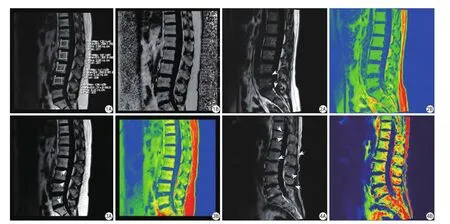
图1A:腰椎T2脂相图像。在L1~L4椎体最正中矢状位手工绘制ROI,测量L1~L4椎体信号强度;B:通过后处理软件计算脂肪分数:[脂相/ (脂相+水相)]×100%,测量图像选择经过腰椎(测量L1~L4)椎体最正中矢状位手工绘制ROI测量脂肪分数图2腰椎骨量正常组T2脂相图像。图像上可见少量稍高信号的脂肪成分,白色箭头所指区域即彩图中橘黄色图3腰椎骨量减少组T2脂相图像。图像上可见散在片状大小不一的高信号的脂肪成分,白色箭头所指区域即彩图中橘黄色区域图4腰椎骨质疏松组T2脂相图像。图像可见诸椎体边缘骨质增生变尖,椎体内可见多发片状高信号脂肪成分,白色箭头所指区域即彩图中橘黄色区域Fig. 1A: Lumbar T2 lipid phase image. In the middle of the L1—L4 vertebral sagittal plane hand drawn ROI, measure the signal intensity; B: Fat fraction calculated by post-processing software: [lipid phase/(fat phase + water phase)]×100%, In the lumbar spine (L1—L4) vertebra most middle sagittal plane, handdrawn ROI, measure the fat fraction.Fig. 2T2 lipid phase image of lumbar spine bone mass in normal group. Image shows a small amount of fat component was slightly high signal intensity, the white arrow indicates the region that is illustrated in the orange region.Fig. 3T2 lipid phase image of lumbar spine bone mass in osteopenia group. Image shows scattered sheet different sizes of fat component showed high, the white arrow indicates the region that is illustrated in the orange region.Fig. 4T2 lipid phase image of lumbar spine bone mass in osteoporosis group. Image shows each vertebral edge bone hyperplasia, and multiple sheet high signal of fat component within the vertebral body, the white arrow indicates the region that is illustrated in the orange region.
磁共振Dixon技术可以无创地测定组织内的脂肪含量[22-23]。骨骼是人体支撑系统,腰椎作为人体中轴骨骼,椎体内的松质骨结构较多,是骨量流失先发病的部位,腰椎骨小梁数目较其它骨质相对较高,因此,椎体BMD下降主要以椎体骨小梁变薄、消失为主,骨质疏松的程度相对较明显。成骨细胞、骨细胞和破骨细胞的功能决定了骨组织发生骨质疏松和骨量减少时,脂肪细胞增加伴随着成骨细胞的减少,脂肪细胞的进一步增加会导致更多的造血干细胞转化为破骨细胞,这样就会导致成骨细胞和破骨细胞失去平衡,最终导致骨质疏松的发生[24],肖越勇等[25]通过对5具新鲜尸体的实验研究表明脂肪首先沉积在骨质疏松的中部,因此本研究以腰1~4 椎体作为研究对象,通过利用3.0 T磁共振化学位移成像技术来测定腰椎松质骨T2脂相信号强度及脂肪分数,以期评价腰椎骨质疏松程度,为临床早期预防及治疗提供依据。
研究共纳入36例患者,共计144个椎体,按照T值进行分组,骨量正常组、骨量减少组和骨质疏松组椎体脂相信号强度分别为100.2±20.1、156.1±56.3、211.9±84.6。组间及组内差异均有统计学意义,脂相信号强度与DXA呈负相关,两者相关系数为r=-0.64,P<0.01,这与雷立存等[13]的研究结果一致,表明通过测量椎体信号强度能够反映椎体骨髓脂肪含量变化。骨量正常组、骨量减少组和骨质疏松组FF分别为:(31.1±6.2)%、(53.3±7.6)%、(77.8±7.2)%,组间及组内比较有显著性差异,FF与DXA呈负相关。该研究结果与陈瑶等[24]通过磁共振水-脂分离技术对大鼠骨质疏松模型进行MRI水-脂成像,脂肪分数测量的研究结果一致。说明磁共振水-脂分离成像通过ROI测定腰椎信号强度并计算FF,可以反映椎体脂肪含量的变化,对骨质疏松做出初步诊断。本研究得出骨密度与椎体脂肪信号强度及FF相关系数分别为r=-0.64、-0.93,证明椎体脂肪信号强度及FF值对椎体骨髓脂肪含量的检测有一定价值,而FF值更准确可靠,对于评估腰椎骨密度具有较广应用前景。

表1不同组别椎体信号强度及脂肪分数比较Tab.1Comparison of the signal strength and fat fraction of different groups of vertebral body
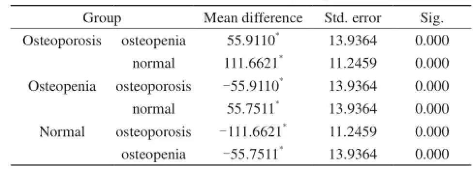
表2腰椎不同骨量组间组内椎体信号强度比较Tab.2Compare the intraclass vertebral signal intensity in different bone mass groups
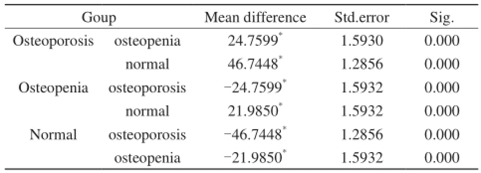
表3腰椎不同骨量组间组内椎体脂肪分数的比较Tab.3Compare the intraclass vertebral fat fraction in different bone mass groups

图5T2脂相信号强度与DXA的线性关系Fig. 5The linear relationship between the lipid phase signal intensity and DXA.
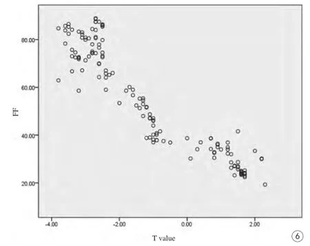
图6椎体脂肪分数与DXA线性关系Fig. 6The linear relationship between fat fraction of vertebral body and DXA.
Youn等[4]使用MRI水-脂分离成像技术对椎体骨髓FF与椎体骨密度的关系进行研究,结果表明椎体骨量减少组的椎体脂肪含量和正常组比较无统计学意义,其结果与本组研究不一致,可能与DXA测量正位腰椎骨密度时存在一定误差有关[27-28]。
本研究的局限性是无病理学的支持,病例数量较少并且缺乏长期随访研究,研究的临界值等需要在更大的研究群体中和外部检验证实。DXA作为参照标准,其特异性较低;3.0 T MR水-脂分离成像可以在腰椎常规检查的基础上无创性测定椎体骨髓的脂肪含量,具有较高的敏感度,椎体信号强度及FF与骨密度值有明显的相关性,通过测量椎体脂相信号强度及计算FF可以作为评价骨质疏松症的有效方法之一。
[References]
[1] Piao JH, Pang LP, Liu ZH, et al. Chinese population status and diagnostic criteria and incidence of primary osteoporosis. Chin J Osteoporos, 2002, 8(1): 1-7.朴俊红, 庞莲萍, 刘忠厚, 等. 中国人口状况及原发性骨质疏松症诊断标准和发生率. 中国骨质疏松杂志, 2002, 8(1): 1-7.
[2] Chinese Society of Osteoporosis and Bone Mineral Research. Primary osteoporosis diagnosis and treatment guidelines (2011). Chinese Journal of Osteoporosis and Bone Mineral Research, 2011, 4(1): 2-17.中华医学会骨质疏松和骨矿盐疾病分会. 原发性骨质疏松症诊治指南(2011年). 中华骨质疏松和骨矿盐疾病杂志, 2011, 4(1): 2-17.
[3] Griffith JF, Yeung DK, Antonio GE, et al. Vertebral bone mineral density, marrow perfusion, and fat content in healthy men and men with osteoporosis: dynamic contrast -enhanced MR imaging and MR spectroscopy. Radiology, 2005, 236(3): 945-951.
[4] Youn Y, Lee HY, Kim JK, et al. Correlation between vertebral marrow fat fraction measured using Dixon quantitative chemical shift MRI and BMD value on dual-energy X-ray absorptiometry. Korean Soc Magn Reson Med, 2012, 16(1): 16-24.
[5] Yang NJ, Song B, Tang HH, et al. In vivo semiquantitative evaluation of liver fat content in alcoholic and nonalcoholic fatty liver rat models: comparison between dual-echo T1-weighted imaging and1H-MR spectroscopy with correlation of histopathology. Chin J Magn Reson Imaging, 2010, 1(3): 208-213.阳宁静, 宋彬, 唐鹤菡, 等.1H-MRS和MR双回波技术活体半定量评价酒精性与非酒精性脂肪肝大鼠模型. 磁共振成像, 2010, 1(3): 208-213.
[6] Hu HH, Kim HW, Nayak KS, et al. Comparison of fat-water MRI and single-voxel MRS in the assessment of hepatic and pancreatic fat fractions in humans. Obesity (Silver spring Md), 2010, 18(4): 841.
[7] Roca M, Mota J, Alfonso P, et al. S-MRI score: A simple method for assessing bone marrow involvement in Gaucher disease. Eur J Radiol, 2007, 62(1): 132.
[8] Lin CL, Jiang GH, Chen CZ, et al. Study on the rapid quantification of nonalcoholic fatty liver fat content based on the mDixon method. Journal of Zhongshan University, 2015, 5(3): 465-471.林楚岚, 江桂华, 陈楚庄, 等. 基于mDixon方法快速量化非酒精性脂肪肝脂肪含量的研究. 中山大学学报, 2015, 5(3): 465-471.
[9] Qin Y, Shang JY, Tang CZ, et al. DXA instrument for the measurement of bone mineral density quality control and effect evaluation. Chinese Journal of Osteoporosis and Bone Mineral Research, 2014, 7(1): 55-61.秦莹, 尚家芸, 唐成志, 等. DXA 仪测量骨密度的质量控制及效果评价. 中华骨质疏松和骨矿盐疾病杂志, 2014, 7(1): 55-61.
[10] Guglielmi G, Van Kuijk C, Li J, et al. Influence of anthropometric parameters and bone size on bone mineral density using volumetric quantitative computed tomographyand dual X-ray absorptiometry at the hip. Acta Radiologica, 2006, 47(6): 574-580.
[11] Kong LY, Ma YM, Wang QQ, et al. Quantitative CT measurement of hip bone mineral density of repeatability and DXA measurement consistency. Chinese Journal of Osteoporosis and Bone Mineral Research, 2013, 6(4): 334-339.孔令懿, 马毅民, 王倩倩, 等. 定量CT测量髋关节骨密度的重复性与DXA 测量的一致性. 中华骨质疏松和骨矿盐疾病杂志, 2013, 6(4): 334-339.
[12] Liu Z, Cai YZ. Application of proton magnetic resonance spectroscopy in the diagnosis of osteoporosis. Chin J Osteoporos, 2014, 20(12): 1417-1420.刘智, 蔡跃增. 氢质子磁共振波谱在骨质疏松诊断中的应用价值.中国骨质疏松杂志, 2014, 20(12): 1417-1420.
[13] Lei LC, He L, Liu Z, et al. Diagnostic value of magnetic resonance chemical shift imaging in the diagnosis of osteoporosis. J Chin Clin Med Imaging, 2014, 25(9): 648-651.雷立存, 何丽, 刘斋, 等. 磁共振化学位移成像对骨质疏松的诊断价值. 中国临床医学影像杂志, 2014, 25(9): 648-651.
[14] Lei LC, Ren QY. Magnetic resonance chemical shift imaging evaluation of vertebral bone marrow fat content in the application of imaging. Diagnosis and interventional radiology, 2015, 2(2): 142-146.雷立存, 任庆云. 磁共振化学位移成像评估椎体骨髓脂肪含量的应用. 影像诊断与介入放射学, 2015, 2(2): 142-146.
[15] Xu Z, Li GW, Gu H, et al. Evaluation of marrow adiposity using twopoint Dixon method in a rat osteoporosis model: comparison withhistopathology. Chin J Osteoporos, 2015, 21(4): 399-402.徐铮, 李冠武, 顾昊, 等. 2点Dixon 技术评价鼠骨质疏松模型骨髓脂肪:与病理对照. 中国骨质疏松杂志, 2015, 21(4): 399-402.
[16] Li GW, Chang SX, Tian F, et al. Bone marrow fat content prediction integrated traditional Chinese and Western medicine for osteoporosis vertebral fracture preliminary application. Combined with the professional committee of the society of medical image. The thirteenth session of the Chinese and Western medicine combined with image Academic Symposium on national traditional Chinese and Western medicine combined with image research learning class, Fujian Province, the eighth of traditional Chinese medicine and Western medicine combined with video conference proceedings. Fuzhou, 2014.李冠武, 常时新, 田芳, 等. 骨髓脂肪含量预测骨质疏松性椎体骨折价值的初步应用. 中国中西医结合学会医学影像专业委员会.全国第十三次中西医结合影像学术研讨会全国中西医结合影像学研究进展学习班福建省第八次中西医结合影像学术研讨会论文汇编. 福州, 2014.
[17] Justesen J, Stenderup K, Ebbesen EN, et al. Adipocyte tissue volume in bone marrow is increased with aging and in patients with osteoporosis. Biogerontology, 2001, 2(3): 165-171.
[18] Meunier P, Aaron J, Edouard C, et al. Osteoporosis and the replacement of cell populations of the marrow by adipose tissue, A quantitative study of 84 iliac bone biopsies. Clin Orthop Relat Res, 1971, 80(2): 147-154.
[19] Zhang WC, Chen ZX, Wang HP, et al. Study on the osteogenic differentiation of rat bone marrow stromal cells in vitro. Chin J Osteoporos, 2005, 11(1): 45-48.张维成, 陈志信, 王和平, 等. 大鼠骨髓基质细胞体外成脂、成骨分化的研究. 中国骨质疏松杂志, 2005, 11(1): 45-48.
[20] Naverias O, Nardi V, Wenzed PL, et al. Bone-marrow adipocytes as negative regulators of the haematopoietic microenviroment. Nature, 2009, 460(7252): 259-263.
[21] Hajek PC, Baker LL, Goobar JE, et al. Focal fat deposition in axial bone marrow, MR characteristics. Radiology, 1987, 162(1): 245-249.
[22] Ojanen X, Borra RJ, Havu M, et al. Comparison of vertebral bone marrow fat assessed by 1H MRS and inphase and out-of phase MRI among family members. Osteoporos Int, 2014, 25(1): 653-662.
[23] Guo QY, Shi Y. Applications and advances of magnetic resonance imaging in diagnosing liver disease. Chin J Magn Reson Imaging, 2014, 5(S1): 10-14.郭启勇, 石喻. 磁共振成像在肝病的应用及进展. 磁共振成像, 2014, 5(S1): 10-14.
[24] Li J, Yin DQ, Zhu L, et al. Analysis of the bone mineral density measurement in normal human lumbar spine. Chin J Osteoporos, 2002, 8(2): 122-121.李瑾, 尹大庆, 朱玲, 等. 正常人腰椎正位骨密度测量结果分析. 中国骨质疏松杂志, 2002, 8(2): 121-122.
[25] Xiao YY, Zhang JS, Hua BX, et al. Experimental study on prediction of vertebral compression fracture by quantitative CT bone mineral density measurement. Chin J Med Imaging Technol, 2002, 18(7): 625-627.肖越勇, 张金山, 华伯勋, 等. 定量CT 骨密度测量预测椎体压缩骨折的实验研究. 中国医学影像技术, 2002, 18(7): 625-627.
[26] Chen Y, Zhou L, Li GW, et al. MRI fat water imaging in the evaluation of primary osteoporosis model of rat bone marrow fat content value. J Clin Radiol, 2014, 33 (10): 1604-1608.陈瑶, 周蕾, 李冠武, 等. MRI水-脂成像评价原发性骨质疏松模型鼠骨髓脂肪含量的价值. 临床放射学杂志, 2014, 33(10): 1604-1608.
[27] Yu W, Qin MW, Zhang Y, et al. Effects of lumbar degenerative osteoarthritis on bone mineral density measurement. Zhonghua Fang She Xue Za Zhi, 2002, 36(3): 245-248.余卫, 秦明伟, 张燕, 等. 腰椎退行性骨关节病对骨密度测定的影响. 中华放射学杂志, 2002, 36(3): 245-248.
[28] Schneider DL, Bettencourt R, Barrett-Connor E. The clinical utility of spine bone density in elderly women. J Clin Densitom, 2006, 9(3): 255-260.
Measurement of fat content in vertebral body by magnetic resonance water fat separation
CHANG Fei-xia1,2, HUANG Gang3*, FAN Dun-hui2, XU Xiang-jiu3, MA Xiao-mei3, WANG Ping3, SHEN Wen-yi1
1Gansu University of Traditional Chinese Medicine, Lanzhou 730000, China
2Dunhuang Hospital of Gansu province, Dunhuang 736200, China
3Department of Radiology, Gansu Provincial People’s Hospital, Lanzhou 730000, China
Objective:Taking DXA as a reference standard, the use of magnetic resonance (NMR) water and fat separation imaging technology (Dixon) can measure the fat signal intensity and calculate the fat fraction (FF) in order to evaluate the application value of vertebral bone mineral density.Materials and Methods:Thirtysix cases of lumbar vertebral body(L1-L4) (144 vertebral body) in which the patients checked in Gansu Provincial People's Hospital were retrospectively collected. All patients who were performed DXA and MR examination of lumbar spine were divided into normal group(T≥-1.0 SD), osteopenia group (-1.0--2.5SD) and osteoporosis group (≤-2.5SD), according to DXA results and the diagnostic criteria of WHO; At the same time, we measured and calculated the waist 1-4 of the vertebral body signal intensity (T2 fat alike) and fat fraction (FF), Marrow fat formula of FF MRI: FF=[Mfat/ (Mfat+Mwater)]×100%(Mwater, Mfatrespectively refers to water and lipid like ROI total pixel signal strength value) to evaluate the magnetic resonance (NMR) water and fat separation imaging techniques to predict the ability of osteoporosis.Results:According to DXA results, in a total of 144 vertebral bodies involved 36 patients, 58 subjects were in normal group, 28 subjects were in osteopenia group and 58 subjects were inosteoporosis group. The Fat signal intensity of the three groups (normal group, osteopenia group and osteoporosis group) were 100.2±20.1, 156.1±56.3, 211.9±84.6, and the fat fraction (FF) were (31.1±6.2)%, (53.3±7.6)%, (77.8±7.2)%, respectively. Among osteoporosis group and osteopenia group and normal group, lumbar fat T2 signal intensity and fat fractions were statistically different (P<0.01). When the fat signal intensity negatively correlated with DXA, the correlation coefficient was r=-0.64, P<0.01; when the fat fraction (FF) negatively correlated with DXA, the correlation coefficient was r=-0.93, P<0.01.Conclusion:Lumbar signal strength and fat fraction through magnetic resonance (NMR) water and fat separation imaging by ROI can reflect the change of the vertebral body fat content in patients with osteoporosis, and is of high value in clinical application. With T value of DXA as the standard, the possibility of having BMD screening for osteoporosis correlation during Dixon check is comparatively high. As a result, it has a certain application prospect to evaluate the bone mineral density of the lumbar spine by simply measuring the fat signal intensity and calculating the fat fraction of ROI.
Magnetic resonance imaging; Water-fat separation technology; Vertebral; Fat fraction; Osteoporosis
Huang G, E-mail: keen0999@163.com
Received 8 Aug 2016, Accepted 20 Sep 2016
1.甘肃中医药大学,兰州 730000
2.甘肃省敦煌市医院,敦煌 736200
3.甘肃省人民医院放射科,兰州 730000
黄刚,E-mail:keen0999@163.com
2016-08-08
接受日期:2016-09-20
R445.2;R589.5
A
10.12015/issn.1674-8034.2016.12.003
常飞霞, 黄刚, 樊敦徽, 等. 磁共振水-脂分离成像技术对椎体脂肪含量的测量.磁共振成像, 2016, 7(12): 902-908.*

