基于磁共振扩散加权成像直肠癌ADC值与其分化程度及神经脉管侵犯相关性研究
吕茜婷,陈勇,李珊玫,葸燕燕,朱凯,马文东,陈宇欣,高知玲*
基于磁共振扩散加权成像直肠癌ADC值与其分化程度及神经脉管侵犯相关性研究
吕茜婷1,陈勇2,李珊玫1,葸燕燕2,朱凯2,马文东2,陈宇欣3,高知玲2*
目的探讨磁共振弥散加权成像中表观扩散系数(apparent diffusion coefficient,ADC)、指数化表观扩散系数(exponential apparent diffusion coefficient,eADC)与直肠癌分化程度及神经脉管侵犯的相关性。材料与方法32例直肠癌患者进行术前常规序列及b值为50、500、1000 s/mm2的MR扩散加权序列检查,测定不同b值下瘤体、正常肠壁的ADC、eADC值并比较其差异。同时比较同一分化程度肿瘤不同b值间ADC、eADC值的差异以及同一b值不同分化程度、神经脉管侵犯是否阳性的肿瘤间ADC、eADC值的差异及相关性。结果直肠癌在不同b值的弥散加权成像(diffusion weighted imaging,DWI)图像上,均表现为稍高或高信号,b值为50、500、1000 s/mm2的瘤体平均ADC值分别为(2.732±0.805)× 10-3mm2/s、(1.226±0.195)×10-3mm2/s、(1.042±0.228)×10-3mm2/s,瘤体eADC值分别为(0.097±0.058) mm2/s、(0.304±0.055) mm2/s、(0.363±0.070) mm2/s,随着b值升高,组织ADC值降低,eADC值则升高。不同b值条件下,瘤体ADC值随着肿瘤分化程度增高而增高,eADC值则相反,b值为500、1000 s/mm2的序列中,差异有统计学意义(b=500 s/mm2时,P=0.03,F=4.123;b=1000 s/mm2时,P=0.001,F=10.797);除b=1000 s/mm2中神经侵犯阳性组平均ADC值较阴性平均ADC值高外,其余组神经脉管侵犯阳性结果组平均ADC值均较阴性组ADC值低。进行不同b值下瘤区ADC值与其分化程度Spearson相关性分析,当b值为1000 s/mm2时,病变ADC值与分化程度呈正相关(rs=0.742,P<0.05),而与eADC值呈负相关(rs=-0.630,P<0.05);b值为1000 s/mm2的病灶的ADC值诊断直肠腺癌效能较佳。结论磁共振弥散加权成像ADC值及eADC值在一定程度上能够反映肿瘤的分化程度及脉管癌栓受侵情况,ADC值可作为早期检测、直肠癌放化疗疗效及预后评价的重要影像学指标。
直肠肿瘤;弥散磁共振成像;磁共振成像;表观扩散系数;脉管侵犯
在美国,结直肠癌是第三大最常见的恶性肿瘤,在男性和女性恶性肿瘤死亡率中位于第3位,且约1/3的结直肠癌起源于直肠[1]。在我国,随着人们生活水平的日益提高,直肠癌的发病率不断上升。直肠解剖位置相对较固定,MRI具有无放射损伤、多方位成像和多参数成像等优势,使得MRI已成为诊断直肠癌并进行分期的重要检查手段。直肠癌最佳治疗方案的制定主要依赖于术前评估,因此术前影像学评估更为重要。磁共振弥散加权成像(diffusion weighted imaging,DWI)是根据体内水分子的随机运动,来反映组织功能状态的一种无创性功能成像技术,可通过测量水分子的弥散效应来反映组织的特性[2]。DWI序列逐步列入磁共振扫描的常规序列,因为它不仅能够提高肿瘤的检出率,同时也能够监测病变的治疗效果[3-5]。本研究采用3.0 T MR扫描仪和体部相控阵线圈对直肠癌患者行弥散加权成像,分析直肠癌瘤体表观扩散系数(apparent diffusion coefficient,ADC)值、指数化表观扩散系数(exponential apparent diffusion coefficient,eADC)值与其组织分化程度及神经脉管侵犯的关系。
1 材料与方法
1.1 病例资料
本研究收集宁夏医科大学总医院2015年1月至2016年6月收治的32例直肠癌患者,其中男23例,女9例,平均年龄64.08岁(35~79岁),未曾接受放、化疗。所有病例均在术前1 w内行直肠常规MRI扫描及DWI扫描,b值分别选择50、500、1000 s/mm2。检查前1晚行清洁肠道准备,对无禁忌证的患者,检查前10 min常规肌注消旋山莨菪碱(654-2) 20 mg,检查前及时经肛门挤入开塞露40 ml。本研究所有受检者均自愿接受盆腔MRI检查并签署知情同意书。本人遵循的程序符合负责人体试验的伦理委员会所制定的伦理学标准,并通过伦理委员会的批准。
1.2 MR扫描设备及扫描方法
采用美国GE公司HD MR 3.0 T超导磁共振机,8通道相控阵体部线圈。扩散加权序列参数:轴位TR/TE,4000 ms/62.3~75.6 ms,FOV 27 cm×27 cm,矩阵320×224,层厚/层距4 mm/ 0 mm,NEX=6,b分别为50、500及1000 s/mm2的DWI 扫描序列。TR=10300 ms,TE=76 ms,FOV:36 cm×36 cm,层厚3 mm,矩阵:128×128,带宽:1502 Hz/pixel,b=50、500、1000 s/mm2,采集次数4次,行全盆腔扫描,包括乙状结肠至肛门,自由呼吸完成扫描。
1.3 DWI数据及病理结果的采集与分析
利用GE 3.0 T MR 机所配备工作站中的GE AW 4.4Functool软件进行ADC值及eADC值的测量,由两名放射科医师共同判断DWI 图像可否用于诊断及ADC值、eADC值的计算,并对不同b值的DWI 图像进行对比、分析。在肿瘤病灶最大层面选择面积约为50 mm2的圆形或椭圆形感兴趣区(regions of interest,ROI)测量ADC 值及eADC值,避开坏死囊变区,测量3次,取其平均值。同样方法测量正常肠壁的ADC值及eADC值。记录瘤体病理结果,有无脉管癌栓(有脉管癌栓为阳性,无为阴性),有无神经侵犯(有神经侵犯为阳性,无为阴性)。
1.4 统计学处理

2 结果
2.1 直肠癌的MRI征像
直肠癌病灶在MRI常规序列上均表现为肠壁局限性均匀或不均匀增厚,呈溃疡型,或向肠腔内或肠腔外突起形成不规则肿块;肿瘤在T1WI上表现为等或稍低信号,信号均匀;在T2WI上表现为均匀或不均匀的低或等信号(图1),当肿瘤内发生坏死时可表现为高信号。
本组直肠癌病例在DWI上信号随着b值的增加逐渐增高,瘤体信号在b=50 s/mm2的DWI中呈等或稍高信号,b值为1000 s/mm2时瘤体信号最高,而图形信噪比(signal noise ratio,SNR)却随着b值增加而减小(图2A、2C、2E)。与T2WI相比,DWI可较直观地显示直肠癌病灶,且容易分辨直肠癌瘤体、正常肠壁及肠内容物。直肠癌ADC图、eADC图均表现为红-黄-绿-蓝逐渐过度的伪彩图,其中红色、黄色区域定义为高ADC值及eADC值区,蓝色区域表现为低ADC值及eADC值,瘤体在ADC图上以蓝绿色为主,在eADC图上以黄绿色为主(图2B、2D、2F)。
2.2 正常肠壁及瘤体ADC值
本组b值=50、500、1000 s/mm2时,正常肠壁与瘤体平均ADC值及正常肠壁与瘤体平均eADC值组间差异有统计学意义;不同b值下正常肠壁ADC值较瘤体高,正常肠壁eADC值较瘤体低。随着b值的增加,ADC值逐渐减小,而eADC值则逐渐增大,两组间差异有统计学意义(表1)。
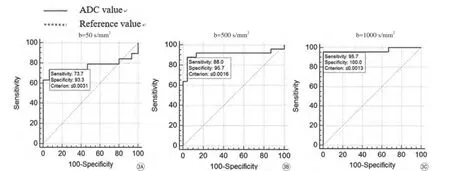
以ADC 值作为检验变量,以不同b值作为状态变量,绘制瘤体ADC 值受试者工作特征曲线(receiver operating characteristic curve,ROC曲线),计算ROC曲线下面积,b值为50、500、1000 s/mm2时曲线下面积分别为0.772、0.910和0.957,可见选取b值为1000 s/mm2时曲线下面积最大,即诊断效能最高,当ADC值为1.075×10-3mm2/s时,其诊断直肠腺癌的敏感度为100%,特异度为92%(图3)。
2.3 不同b值不同分化程度组间ADC 值及eADC值比较
本组除b值=50 s/mm2时,3个分化级别组间ADC值差异无统计学意义外,b=500、1000 s/mm2时3个分化级别组间ADC值差异均有统计学意义(P<0.05);当b=1000 s/mm2时3个分化级别组间eADC值差异有统计学意义(P<0.05),其余当b值=50、500 s/mm2时,不同分化级别组间差异无统计学意义;随着分化程度增高,瘤体ADC值逐渐增大,而eADC值则逐渐减小(表2)。本组数据显示各b值组与肿瘤分化相关性中,肿瘤ADC值与分化程度呈正相关,肿瘤eADC值与分化程度呈负相关(表3)。
2.4 ADC值与直肠腺癌神经脉管侵犯相关性
在b=50、500、1000 s/mm2的DWI序列中神经脉管侵犯阳性与阴性瘤体平均ADC值及两者的相关性如表4所示,除b=1000 s/mm2中神经侵犯阳性平均ADC值较阴性平均ADC值高外,其余组神经脉管侵犯阳性结果组平均ADC值均较阴性组ADC值低,b=50 s/mm2的神经侵犯组与b=500 s/mm2的脉管癌栓组差异有统计学意义(表4)。
3 讨论
DWI是一种无创性MR功能成像技术,已经广泛应用于各个部位的MRI检查中,并对于脑梗死、肿瘤的良恶性判断等有很高的价值。其通过检测活体组织内水分子扩散运动来反映组织内部结构变化及病理生理状态,如细胞膜完整性、细胞密度等[6],在正常组织中,水分子做布朗运动,水分子运动呈各项同性,在DWI序列上呈低信号,在肿瘤组织中,水分子运动呈各向异性,在DWI序列上呈高信号[7]。通过定量测量ADC值,定量反映组织器官结构和功能的改变。

表1瘤体与正常肠壁在不同b值下ADC值、eADC值比较Tab.1Compare the ADC values and eADC values of different b values of the rectal wall and rectal tumor

表2不同分化肿瘤组在不同b值下ADC、eADC值比较Tab.2Compare the ADC value and eADC value of differential differentiation of rectal tumor in diffrernt b value of MR diffusion weighted imaging

表3ADC、eADC值与直肠癌分化程度之间的相关性Tab.3The correlation of the ADC value, eADC value of the tumors and pathological classification of each tumor

表4神经脉管侵犯与瘤体ADC值相关性Tab.4The correlation of the ADC value of the tumors and perineural invasion, peritumor-intravascular cancer emboli of each tumor
瘤体在DWI序列上均呈稍高或高信号,不同b值及不同分化程度组间瘤体平均ADC值、eADC值均有差异。本研究的初步结果表明,在同一b值下除低b值组瘤体ADC值各分化程度组间差异无统计学意义外,其余中、高b值组瘤体ADC值组间差异有统计学意义(P<0.05)。这可能是由于肿瘤细胞增殖速度较正常细胞快,细胞密度较正常组织细胞高,水分子在瘤体组织内扩散受限,并且随着肿瘤组织分化程度降低,水分子在瘤体组织内扩散受限越明显,瘤体ADC值较分化程度好的瘤体ADC值低。本研究结果显示,随着b值增大,图像扩散权重相应增加,病变与正常组织间的对比度增加,可以提高DWI检测瘤体的敏感度,使ADC值测量更准确。当b值为1000 s/mm2时ROC曲线下面积大于b值等于50、500 s/mm2的DWI序列,说明b=1000 s/mm2时诊断效能最高。在不同b值下,各分化程度组间瘤体ADC值差异均有统计学意义(P<0.05),这与高永伟等[8]、亓俊霞等[9]研究结果基本一致。而同一b值下低b值组瘤体ADC值各分化程度组间差异无统计学意义,分析其原因可能有:(1)本组高、低分化直肠癌的病例数较少,影响统计结果;(2)恶性肿瘤不同分化程度的判定存在一定重叠,例如本组将中-低分化腺癌、中-高分化腺癌均归类为中分化腺癌;(3)测量瘤体ADC值划定ROI时,测量3次范围均约为50 mm2,后取平均值,并不是沿肿瘤边缘测量瘤体ADC值。

图1男,50岁,直肠高分化腺癌,T3a期。A:轴位T2WI序列显示瘤体低信号(白箭),病变突破直肠固有肌层;B:轴位T1增强序列显示瘤体明显强化,瘤体外缘呈棘突样改变(白箭)图2与图1为同一患者。A:b=50 s/mm2的DWI序列,显示瘤体为稍高信号(白箭),直肠固有肌层不连续。C:b=500 s/mm2的DWI序列,显示瘤体为高信号,瘤体信号较2A DWI序列中信号高。E:b=1000 s/mm2的DWI序列,显示瘤体为高信号(白箭),瘤体信号在A、C、E中最高,但信噪比最低,随着b值得增高,直肠系膜内淋巴结信号逐渐增高(白箭头)。B、D、F:伪彩图中瘤体呈黄色为高ADC值区,绿色区域为低ADC值区,随着b值得增大,瘤体ADC值逐渐减小Fig. 1Male, 50-year-old, well-differentiated adenocarcinoma of rectum, T3a stage. A: The image of AXI-T2WI images of the rectum, the rectum was fully expanded. The lesion displayed low signal (white arrow); B: The image of AXI-T1WI contrast- enhanced images of the rectum, which showed that lesion was intense enhancement, and on the outer edge of the lesion as spina (white arrow).Fig. 2The same patient with Fig.1. A: MR diffusion weighted imaging (DWI) with b=50 s/mm2, The annular thread-like low signal of the muscular layer was not continuous. The lesion displayed slightly high signal (white arrow) . C: MR diffusion weighted imaging (DWI) with b=500 s/mm2. The signal of lesion was higher than DWI imaging with b=50 s/mm2(white arrow). E: MR diffusion weighted imaging (DWI) with b=1000 s/mm2. The signal of lesion was the highest of the three images (A, C, E) ( white arrow), but the signal to nosie ratio was the worst. The signal of lymphaden in mesorectum increased following the ascending of b values (white arrow head). B, D, F: The ADC pseudo color maps. The yellow part of the ADC pseudo color map showed high ADC value, and the green showed the low ADC value. The mean ADC values of tumor decreased following the ascending of b values.

图3A~C分别为 b值为50、500、1000 s/mm2时ADC值检测直肠腺癌的ROC曲线。设正常肠壁ADC值为参考值。随着b值的增高,曲线下面积逐渐增大,当b值=1000 s/mm2时,曲线下面积最大,即诊断效能最高Fig. 3A—C: b value is 50, 500, 1000 s/mm2from left to right, respectively, are ROC curves of ADC value diagnosing rectal tumor of different b value. The ADC value of rectal wall was formed reference value, the area under the ROC curves increase following the ascending of b values. With b value of 1000 s/mm2there was the best diagnostic efficiency in rectal carcinoma.
DWI作为一种应用广泛的功能成像序列,弥补了T2WI单纯从形态学的观察来诊断直肠癌的不足,对直肠癌的诊断具有重要价值[10]。DWI序列不仅能够更准确的发现、定位瘤体,对直肠周围脂肪内及髂血管周围的转移淋巴结的显示亦优于T2WI。由于直肠周围脂肪内及髂血管周围的转移淋巴结在T2WI上呈低信号,与血管断面相似,易被漏诊。而DWI上血管等正常组织信号被抑制,转移的淋巴结呈明显高信号,较易被检出。因此,对于那些无明显肠周浸润而已经有淋巴结转移的直肠癌患者,DWI可更准确地判断淋巴结的转移情况,进一步指导外科医生制定手术范围。与单纯应用T2WI诊断直肠癌相比,结合DWI后不仅提高了直肠癌诊断的准确性,同时对病变周围软组织是否受侵及可疑淋巴结转移做出全方位判断。随着新辅助治疗的应用,对于周围器官受侵犯、T3分期以上的患者可先选择辅助放化疗,待病灶体积缩小或降期后再行手术治疗。多篇文献报道DWI序列中ADC值在直肠癌放化疗疗效评价中有潜在的应用价值,可作为有效的时间监测点[11-12]。作为一种无创性的检查,在磁共振扩散加权成像中,对患者术前瘤体与辅助治疗后瘤体ADC值进行比较分析,可帮助临床医生对患者的术前疗效做出评估,对预后做出判断。
直肠癌瘤体各项病理免疫组化指标,对临床后续治疗方案制定有很大帮助,也成为影响患者预后的独立指标。由于肿瘤细胞的生长方式和术中操作的原因,癌栓从原发病灶脱落进入脉管很可能导致转移。一般认为,与同一分期患者相比,神经侵犯及脉管癌栓阳性较结果阴性的患者,术后复发率可能更高,预后更差,杜长征等[13]研究发现,直肠癌根治术后,有22%的患者具有脉管浸润,其5年生存率明显下降。本研究也将瘤体ADC值与神经脉管侵犯做了相关性研究,本组神经脉管侵犯阳性组ADC值几乎均较阴性组低,这与其瘤体本身恶性程度较高,预后较差结果一致,并非每组结果差异均有统计学意义的原因可能为:(1)本组病例数较少,影响数据结果;(2)b=50 s/mm2的DWI图像中信噪比较高,但瘤体与正常肠壁差异较小,测量瘤体ADC值较易出现误差。本组病例脉管癌栓,神经侵犯阳性患者术后均积极行辅助放化疗,以期提高患者的生存率。
对于直肠癌患者,DWI序列为无创性操作,不但扫描时间短,且后处理简单,应作为直肠癌检出、治疗前后监测疗效及转归的必要序列。借助DWI序列,通过测量ADC值、eADC值还可取得肿瘤组织病变扩散的量化指标,可为直肠癌患者治疗方案的选择及预后评价提供有利的依据。
[References]
[1] Siegel R, Deepa MS, Dvm AJ. Cancer statistics, 2013. Ca A Cancer Journal for Clinicians, 2013, 63(1): 11-30.
[2] Kang YJ, Zhang H. The value of magnetic resonance diffusion weighted imaging in rectal cancer diagnosis. Chin J Magn Reson Imaging, 2015, 6(2): 155-160.康英杰, 张皓. 磁共振弥散加权成像在直肠癌检查中的价值. 磁共振成像, 2015, 6(2): 155-160.
[3 ] Koh DM, Padhani AR. Diffusion weighted MRI: a new functional clinical technique for tumor imaging. Br J Radiol, 2006, 79(944): 633-635.
[4] Patterson DM, Padhani AR, Collins DJ. Technology insight: water diffusion MRI-a potential new biomarker of response to cancer therapy. Nat Cli Pract Oncol, 2008, 5(4): 220-233.
[5] Padhani AR, Liu G, Koh DM, et al. Diffusion-weighted magnetic resonance imaging as a cancer biomarker: consensus and recommendations. Neoplasia, 2009, 11(2): 102-125.
[6] Zhang DX, Zhu SC, Guan S, et al. The value of MR intravoxel incoherent motion diffusion weighted imaging in T stage and differentiated degree of rectal adenocarcinoma. Chin J Magn Reson Imaging, 2016, 7(8): 561-566.张单霞, 朱绍成, 管枢, 等.MR体素内不相干运动扩散加权成像对直肠腺癌T分期及分化程度的应用价值研究. 磁共振成像, 2016, 7(8): 561-566.
[7] Chavhan GB, Alsabban Z, Babyn PS. Diffusion-weighted imaging in pediatric body MR imaging: principles, technique, and emerging applications. Radio Graphics, 2014, 34(3): 73-88.
[8] Gao YW, Niu GM, Han XD. Study on the relationship between ADC value and malignant degree of rectal cancer using 3.0 T MR scanner. National Medical Frontiers of China, 2012, 7(6): 7-8.高永伟, 牛广明, 韩晓东. 3.0 T磁共振诊断直肠癌ADC 值与肿瘤分化程度的相关性分析. 中国医疗前沿, 2012, 7(6): 7-8.
[9] Qi JX, Bai RJ, Yu CL, et al. Preliminary study of diffusion weight imaging in detecting and evaluating the prognosis of rectal carcinoma. J Pract Radiol, 2013, 29(3): 400-404.亓俊霞, 白人驹, 于长路, 等.磁共振扩散加权成像对直肠癌的显示及恶性程度评估的初步研究. 实用放射学杂志, 2013, 29(3): 400-404.
[10] Liao XR, Zhang XS, Zhang HX. Correlation between ADC value of MR diffusion weighted imaging and clinicopathological factors in rectal cancer. China Cancer, 2016, 25(1): 76-80.廖雪芮, 张修石, 张红霞. MR 弥散加权成像ADC 值与直肠癌临床病理因素的相关性. 中国肿瘤, 2016, 25(1): 76-80.
[11] Dzik-Jurasz A, Domenig C, George M, et al. Diffusion MRI for prediction of response of rectal cancer to chemoradiation. Lancet, 2002, 360(9329): 307-308.
[12] Sun YS, Tang L, Li J, et al. A comparative study in rectal carcinoma between diffusion weighted MR imaging and histopathologic response with preperative chemoradiotherapy. Chinese Journal of Oncoradiology, 2010, 16(8): 58-63.孙应实, 唐磊, 李洁, 等. 磁共振扩散成像与直肠癌术前放化疗病理反应性的对照研究. 当代医学, 2010, 16(8): 58-63.
[13] Du CZ, Wang XC, Xue WC, et al. Clinical significance of lymphovascular invasion in rectal cancer following neoadjuvant therapy. Chinese Journal of Digestive Surgery, 2010, 9(4): 265-268.杜长征, 王晓春, 薛卫成, 等. 新辅助治疗在直肠癌脉管癌栓中的临床意义. 中华消化外科杂志, 2010, 9(4): 265-268.
Correlation research of the ADC value of MR diffusion weighted imaging in differential differentiation of rectal tumor, perineural invasion and peritumor-intravascular cancer emboli
LV Qian-ting1, CHEN Yong2, LI Shan-mei1, XI Yan-yan2, ZHU Kai2, MA Wen-dong2, CHEN Yu-xin3, GAO Zhi-ling2*
1Ningxia Medical University, Yinchuan 750004, China
2General Hospital of Ningxia Medical University, Yinchuan 750004, China
3Guiyang Medical University, Guiyang 550004, China
ACKNOWLEDGMENTSThis work was part of Science and Technology Key Projects of Ningxia (No. 2016KJHM63).
Objective:To observe the correlation of the apparent diffusion coefficient (ADC) value, exponential apparent diffusion coefficient (eADC) value of MR diffusion weighted imaging (DWI) in differential differentiation of rectal tumor with different b values, and the correlation of ADC value and perineural invasion, peritumorintravascular cancer emboli in tumor.Materials and Methods:Thirty-two patients with operable rectal cancer underwent preoperative MR imaging with single-shot echo plannar imaging (EPI) diffusion-weighted sequences (b=50, 500, 1000 s/mm2). Measure and compare the apparent diffusion coefficient (ADC) value and exponential apparentdiffusion coefficient (eADC) value of MR diffusion weighted imaging (DWI) in rectal wall and differential differentiation of rectal tumor, and the correlation of ADC value and perineural invasion, peritumor-intravascular cancer emboli in tumor. All of them were compared with pathologicstaging.Results:The rectal carcinomas were slightly high signal or high signal on DWI. When b value is 50, 500, 1000 s/mm2, the mean ADC value of tumor is (2.732±0.805)×10-3mm2/s, (1.226±0.195)×10-3mm2/s, (1.042±0.228)× 10-3mm2/s, the mean eADC value of tumor is (0.097±0.058) mm2/s, (0.304±0.055) mm2/s, (0.363±0.070) mm2/s, respectively. With each b value, the mean ADC value of tumor increases following the ascending of differentiated degree of rectal carcinoma, and the mean eADC value decreases. When b value is 500, 1000 s/mm2, the difference is statistically significant (b=500 s/mm2, P= 0.03, F= 4.123; b=1000 s/mm2, P=0.001, F=10.797). In most of group, the mean ADC value of peritumor-intravascular cancer emboli and perineural invasion are lower than the negative, and the difference is statistically significant in b=500 s/mm2of MR diffusion weighted imaging. And analyze the spearson correlation coefficient of differential differentiation of rectal tumor in different b values.The ADC value of tumor and differentiation have positive correlation (rs=0.742, P<0.05), and have negative correlation with eADC values (rs=-0.630, P<0.05) with b value of 1000 s/mm2. ADC values have the better diagnostic efficiency for diagnosis of rectal adenocarcinoma with b value of 1000 s/mm2.Conclusion:The diffusion-weighted imaging is a rapid and feasible method in detecting rectal cancer. The correlation can be pointed out between ADC and pathological classification of each tumor, and the relationship of the ADC value and perineural invasion, peritumor-intravascular cancer emboli in tumor. Therefore, the ADC value can respresent an important imaging biomarker for assessing the response to radiochemotherapy and the prognose of the tumor.
Rectal neoplasms; Diffusion magnetic resonance imaging; Magnetic resonance imaging; Apparent diffusion coefficient; Peritumor-intravascular cancer emboli
Gao ZL, E-mail: gzl6988@163.com
Received 18 Sep 2016, Accepted 20 Nov 2016
宁夏回族自治区重点研发计划(科技惠民)(编号:2016KJHM63)
1.宁夏医科大学,银川 750004
2.宁夏医科大学总医院,银川 750004
3.贵州医科大学,贵阳 550004
高知玲,E-mail:gzl6988@163.com
2016-09-18
接受日期:2016-11-20
R445.2;R735.37
A
10.12015/issn.1674-8034.2016.12.005
吕茜婷, 陈勇, 李珊玫, 等. 基于磁共振扩散加权成像直肠癌ADC值与其分化程度及神经脉管侵犯相关性研究. 磁共振成像, 2016, 7(12): 915-920.*

