多参数MRI前列腺影像报告和数据系统评分与经直肠超声引导下穿刺病理的相关性分析
李拔森,王良*,邓明,闵祥德,蔡杰,冯朝燕,可赞,王国平
多参数MRI前列腺影像报告和数据系统评分与经直肠超声引导下穿刺病理的相关性分析
李拔森1,王良1*,邓明1,闵祥德1,蔡杰1,冯朝燕1,可赞1,王国平2

目的旨在探讨多参数MRI (multi-parametric MRI, Mp-MRI)前列腺影像报告和数据系统(prostate imaging reporting and data system version 2, PI-RADS V2)评分与经直肠超声引导下穿刺病理的相关性。材料与方法回顾性分析经病理证实的128例前列腺病变患者的MRI资料,其中前列腺癌75例,良性前列腺增生48例、前列腺炎5例,所有患者均行3.0 T MRI扫描,获取完整的T2WI、DWI及DCE图像;由2名前列腺诊断医师在不知患者临床资料及病理的情况下采用PIRADS V2评分标准进行评分,评分结果分别记录;所有患者均行经直肠超声引导下病理穿刺,并由泌尿专业病理诊断医师进行诊断,对前列腺癌则进行Gleason评分。采用Spearman相关分析PI-RADS V2评分与穿刺病理的相关系数,并采用ROC曲线分析PI-RADS V2评分诊断前列腺癌的敏感性、特异性和准确性。结果PI-RADS V2评分与穿刺病理呈正相关,r=0.887。PI-RADS V2评分诊断前列腺癌的ROC曲线下面积0.975,其敏感性为93.33%,特异性为96.23%,准确性为94.51%,阳性预测值97.22%,阴性预测值91.07%。Gleason评分≥8分的前列腺癌的PI-RADS V2评分为5分。结论PI-RADS V2评分与经直肠超声引导下穿刺病理的相关性高,PI-RADS V2评分对前列腺疾病的诊断准确性高。
前列腺肿瘤;磁共振成像;病理学
Received 28 Jan 2016, Accepted 26 Feb 2016
ACKNOWLEDGMENTSThis study was supported by Grants the National Natural Science Foundation of China (No. 81171307).
近年来,由于多参数MRI (Mp-MRI)兼备常规T2WI和功能成像,在前列腺癌、前列腺增生及前列腺炎的鉴别诊断中优势明显[1]。实际工作中,由于不同影像医师的诊断水平和阅片习惯的差异,且在前列腺报告的书写中使用的较为模糊的描述词语也对前列腺疾病的诊断产生一定影响。加之我国的就诊文化、医疗报销制度和医疗环境的变化等综合因素的影响,针对绝大部分中老年患者需要明确鉴别其性质并及时提示其风险,尤其是针对泌尿外科医师与影像科医师的沟通都需要明确的数据报告。2012年欧洲泌尿生殖放射协会推出的前列腺影像报告和数据系统(prostate imaging reporting and data system, PI-RADS)是欧洲前列腺诊断报告书写和定量评估指南[2-3],受到了全球前列腺诊断医师重视,2014年北美放射学年会上公布了第2版PI-RADS,即PI-RADS V2[4-5]。目前,PI-RADS V2的临床适用性还有待于进一步在临床工作中证实,且国内尚无前列腺报告的规范指南作参考,因此笔者试探究PI-RADS V2在我国前列腺癌的诊断与风险评估中的价值,并进一步评估PI-RADS V2与穿刺病理的相关性。
1 材料与方法
1.1 病例资料
回顾性分析2014年1月至2015年6月期间,于我院行前列腺MRI检查的患者。因是回顾性研究,故无需签署知情同意书。纳入标准:(1)前列腺MRI检查序列完整,包括T2WI、DWI及DCE序列;(2)经直肠超声引导下进行了12针病理穿刺活检;(3) MRI检查均在穿刺活检前进行;(4)前列腺MRI检查前未行过内分泌治疗、冷冻及放疗。排除标准:(1)有MRI检查或者病理穿刺活检的禁忌;(2)前列腺MRI图像质量较差或部分序列缺失,无法进行PI-RADS V2评分。
128例患者纳入了本研究,年龄44~86岁,平均(69±5)岁。实验室检查前列腺特异性抗原为0.23~1 000.00 ng/ml,中位值为78.31 ng/ml。诊断均经超声引导下直肠穿刺活检病理证实,标本以10%福尔马林固定,用石蜡包埋切片。常规采用6区12针系统穿刺法,对可疑病变,先在Mp-MRI上按照六分区法确定病变所在区域,后在经直肠超声引导下,对准相应区域进行穿刺,并由操作医师记录活检位置。
1.2 图像采集
前列腺MRI检查采用德国Siemens公司3.0 T Skyra磁共振扫描仪,射频发射线圈采用体线圈,射频接收线圈为腹部相控阵线圈(18通道)。磁共振扫描前保持膀胱适度充盈,取仰卧位,扫描中心为耻骨联合上方2.0 cm处。前列腺局部行轴面、矢状面和冠状面快速自旋回波(turbo spin echo, TSE) T2WI、轴面T1WI、轴面单次激发平面回波成像(single-shot echo-planar imaging, SS-EPI) DWI、轴面磁共振动态增强扫描(dynamic contrast enhanced MRI, DCE-MRI)。扫描序列包括:(1) TSE T2WI扫描参数为:TR 6 750 ms,TE 104 ms,层厚 3 mm,层间距0 mm,FOV 180 mm×l80 mm,激励次数2次,矩阵384×384。(2) DWI SS-EPI序列,扫描参数为:TR 4 500 ms,TE 85 ms,FOV 214 mm×171 mm,矩阵90×90,层厚3 mm,层间距0 mm,回波间隙0.75 ms。扩散敏感系数(b)值分别为100 s/mm2、800 s/mm2、1500 s/mm2。(3) DCE-MRI采用梯度回波容积内插法(volumetric in terpolated breath-hold examination,VIBE)序列TR 3.40 ms,TE 1.23 ms,层厚3 mm,层间距0 mm,FOV 260 mm×260 mm。采用MED TRON高压注射器,对比剂为马根维显(Gd-DTPA),注射速率2 ml/s,剂量0.2 mmol/Kg,注射完毕后用生理盐水20 ml进行冲洗。
1.3 数据采集与PI-RADS V2评分
将MRI扫描所得图像传入图像存储与传输系统(picture archiving and communication system,PACS),由2名分别有5年、10年前列腺MRI工作经验的医师采用双盲法进行T2WI、DWI、DCE的PI-RADS V2评分,并记录评分所在病灶的所在区域。对照PI-RADS V2评分病灶所在区域与穿刺活检所记录病变位置,以保证二者一一对应,评分完毕意见不一致由双方共同协商达成一致意见。根据2014年版PI-RADS V2评分标准[4],对出现有临床意义前列腺癌[6]的可能性进行PI-RADS V2评分。1分:非常低,极不可能存在;2分:低,不可能存在;3分:中等,可疑存在;4分:高,可能存在;5分:非常高,极有可能存在。对于前列腺外周带疾病PI-RADS V2评分以DWI结果为主,而移行带疾病则以T2WI结果为主。例如外周带病变DWI评分为5分,T2WI评分为3分,则PI-RADS V2评分为5分。DCE阳性主要对PI-RADS V2评分为3分的结果有影响,若阳性则相应整体评分加1分,对1、2、4、5分则无影响。PI-RADS V2评分标准见表1,2。
1.4 病理分析
由泌尿专业病理医师观察病理切片并进行报告,报告需报告病变的位置及诊断结果,若为前列腺癌,则需要报告其Gleason分级,若一个区内两针皆为癌灶,但Gleason分级不一致,则取Gleason分级高者作为最终病理结果。
1.5 统计学方法
采用SPSS 19.0及MedCalc Version 11.4.2.0软件包数据处理,分析PI-RADS V2评分与穿刺病理的相关性。分析PI-RADS V2评分对诊断前列腺癌的受试者工作特征曲线(receiver operating characteristic curve, ROC),并计算其敏感度、特异性、准确度、阳性预测值、阴性预测值。

表1 PI-RADS V2在前列腺移行带和外周带病灶中的评分标准Tab. 1 PI-RADS V2 score for Peripheral zone and Transition zone

表2 PI-RADS V2的DCE评分标准Tab. 2 PI-RADS V2 assessment for DCE
2 结果
2.1 PI-RADS V2评分及穿刺病理结果
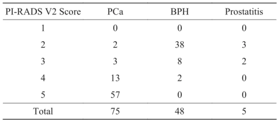
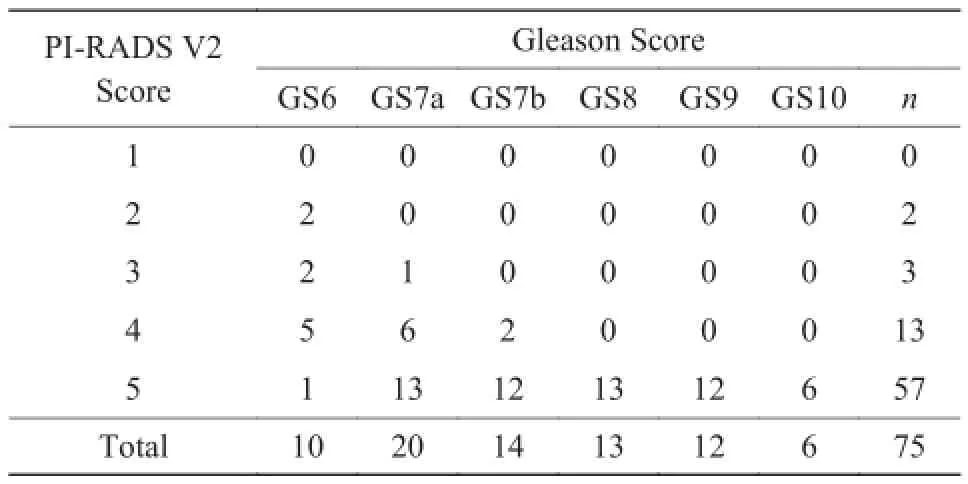
128例患者中前列腺癌75例,前列腺增生48例,前列腺炎5例。75例前列腺癌患者Gleason评分均≥6分,分别为Gleason 6、Gleason 7a (3+4)、Gleason 7b (4+3)、Gleason 8、Gleaso 9、Gleason 10,其PI-RADS V2评分及前列腺癌Gleason评分见表3,4和图1。
2.2 PI-RADS V2评分与穿刺病理的相关性分析
对T2WI+DWI+DCE的整体PI-RADS V2评分与经直肠超声引导下病理穿刺结果进行Spearman相关分析,r=0.887,P<0.01,PI-RADS V2评分与经直肠超声引导下穿刺病理呈明显正相关。
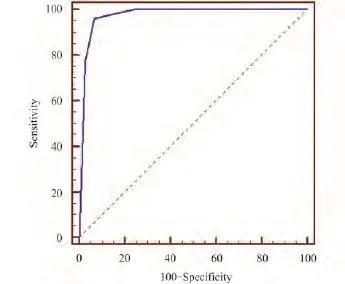
2.3 ROC诊断前列腺癌的敏感性、特异性及准确性(图2)
由ROC曲线算出约登指数,该指数最大值所对应的PI-RADS V2评分3为界值。PI-RADS V2评分诊断前列腺癌的ROC曲线下面积0.975,其敏感性为93.33% (70/75),特异性为96.23% (51/53),准确性为94.51% (71/75),阳性预测值为97.22% (70/72),阴性预测值为91.07% (51/56)。

表3 128例患者的PI-RADS V2整体评分Tab. 3 PI-RADS V2 score of 128 patients

表4 75例前列腺癌患者的PI-RADS V2评分与Gleason分级Tab. 4 PI-RADS V2 score and Gleason score of 75 patients with prostate cancer

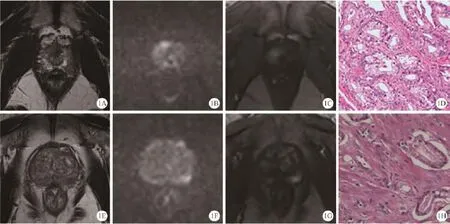
图1 A~D为同一患者,59岁,PSA 25.199 ng/ml。A:轴面T2WI示右侧尖部移行带见边界不清低信号灶,大小约为8 mm×5 mm。B:DWI示该结节状病灶呈明显高信号。C:DCE示病灶早期明显强化。据PI-RADS V2 评分标准,该病灶在T2WI序列评分为4分,DWI序列评分为4分,DCE (+),PI-RADS V2 整体评分为4分。D:穿刺活检病理证实为前列腺移行带癌,Gleason (3+4)(HE ×200)。E~H:为一77岁患者,PSA 11 ng/ml。轴面T2WI示左侧外周带见边界不清低信号灶,大小约为10 mm×5 mm (E)。DWI示病灶扩散受限,呈结节状明显高信号(F)。DCE示病灶早期明显强化(G)。据PIRADS V2 评分标准,该病灶在T2WI序列评分为3分,其轴面最大径<1.5 cm,DWI序列评分为4分,DCE (+),PI-RADS V2 整体评分为4分。穿刺活检病理证实为前列腺外周带癌(H),Gleason (3+3)(HE ×200)Fig. 1 A 59-year-old male patient with PSA 25.199 ng/ml (A—D). A: On axial T2WI there was an ill-defined lower signal 8 mm× 5 mm lesion in the right transition zone. B: The lesion showed focal markedly hyperintense on DWI. C: The lesion showed focal and earlier enhancement. The PI-RADS V2 score of the lesion on T2WI, DWI, T2WI+DWI+DCE, respectively, was 4, 4, 4, DCE (+), according to the PI-RADS V2 criteria. D: This case was confirmed prostate cancer (Gleason (3+4)) by pathology (HE ×200). A 77-year-old male patient with PSA 11 ng/ml (E—H). E: On axial T2WI there was an ill-defined lower signal 10 mm×5 mm lesion in the left peripheral zone. F: The lesion showed focal markedly hyperintense on DWI. G: The lesion showed focal and earlier enhancement. The PI-RADS V2 score of the lesion (axial size<1.5 cm) on T2WI, DWI, T2WI+DWI+DCE, respectively, was 3, 4, 4, DCE (+), according the PI-RADS V2 criteria. H: This case was confirmed prostate cancer [Gleason (3+3)] by pathology (HE ×200).

图2 PI-RADS V2评分对前列腺癌诊断的ROC曲线图Fig. 2 The ROC carve of PI-RADS V2 score in diagnosing prostate cancer.
3 讨论
自PI-RADS提出以来,规范了前列腺MRI报告,对前列腺病变按照5分制标准进行评分,提高了前列腺癌的检出率,其临床有效性和适用性得到了进一步验证[7-10]。PI-RADS V2中将有临床意义的前列腺癌定义为Gleason评分≥7分,伴或不伴体积≥0.5 cm3、包膜外侵犯。依据前列腺T2WI、DWI及DCE序列的综合表现,对出现有临床意义前列腺癌的可能性依然按照5分制评分标准进行。在第一版PI-RADS的基础上进行了补充、完善及删减。PI-RADS V2对MRI检查设备和技术要求提出了指导性建议,对评估分类标准、技术规范及扫描参数进行了重新规范。
本研究显示良性前列腺增生或前列腺炎的PI-RADS V2评分为2分或3分,本研究中8/48例前列腺增生的PI-RADS V2评分为3分,2/48评分为4分,究其原因可能是前列腺增生发生在移行带,其内含有的细胞以及间质成分有所不同而导致在T2WI序列的PI-RADS V2评分可为2~4分,而移行带的PI-RADS V2评分主要以T2WI为主,故造成其特异性减低;PI-RADS V2整体评分为2分或3分的前列腺癌,其对应为低级别,Gleason评分为6或7a;PI-RADS V2整体评分为4分的前列腺癌,其对应级别为≤7b;高级别前列腺癌Gleason评分≥8分的,其PI-RADS V2评分为5分,但PIRADS V2评分为5分的前列腺癌的Gleason评分不一定≥8分。对PI-RADS V2整体评分与12针穿刺病理结果进行相关性分析,发现存在明显正相关。本研究中将Gleason (4+3)与Gleason (3+4)单独分出,即Gleason 7b和Gleason 7a,因前者的恶性程度高于后者,并且前者易包膜外侵犯[11]。Junker等[12]利用 PI-RADS对50例经活检证实的前列腺癌患者进行评分,显示出PI-RADS具有良好的诊断准确性,高级别前列腺癌仅在PI-RADS评分为4分或5分时出现,进行整体评分时其检出前列腺癌的AUC为0.97,95%可信区间(0.95-0.99)。
联合使用T2WI、DWI、DCE序列能提高前列腺癌的检出率[13-16],并能对前列腺病变进行鉴别诊断。国内沈钧康等[17]利用1.5 T MR功能成像序列联合T2WI对前列腺癌进行筛查,发现T2WI+ DWI+DCE和T2WI+DWI+DCE +MRS为最佳诊断方案,二者诊断效能接近,前者的敏感性、特异性、准确度分别为95.35%、84.00%、89.25%,这也与本研究结果是一致,但MRS未写入PI-RADS V2中。T2WI能清晰显示前列腺解剖结构,评估腺体内异常、精囊浸润、包膜外侵犯以及淋巴结受累情况[18]。DCE扫描经静脉注射含钆对比剂,反映前列腺正常及病变组织微循环的改变情况[19]。前列腺癌常呈明显早期强化。DWI反应细胞内外水分子的扩散运动,前列腺癌弥散受限而呈高信号,ADC值与Gleason评分呈负相关[20]。
本研究主要是评估PI-RADS V2评分与穿刺病理的相关性,临床中使用的大部分为12点穿刺获取病理标本,尚无大标本与影像作对照,均为重复穿刺,因此也存在穿刺漏诊的可能;而对于PIRADS V2评分较低的病例,临床未对其采取靶向病理穿刺,也对评估低分值的PI-RADS V2的准确性产生一定影响。另外,由于PI-RADS V2主要是主观评分,虽然最终两名观察者的结果进行协商一致,但未对其一致性做出评估,且未对观察者之间的评分能力作统计分析,因此也存在一定的影响。
总之,PI-RADS V2评分与经直肠超声引导下穿刺病理的相关性高,PI-RADS V2能鉴别前列腺良恶性病变,提高有临床意义前列腺癌的检出率,准确评估可疑前列腺癌的风险以及肿瘤的侵袭性,准确和严格执行PI-RADS V2评分标准在一定程度上提高诊断信心。
参考文献 [References]
[1]Wang L. The advantages and limitations of magnetic resonance imaging in the diagnosis of prostate cancer. Radiologic Practice, 2014, 29(5): 466-468.
王良. 前列腺癌磁共振诊断的优越性和局限性. 放射学实践, 2014, 29(5): 466-468.
[2]Barentsz JO, Richenberg J, Clements R, et al. ESUR prostate MR guidelines 2012. Eur Radiol, 2012, 22(4): 746-757.
[3]Rosenkrantz AB, Kim S, Lim RP, et al. Prostate cancer localization using multiparametric MR imaging: comparison of prostate imaging reporting and data system (PI-RADS) and likert scales. Radiology, 2013, 269(2): 482-492.
[4]Weinreb JC, Barentsz JO, Choyke PL, et al. PI-RADS prostate imaging - reporting and data system: 2015, Version 2. Eur Urol, 2016, 69(1): 16-40.
[5]Li BS, Wang L. The interperation of prostate imaging reporting and data system (PI-RADS) version 2. Chin J Radiol, 2015, 49(10): 798-800.
李拔森, 王良. 第二版前列腺影像报告和数据系统(PIRADS)解读. 中华放射学杂志, 2015, 49(10): 798-800.
[6]Vargas HA, Akin O, Shukla-Dave A, et al. Performance characteristics of MR imaging in the evaluation of clinically low-risk prostate cancer: a prospective study. Radiology, 2012, 265(2): 478-487.
[7]Baur AD, Maxeiner A, Franiel T, et al. Evaluation of the prostate imaging reporting and data system for the detection of prostate cancer by the results of targeted biopsy of the prostate. Invest Radiol, 2014, 49(6): 411-420.
[8]Muller BG, Shih JH, Sankineni S, et al. Prostate cancer: interobserver agreement and accuracy with the revised prostate imaging reporting and data system at multiparametric MR imaging. Radiology, 2015, 277(3): 741-750.
[9]Grey AD, Chana MS, Popert R, et al. Diagnostic accuracy of magnetic resonance imaging (MRI) prostate imaging reporting and data system (PI-RADS) scoring in a transperineal prostate biopsy setting. BJU Int, 2015, 115(5): 728-735.
[10]Cash H, Maxeiner A, Stephan C, et al. The detection of significant prostate cancer is correlated with the prostate imaging reporting and data system (PI-RADS) in MRI/transrectal ultrasound fusion biopsy. World J Urol, 2016, 34(4): 525-532.
[11]Zhou Q. Gleason grading of prostate cancer. Chin J Pathology, 2005, 34(4): 240-243.
周桥. 前列腺癌Gleason分级. 中华病理学杂志, 2005, 34(4): 240-243.
[12]Junker D, Schafer G, Edlinger M, et al. Evaluation of the PIRADS scoring system for classifying mpMRI findings in men with suspicion of prostate cancer. Biomed Res Int, 2013, 2013: 252939.
[13]Delongchamps NB, Rouanne M, Flam T, et al. Multiparametric magnetic resonance imaging for the detection and localization of prostate cancer: combination of T2-weighted, dynamic contrast-enhanced and diffusion-weighted imaging. BJU Int, 2011, 107(9): 1411-1418.
[14]Turkbey B, Mani H, Aras O, et al. Prostate cancer: can multiparametric MR imaging help identify patients who are candidates for active surveillance? Radiology, 2013, 268(1): 144-152.
[15]Rais-Bahrami S, Siddiqui MM, Turkbey B, et al. Utility of multiparametric magnetic resonance imaging suspicion levels for detecting prostate cancer. J Urol, 2013, 190(5): 1721-1727.
[16]Min XD, Wang L, Feng ZY, et al. Evaluation of T2WI and readout-segmented echo-planar imaging in diagnosing early prostate cancers: a study based on PI-RADS system. Chin J Magn Reson Imaging, 2014, 6(4): 294-298.
闵祥德, 王良, 冯朝燕, 等. 基于前列腺影像报告和数据系统评估T2WI联合分段读出弥散加权成像诊断早期前列腺癌的价值. 磁共振成像, 2014, 6(4): 294-298.
[17]Shen JK, Lu YL, Yang Y, et al. Application evaluation of M R diffusion weighted imaging in the diagnosis and differential diagnosis of early prostate cancer. Chin J Radiol, 2014. 48(2): 114-118.
沈钧康, 卢艳丽, 杨毅, 等. MR扩散加权成像在早期前列腺癌诊断和鉴别诊断中的应用价值. 中华放射学杂志, 2014, 48(2): 114-118.
[18]The work group of Chinese Journal of radiology in the diagnosis and treatment of prostate disease, the Chinese Journal of Radiology editorial board. The Consensus of MRI examination and diagnosis of prostate cancer. Chin J Radiol, 2014, 48(7): 531-534.
中华放射学杂志前列腺疾病诊疗工作组, 中华放射学杂志编辑委员会. 前列腺癌MR检查和诊断共识. 中华放射学杂志, 2014, 48(7): 531-534.
[19]Li CM, Chen M, Li SY, et al. Quantitative analysis and its application of dynamic contrast-enhanced MRI on prostate cancer. Chin J Radiol, 2011, 45(5): 508-510.
李春媚, 陈敏, 李飒英, 等. 前列腺癌MR动态增强扫描定量分析及其应用. 中华放射学杂志, 2011, 45(5): 508-510.
[20]Hambrock T, Somford DM, Huisman HJ, et al. Relationship between apparent diffusion coefficients at 3.0-T MR imaging and Gleason grade in peripheral zone prostate cancer. Radiology, 2011, 259(2): 453-461.
The correlation between multi-parametric MRI of prostate imaging reporting and data system score and transrectal ultrasound guided needle biopsy
LI Ba-sen1, WANG Liang1*, DENG Ming1, MIN Xiang-de1, CAI Jie1, FENG Zhaoyan1, KE Zan1, WANG Guo-ping21Department of Radiology, Tongji Hospital of Tongji Medical College, Huazhong University of Science and Technology, Wuhan 430030, China
2Department of Pathology, Tongji Hospital of Tongji Medical College, Huazhong University of Science and Technology, Wuhan 430030, China
Objective:To explore the correlation between prostate imaging reporting and data system score version 2 (PI-RADS V2) on multi-parametric MRI and transrectal ultrasound (TRUS) guided prostate biopsy.Materials and Methods:A retrospective analysis of multi-parametric MRI (Mp-MRI) was performed in 128 patients with pathologically proven prostate diseases including prostate cancer (n=75), benign prostatic hyperplasia (n=48) and prostatitis (n=5). All the patients underwent 3.0 T Mp-MR imaging and subsequently TRUS guided prostate biopsy. Two readers independently assessed T2WI, DWI, DCE for each examination by using the PIRADS V2 score, blinded to the indication for the MR imaging. The location of one specific lesion per case was denoted. Spearman correlation analysis was used to compare differences between PI-RADS V2 score and pathological findings. ROC curve analysis was performed to determine sensitivity, specificity and accuracy. Results: PI-RADS V2 score showed a significant positive correlation with needle biopsy (Spearman's coefficient 0.887, P<0.01). PI-RADS V2 had an accuracy of94.51%, a PPV of 97.22%, and a NPV of 91.07%. Sensitivity was 93.33%, and specificity was 96.23%. Conclusion: The study demonstrates the value of PI-RADS V2 to improve detection and risk stratification in patients with suspected cancer in treatment of prostate glands diseases.
Prostatic neoplasms; Magnetic resonance imaging; Pathology
国家自然科学基金项目(编号:81171307)
1. 华中科技大学同济医学院附属同济医院放射科,武汉 430030
2. 华中科技大学同济医学院附属同济医院病理科,武汉 430030
王良,E-mail: wang6@tjh.tjmu.edu.cn
2016-01-28
接受日期:2016-02-26
R445.2;R697.3
A
10.12015/issn.1674-8034.2016.05.001
李拔森, 王良, 邓明, 等. 多参数MRI前列腺影像报告和数据系统评分与经直肠超声引导下穿刺病理的相关性分析.磁共振成像, 2016, 7(5): 321–326.
*Correspondence to: Wang L, E-mail: wang6@tjh.tjmu.edu.cn

