
Chondromyxoid fibroma of the distal one-third of the fibula: a rare tumour at rare site
2016-04-09 07:36:14KaranManeShraddhaSinghaniaSohaelKhanMahendraGudheSaherishKhanPradeepSingh
Karan Mane, Shraddha Singhania, Sohael M. Khan, Mahendra Gudhe Saherish Khan, Pradeep Singh
1 Department of Orthopaedics, Latamaugeshkar Medical College, Nagpur, India
2 Department of Radio Diagnosis, Jawaharlal Nehru Medical College, Wardha, India
3 Department of Orthopaedics, Jawaharlal Nehru Medical College, Wardha, India
4 Hiranandani Hospital, Mumbai, India
INTRODUCTION
Chondromyxoid fibroma is a rare benign bone tumour accounting for < 1% of primary bone neoplasms (Jaffe and Lichtenstein, 1948). Patients most commonly affected tend to be in their second or third decades of life (Stoller et al.,2004; Adam and Dixon, 2008). The most common site is around the knee (metaphysis of the tibia or femur) (40%),followed by the foot (17%) (Fahmy et al., 1998). Chondromyxoid fibroma of lower end of fibula accounts for only 0.186% of all bone tumours (Campanacci, 1999). Other frequent sites are the pelvis, spine, and sternum (Campanacci, 1999). It presents as a local swelling, persistent pain,and eventually results in pathological fracture.
Initially, it manifests as a purely osteolytic lesion with a general oval and eccentric form. This area shows sharp outlines and tends to extend to the cortical bone, which may also be scalloped, with no visible signs of periostal reaction (Campanacci, 1999). Histopathologically, the tumour is characterized by slow growth and is generally made up of compact and elastic tissue with a whitish colour, with lobules containing chondroid, myxoid, and fibroid material.
CASE REPORT
A 15-year-old female came with complaints of pain in the left lower leg since 3 months, which was moderate to severe in nature. Clinical examination revealed swelling and tenderness in anterolateral aspect of the left lower leg. A diffuse oval swelling of 6 cm × 4 cm was present over the left lower leg, lateral aspect, the overlying skin was normal non-tender and there was no local rise in temperature. Swelling was fixed to the underlying bone smooth surface, hard and bony in consistency. There were no palpable or audible bruits but distal pulsations were palpable. Radiographs showed an osteolytic defect in the lower end of fibula (Figure 1).MRI was done to make a con firmatory diagnosis and report said aneurismal bone cyst/simple bone cyst (Figure 2).Histopathology was done. It consisted of mixture of fibroid myxoid and chondroid areas of varying maturity with increased cellularity at periphery. There were occasional foci of giant cell calci fication and irregular nuclei. The patient was planned for operative procedure; lower one-third fibulectomy was done with fibular turnoplasty from the middle 3rdfibula with 1/3rdtubular plate fixation for stabilization(Figure 3). After 6 months, the graft was incorporated and after 1 year the implant was removed (Figure 4). After 3 years, the patient was asymptomatic. Repetitive radiographs showed that there was no sign of recurrence and the patient was permitted full weight-bearing mobilization without any restrictions (Figure 5).
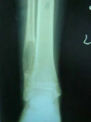
Figure 1: Anteroposterior radiograph of the ankle showing osteolytic lesion in the distal one-third of the fibulal.
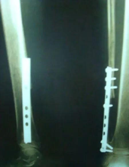
Figure 3: Post-operative X-ray after tumor excision.
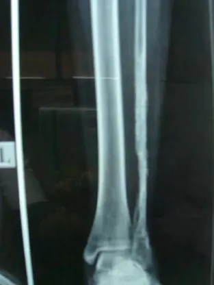
Figure 4: X-ray after implant removal.
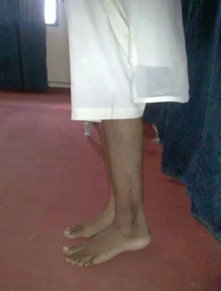
Figure 5: Clinical observation of the ankle in the patient when walking with full weight-bearing.
DISCUSSION
Chondromyxoid fibroma is a rare benign tumour that should be considered in differential diagnosis. Some of the tumours mimicking chondromyxoid fibroma include -
1) Simple bone cyst (Greenspan, 2004; Dähnert, 2007;Adam and Dixon, 2008)
2) Aneurysmal bone cyst - usually demonstrates fluidfluid levels and periosteal new bone formation without matrix mineralisation (Stoller et al., 2004)
3) Non-ossifying fibroma - usually no cortical ballooning or cortical erosion (Stoller et al., 2004)
4) Fibrous dysplasia - usually at a central location without internal septations. The peak incidence of osteo fibrous dysplasia occurs in the first decade of life and the lesions typically demonstrate more sclerosis (Stoller et al., 2004)
5) Giant cell tumour - expansile, lytic tumour that usually extends to the subchondral bone (Stoller et al., 2004)
6) Enchondroma - more classically involves the hands and feet (Greenspan, 2004)
7) Chondroblastoma - usually epiphyseal lesions with calci fied matrix in approximately 50% of tumours (Stoller et al., 2004)
Hence, de finitive diagnosis was made by clinical, radiological and pathological evaluation of tumour characteristics. Histologically the tumour consists of primitive cartilaginous tissue, fibrous tissue as well as immature myxoid tissue that may, histologically, mimic a chondrosarcoma thus necessitating imaging modalities (X-ray, CT and MRI)to aid in the final diagnosis. With above discussion, we are stressing upon the need to include this lesion in painful radiologically lytic lesion of the bone even though these tumours are very rare and often misdiagnosed radiologically.
Adam A, Dixon AK (2008) Grainger & Allison’s Diagnostic Radiology. 5thed. In: Bone Tumors: General Characteristics and Benign Lesions (Stoker DJ, Saiffudin A, eds), pp1039-1041. Philadelphia: Elsevier Churchill Livingstone.
Campanacci M. Bone and soft tissue tumors (1999). Padova: Piccin Nuova Libraria 265-277.
Dähnert W (2007) Radiology Review Manual. 6thed. Philadelphia:Lippincott Williams & Wilkins.
Fahmy ML, AlRayes M, Iskaf W, Hammouda A (1998) Chondromyxoid fibroma of the foot: case report and literature review. Foot 8:106-108.
Greenspan A (2004) Orthopedic Imaging. 4thed. Philadelphia: Lippincott Williams & Wilkins.
Jaffe HL, Lichtenstein L (1948) Chondromyxoid fibroma of bone:a distinctive benign tumor likely to be mistaken especially for chondrosarcoma. Arch Pathol (Chic) 45:541-551.
Stoller DW, Tirman PFJ, Bredella MA (2004) Diagnostic Imaging Orthopaedics. Salt Lake City: Amirsys 8/30-8/33.
 Clinical Trials in Orthopedic Disorder2016年3期
- Clinical Trials in Orthopedic Disorder的其它文章
- Traction apophysitis of the medial malleolus
- Effects of a combined nerve block on intraoperative stress and postoperative immune function in elderly patients subjected to total hip replacement: study protocol for a randomized controlled trial
- Digital navigation enhances cervical pedicle screw placement accuracy and safety: study protocol of a randomized controlled trial
- Porous tantalum rods improve the hip joint function of patients with avascular necrosis of the femoral head after femoral neck fracture surgery: study protocol of a randomized controlled trial
- Safety and effectiveness of proximal femoral nail antirotation for the treatment of intertrochanteric femoral fracture: study protocol for a prospective case series
- Commentary on "clinical efficacy of tranexamic acid administration via different routes during total hip arthroplasty:study protocol for a prospective randomized controlled trial”