丁酸钠联合顺铂对卵巢透明细胞癌ES-2细胞活性及增殖的影响
2016-01-20 13:54:00苏强马妮娜李琴俞静张晨光首都医科大学附属北京友谊医院北京100050
山东医药 2015年32期
苏强,马妮娜,李琴,俞静,张晨光(首都医科大学附属北京友谊医院,北京100050)
丁酸钠联合顺铂对卵巢透明细胞癌ES-2细胞活性及增殖的影响
苏强,马妮娜,李琴,俞静,张晨光
(首都医科大学附属北京友谊医院,北京100050)
摘要:目的观察丁酸钠联合顺铂对卵巢透明细胞癌ES-2细胞活性及增殖的影响。方法将对数生长期的卵巢透明细胞癌ES-2细胞随机分为观察组和对照组。观察组分别加入1、2、4、6 mmol/L的丁酸钠和5 μg/mL顺铂,对照组分别加入与观察组相同浓度的丁酸钠。采用MMT法检测两组细胞活性,Western blotting法检测两组凋亡蛋白( c-PAPP、c-Caspase-3)和增殖蛋白( ERK、p-ERK)相对表达量。结果观察组加入1、2、4、6 mmol/L丁酸钠和顺铂细胞活性分别为51%±3%、39%±2%、22%±2%、11%±1%,多组间及组内两两比较,P均<0.05;对照组加入1、2、4、6 mmol/L丁酸钠细胞活性分别为88%±4%、76%±3%、69%±2%、54%±2%,多组间及组内两两比较,P均<0.05;两组加入相同浓度丁酸钠细胞活性比较,P均<0.05。随着丁酸钠浓度增加,两组凋亡蛋白c-PAPP、c-Caspase-3相对表达量逐渐升高,增殖蛋白ERK、p-ERK相对表达量逐渐降低。与对照组同浓度丁酸钠比较,观察组1、2、4、6 mmol/L丁酸钠和顺铂c-PAPP、c-Caspase-3蛋白表达均升高,1、6 mmol/L丁酸钠和顺铂ERK、p-ERK蛋白表达均降低;两组比较,P均<0.05。结论丁酸钠联合顺铂可降低卵巢透明细胞癌ES-2细胞活性,抑制其增殖、促进其凋亡;丁酸钠浓度越高,上述作用越明显。
关键词:卵巢肿瘤;卵巢透明细胞癌;丁酸钠;顺铂;细胞活性;增殖蛋白;凋亡蛋白
Effect of sodium butyrate combined with cisplatin on cell viability and proliferation of ovarian clear cell carcinoma ES-2 cells
SU Qiang,MA Ni-na,LI Qin,YU Jing,ZHANG Chen-guang
( Beijing Friendship Hospital Affiliated to Capital Medical University,Beijing 100050,China)
Abstract:Objective To observe the effect of sodium butyrate combined with cisplatin on cell viability and proliferation of ovarian clear cell carcinoma ES-2 cells.Methods The ovarian clear cell carcinoma ES-2 cells in the logarithmic phase were randomly divided into the observation groups and control groups.The observation groups were added 1,2,4 and 6 mmol/L sodium butyrate ( NaB) and 5 μg/mL cisplatin.The control groups were added the same concentrations of NaB.The cell activities were detected by MMT,and the relative expression levels of c-PAPP,c-Caspase-3,ERK and p-ERK were detected by Western blotting.Results The cell viabilities of the observation groups which were treated with 1,2,4,6 mmol/L NaB and 5 μg/mL cisplatin were 51%±3%,39%±2%,22%±2% and 11%±1%,respectively; and significant difference was found between groups ( all P<0.05).The cell viabilities of the control groups were 88%± 4%,76%±3%,69%±2% and 54%±2%,respectively; and significant difference was found between groups ( all P<0.05).Meanwhile,significant difference was found in the cell viability between the two groups which were added with the same concentrations of NaB ( all P<0.05).With the increased concentrations of NaB,the relative expression levels of c-PAPP and c-Caspase-3 in the two kinds of groups were gradually increased,and the levels of ERK and p-ERK were significantly decreased.Compared with the control groups,the relative expression levels of c-PAPP and c-Caspase-3 in the observation groups which were added 1,2,4 and 6 mmol/L NaB and 5 μg/mL cisplatin were all increased and the levels of ERK and P-ERK were decreased in the observation groups which were added 1 and 6 mmol/L NaB and 5 μg/mL cisplatin.Significant difference was found between the two kinds of groups ( all P<0.05).Conclusion NaB combined with cispla-
tin may decrease the cell viability of ovarian clear cell carcinoma ES-2 cells,inhibit the proliferation and promote the apoptosis.The effect can be stronger with the higher concentrations of NaB.
Key words:ovarian neoplasms; ovarian clear cell carcinoma; sodium butyrate; cisplatin; cell viability; proliferin; apoptin
卵巢透明细胞癌发病率呈逐渐升高趋势,其术后复发率高,对铂类化疗药物不敏感;化疗耐药是影响患者预后的主要因素;增强患者的顺铂化疗敏感性是近年来国内外研究的热点。丁酸钠( NaB)是组蛋白去乙酰化酶抑制剂,可通过抑制组蛋白去乙酰化酶松弛染色质结构,促进肿瘤细胞凋亡,并抑制其生长[1],但其是否可增加卵巢透明细胞癌患者对顺铂化疗的敏感性鲜见报道。2014年8~12月,我们观察了NaB联合5 μg/mL顺铂对卵巢透明细胞癌ES-2细胞活性及增殖的影响。
1 材料与方法
1.1材料人卵巢透明细胞癌ES-2细胞由首都医科大学附属遗传学实验室传代培养。顺铂购自山东齐鲁制药有限公司,NaB、ERK、P-ERK、Caspase-3、PARP一抗、二抗均购自美国Sigma-Aldrich公司。酶联免疫检测仪购于美国BioTek公司,全套聚丙烯酰胺电泳设备购于美国Bio-Rad公司,自动洗片机购于美国ALL PRO公司。
1.2细胞培养及分组处理将人卵巢透明细胞癌ES-2细胞置于含10% FBS及青链霉素的RPMI-1640培养基中,于37℃、5% CO2的无菌培养箱中培养,3 d传代1次。将对数生长期的人卵巢癌ES-2细胞随机分为观察组和对照组,按每孔1×10⁴个接种至96孔板中,每孔设3个复孔。加入100 μL的培养液后,观察组分别加入1、2、4、6 mmol/L的NaB和5 μg/mL顺铂,对照组分别加入1、2、4、6 mmol/L的NaB。
1.3相关指标观察
1.3.1细胞活性采用MTT法。两组置于37℃、5% CO2的无菌培养箱中培养72 h,同时以加入二甲基亚砜( DMSO)的细胞作为对照。吸弃培养基后PBS冲洗3次,加入完全培养基100 μL。各孔加入MTT溶液10 μL,37℃孵育2 h,脱色摇床振荡10 min。于酶标仪上测定波长570 nm处各孔吸光度( OD值),细胞活性= ( OD细胞-OD对照)/OD对照×100%。
1.3.2细胞凋亡蛋白( c-PAPP、c-Caspase-3)及增殖蛋白( ERK、p-ERK)表达采用Western blotting法。收集两组培养72 h后的细胞,接种至培养瓶内培养,弃培养液后用冰冷PBS冲洗2次。细胞刮刀收集细胞,裂解液重悬细胞,12 000 r/min离心30 min,取上清,于-80℃保存。采用考马斯亮蓝法测定蛋白浓度,聚丙烯酸胺凝胶电泳,电转移至硝酸纤维素膜上,5%脱脂奶粉封闭。分别加入一抗室温孵育2 h,加入二抗室温孵育45 min。以Odyssey荧光扫描仪扫描显影,Quantity one软件计算灰度比值以β-actin为内参,采用Image J软件计算凋亡蛋白( c-PAPP、c-Caspase-3)及增殖蛋白( ERK、p-ERK)的相对表达量。
1.4统计学方法采用SPSS13.0统计软件。计量资料以珋x±s表示,多组间比较采用秩和检验,两组间比较采用t检验。P<0.05为差异有统计学意义。
2 结果
2.1两组细胞活性比较观察组加入1、2、4、6 mmol/L NaB和顺铂的细胞活性分别为51%±3% 39%±2%、22%±2%、11%±1%,多组间及组内两两比较,P均<0.05;对照组加入1、2、4、6 mmol/L NaB细胞活性分别为88%±4%、76%±3%、69% ±2%、54%±2%,多组间及组内两两比较,P均<0.05;两组相同浓度NaB细胞活性比较,P均<0.05。
2.2两组凋亡蛋白及增殖蛋白表达比较随着NaB浓度的增加,两组凋亡蛋白c-PAPP、c-Caspase 3相对表达量逐渐升高,增殖蛋白ERK、p-ERK相对表达量逐渐降低。两组凋亡蛋白及增殖蛋白表达比较见表1。
3 讨论
卵巢透明细胞癌临床少见,恶性程度极高,占卵巢恶性肿瘤的5%~13%[2]。铂类是临床上治疗卵巢恶性肿瘤最常用的化疗药物,顺铂主要作用于DNA链间及链内绞链,形成DDP-DNA复合物而干扰DNA复制;或与核蛋白及胞浆蛋白结合,抑制肿瘤细胞生长而导致其死亡。但卵巢透明细胞癌对于铂类化疗不敏感[3]。研究表明,卵巢上皮性癌的化疗有效率(含铂类)为73%~81%,而卵巢透明细胞癌的一线化疗有效率(含铂类)为11%~27%[4~6];对于复发的卵巢透明细胞癌,二线化疗有效率(含铂类)<10%[7,8]。因此,如何增强卵巢透明细胞癌对于顺铂的化疗敏感性成为国内外研究的热点。
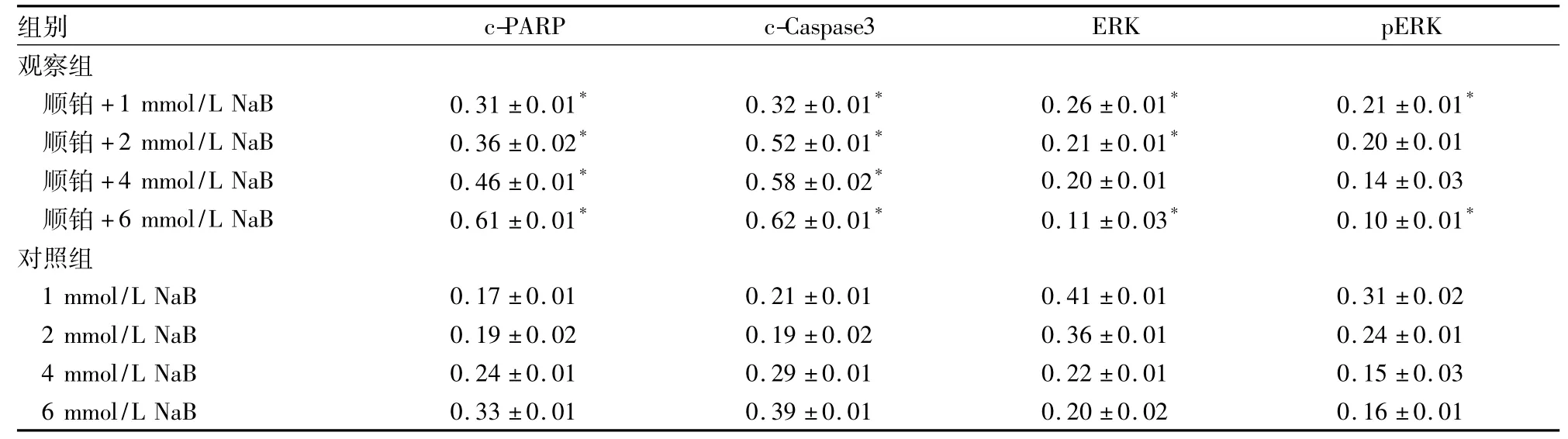
表1 两组凋亡蛋白c-PAPP、c-Caspase-3及增殖蛋白ERK、p-ERK表达比较(相对表达量,珔x±s)
研究表明,肿瘤发生与组蛋白N端赖氨酸残基的乙酰化和去乙酰化过程失衡密切相关,其中组蛋白乙酰转移酶和组蛋白去乙酰基酶( HDAC)共同控制着组蛋白的乙酰化水平,两者的动态平衡控制着染色质结构和基因表达,其动态失衡与多种肿瘤发生有关[9,10]。组蛋白去乙酰基酶抑制剂( HDACI)可通过促进组蛋白的乙酰化以及促进非组蛋白底物的乙酰化发挥抗癌活性;其基因转录异常可阻滞细胞周期,抑制DNA修复,从而诱导细胞凋亡[11]。有研究表明,HDACI与一些传统化疗药物共同应用可能会降低化疗药物介导的肿瘤细胞凋亡的阈值,具有协同抗肿瘤作用[12,13]。NaB属于HDACI的短链脂肪酸盐,可通过抑制HDAC松弛染色质结构,促进抑癌基因表达,具有抑制肿瘤细胞生长的作用[14]。
前期研究显示,5 μg/mL的顺铂作用于卵巢透明细胞癌ES-2细胞后,其细胞活度约为50%,因此本实验选择此浓度进行研究。本研究结果显示,与对照组加入相同浓度NaB比较,观察组细胞活性和凋亡蛋白c-PAPP、c-Caspase-3相对表达量均显著升高,增殖蛋白ERK、p-ERK相对表达量均显著降低;说明NaB联合顺铂可降低卵巢透明细胞癌ES-2细胞活性,并抑制其增殖、促进其凋亡;且观察组上述指标变化随NaB浓度的增加而变化更明显,说明其具有一定的浓度依赖性。本实验属于细胞水平的初步研究,NaB联合顺铂对卵巢透明细胞癌ES-2细胞增殖的抑制作用及其机制仍需要进一步的动物、分子甚至临床水平的深入研究。
参考文献:
[1]Minucci S,Pelicci PG.Histone deacetylase inhibitors and the promise of epigenetic and more treatments for cancer[J].Nat Rev Cancer,2006,6( 1) :38-51.
[2]修敏,郭玲,刘丝荪.卵巢透明细胞癌发病与耐药分子机制的研究进展[J].肿瘤,2014,34( 10) : 963-968.
[3]Jemal A,Siegel R,Xu J,et al.Cancer statistics[J].CA Cancer J Clin,2010,60( 5) : 277-300.
[4]Sugiyama T,Yakushiji M,Nishida T,et al.Irinotecan ( CPT-11) combined with cisplatin in patients with refractory or recurrent o varian cancer[J].Cancer Lett,1998,128( 2) : 211-218.
[5]Ho CM,Huang YJ,Chen TC,et al.Pure-type clear cell carcino ma of the ovary as a distinct histological type and improved surviva in patients treated with paclitaxel-platinum-based chemotherapy in pure-type advanced disease[J].Gynecol Oncol,2004,94( 1) : 197-203.
[6]Utsunomiya H,Akahira J,Tanno S,et al.Paclitaxel-platinum combination chemotherapy for advanced or recurrent ovarian clea cell adenocarcinoma: a multicenter trial[J].Int J Gynecol Canc er,2006,16( 1) : 52-56.
[7]Takano M,Kikuchi Y,Kudoh K,et al.Weekly administration o temsirolimus for heavily pretreated patients with clear cell carcino ma of the ovary: a report of six cases[J].Int J Clin Oncol,2011 16( 5) : 605-609.
[8]Takano M,Kikuchi Y,Yaegashi N,et al.Clear cell carcinoma o the ovary: aretrospective multicentre experience of 254 patient with complete surgical staging[J].Br J Cancer,2006,94( 10) : 1369-1374.
[9]Kristensen LS,Nielsen HM,Hansen LL.Epigenetics and cance treatment[J].Eur J Pharmacol,2009,625( 1) : 131-142.
[10]崔路佳,高善玲,裴凤华.丁酸钠抗肿瘤作用的新进展[J].世界华人消化杂志,2015,13( 14) : 1744-1746.
[11]Lakshmaiah KC,Jacob LA,Aparna S,et al.Epigenetic therapy o cancer with histone deacetylase inhibitors[J].J cancer Res Ther 2014,10( 3) : 469-478.
[12]Arnold NB,Arkus N,Gunn J,et al.The histone deacetylase in hibitor suberoylanilidehydroxamic acid induces growth inhibition and enhances gemcitabine-induced cell death in pancreatic cance [J].Clin Cancer Res,2007,13( 1) : 18-26.
[13]Dowdy SC,Jiang S,Zhou XC,et al.Histonedeacetylase inhibitor and paclitaxel cause synergistic effects on poptosis and microtubule stabilization in papillary serous endometrial cancer cells[J].Mo Cancer Ther,2006,5( 11) : 2767-2776.
[14]董梅,胡兴胜,陈闪闪,等.组蛋白去乙酰化酶抑制剂在肿瘤治疗领域的进展[J].中华肿瘤学杂志,2013,35( 7) : 481-485.
收稿日期:( 2015-06-13)
通信作者简介:张晨光( 1980-),男,高级讲师,研究方向为实体恶性肿瘤的基础。E-mail: chzhang@ ccmu.edu.cn
作者简介:第一苏强( 1978-),男,主治医师,研究方向为实体恶性肿瘤的临床与基础。E-mail: BJSQ1978@163.com
基金项目:国家自然科学基金青年基金资助项目( 81201816) ;首都医科大学临床基础合作研究基金资助项目( 15JL33) ;首都医科大学附属北京友谊医院院启动基金资助项目( yyqdkt2014-12)。
文章编号:1002-266X( 2015) 32-0017-03
文献标志码:A
中图分类号:R737.31
doi:10.3969/j.issn.1002-266X.2015.32.006
猜你喜欢
中国实用医药(2016年30期)2016-12-28 14:56:41
中国医药导报(2016年25期)2016-11-30 07:20:06
医学信息(2016年29期)2016-11-28 10:10:49
医学信息(2016年29期)2016-11-28 09:52:48
中外医学研究(2016年28期)2016-11-28 07:12:34
今日健康(2016年12期)2016-11-17 19:25:57
中国实用医药(2016年24期)2016-10-17 05:10:07
中国实用医药(2016年24期)2016-10-17 04:52:27
中国实用医药(2016年13期)2016-07-05 13:46:44
上海医药(2016年9期)2016-06-03 23:22:03