颅内动脉瘤破裂的血流动力学和形态学因素
江国权,方兴根,徐善水,张连富
·综述General review·
颅内动脉瘤破裂的血流动力学和形态学因素
江国权,方兴根,徐善水,张连富
随着计算机断层摄影血管成像(CTA)、磁共振血管成像(MRA)等影像技术的广泛应用,越来越多的无症状性颅内动脉瘤被检测出来,如何从中鉴别出高破裂风险的动脉瘤成为治疗的关键,本文就近年来动脉瘤破裂的危险因素做一总结。
颅内动脉瘤;血流动力学;形态学
颅内未破裂动脉瘤的发病率为2%~3%[1],而我国35~75岁年龄段成年人的发病率则高达7%[2]。这些未破裂动脉瘤中大部分不发生破裂,只有一小部分发生破裂。有学者统计破裂的动脉瘤仅占全部动脉瘤的1%~2%[3]。但是一旦动脉瘤破裂,后果非常严重,病死率可高达50%[4]。因此如何从中判断出具有高破裂风险的动脉瘤并及时对其进行干预成为动脉瘤治疗的关键。本文就如何判别高破裂风险的动脉瘤,从血流动力学因素、形态学因素及其他因素等方面进行阐述。
1 血流动力学因素
血流动力学与动脉瘤的发生、发展以及破裂密切相关。目前了解,影响动脉瘤发生发展的血流动力学因素主要有:壁面切应力(wall shear stress,WSS)、切应力震荡指数(oscillatory shear index,OSI)、壁面切应力梯度(wall shear stress gradient,WSSG)、冲击域大小、血流模式以及血流速度。
1.1 WSS
血流的冲击对血管壁会形成3种不同方向的生理压力:WSS、跨壁压(transmural pressure)、机械性伸缩力(mechanical stretch)。其中WSS被认为是血流动力学因素中影响颅内动脉瘤形成、发展和破裂的主要因素[5-6]。由于血液具有黏性,当血流通过血管壁时便会产生摩擦形成WSS。WSS对维持细胞的正常形态具有重要作用。研究认为维持细胞正常形态的WSS需要大约2 Pa左右,过低的WSS会导致上皮细胞间隙破坏、抗氧化和抗炎介质调节失常,使得内皮细胞壁重塑,甚至细胞变性、凋亡[7]。但是过高的WSS对动脉瘤也是不利。总结起来,目前WSS在动脉瘤的破裂中的作用机制有2种观点:高血流理论和低血流理论。前者认为过高的WSS会导致局部血管壁退化、扩张甚至膨胀,进而形成动脉瘤或者导致动脉瘤的生长[8-9]。后者认为低WSS可导致动脉瘤壁发生退行性变,局部壁面WSS的不足导致动脉瘤破裂[10-11]。
1.2 OSI和WSSG
OSI代表一个区域随时间变化与WSS相对的方向改变的速度,它是预测血管疾病的有用的血流动力学参数。OSI值的大小代表流动振荡水平的大小,即流动方向和强度改变的程度。OSI越高,该处流体在周期内的流动方向越不稳定,随时间越频繁变化甚至出现回流;相反,OSI越低,该处流动越稳定,变化越小。研究认为高OSI与动脉瘤破裂明显相关[12]。
WSSG是表示壁面切应力变化大小的系数。Valencia等[13]发现,高WSSG下高振幅WSS变化会引起内皮细胞疲劳和损伤,从而导致内皮细胞功能紊乱和血管病理性重塑,并进一步诱发动脉瘤。
1.3 冲击域大小
Hoi等[14]首先定义了“冲击域”这一概念,它是指射入的血流作用于动脉瘤壁的主要区域。冲击域的大小与动脉瘤的破裂密切相关。原因在于冲击域内血流速度会发生明显改变,同时血流方向也会发生明显变化,造成该区域动脉壁压力明显高于其他区域,从而导致该区域管壁更易发生破裂。研究认为未破裂动脉瘤内有相对广的血流冲击域,破裂前的动脉瘤则有更为局限的冲击区域[15-16]。
1.4 血流模式
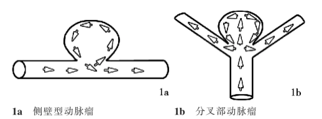
血流模式是影响动脉瘤发展变化的重要因素。它主要分为侧壁性动脉瘤血流模式和顶端动脉瘤血流模式两种。侧壁性动脉瘤:血流从瘤颈口下游侧流向动脉瘤瘤内,并沿着瘤周壁回流至载瘤动脉中,再从瘤颈口上游流出,形成一个主要涡流。该模式造成血流动力学的特点是:流入道血流速度、动压及流入道侧壁的壁面WSS最高,流出道次之,瘤顶部最低,且动脉瘤壁面WSS、动压与血流速度呈正相关[17]。顶端动脉瘤:血流从流入动脉直接冲击动脉瘤,然后沿动脉瘤周壁回流至两条流出动脉,形成两侧两个主要涡流(图1)。由于血流由入口动脉直接进入动脉瘤,造成血流直接冲击瘤顶。此外由于血流阻力骤然增加,血流动能转换瘤内压力,导致瘤内压增加,血流速度减慢,瘤内WSS下降。因此该模式的血流动力学特点是:瘤内低血流速度,高压力和低WSS[11]。
1.5 血流速度

图1 动脉瘤的血流模式(图中箭头方向为血流的方向)
血流速度作为血流动力学上的重要参数,对动脉瘤的影响是多方面的,主要从2方面影响动脉瘤。一方面可通过影响WSS影响动脉瘤的发展: Ujiie等[18]对大脑前动脉-前交通动脉交界处血流动力学的研究发现,均一流速的血流对前交通动脉管壁的切应力几乎为零,而血流速度不均一时可观察到交叉血流和血流停滞点,停滞点附近的WSS显著增高。另一方面它可通过影响冲击域的大小影响动脉瘤的进展:Castro等[19]发现动脉瘤瘤体内血流速度越快,瘤体WSS越高,受冲击的区域越小,动脉瘤破裂的风险也越高。
2 形态学因素
形态学因素是影响动脉瘤发生、发展及破裂的重要因素。目前描述形态学因素指标较多,本文将其分为2方面——二维形态学参数和三维形态学参数进行归纳。二维形态学参数是指那些能被直接采集到,不需要进行三维重建的参数,主要包括动脉瘤的大小、位置、AR值、SR值、载瘤动脉曲度半径、血管角以及动脉瘤角度。三维形态学参数是指通过三维重建得到的参数,主要有:波动指数、椭圆度指数、非球形指数以及体积瘤颈比。
2.1 二维形态学参数
动脉瘤的大小是影响动脉瘤发生、发展、破裂的重要因素。以前认为5~10 mm的动脉瘤容易破裂,>10mm或<5mm的不易破裂。但是越来越多的研究表明,动脉瘤越大其破裂风险越高。UCAS[20]计算不同大小的动脉瘤年破裂率,比较动脉瘤破裂风险的大小。结果如下:<7 mm的动脉瘤不易破裂,5~6mm的动脉瘤与3~4mm动脉瘤破裂风险无明显差别,平均年破裂率为1.13%,但是动脉瘤>7mm时,动脉瘤的破裂风险与动脉瘤的大小呈正相关。7~9 mm的动脉瘤平均年破裂率为3.35%;10~24 mm的动脉瘤平均年破裂率为9.09%;25 mm或者更大的动脉瘤平均年破裂率更高为76.26%。另外,动脉瘤越大越容易生长:<8 mm的动脉瘤,年生长率为6.9%,8~12 mm为25%,>12 mm为83%[21]。增长的动脉瘤破裂风险为2.4%,比不增长的动脉瘤0.2%的破裂风险高出20多倍[22]。
动脉瘤的位置也是影响动脉瘤变化的重要因素。尽管有学者认为后循环动脉瘤破裂风险高于前循环,但越来越多的研究证实前交通动脉瘤跟其他位置动脉瘤相比更易发生破裂[23-25]。
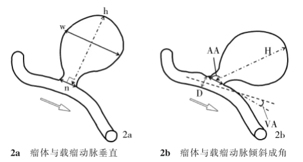
AR值(aspect ratio,AR)是动脉瘤的瘤体高与瘤颈宽的比值,瘤体高是指瘤顶到瘤颈的最大垂直距离[26]。动脉瘤破裂风险与AR值呈正相关性,AR值越高,动脉瘤破裂的风险就越大。Tateshima等[27]研究认为AR的阈值应为1.6,因为当AR>1.6后,动脉瘤破裂的风险显著增加。
SR值(size ratio,SR)是瘤体长与载瘤动脉血管直径的比值,瘤体长是指瘤顶至瘤颈的中心点最长距离,不一定是垂直距离(图2)。它最先由Dhar等[23]提出,也与动脉瘤的破裂成正相关性。但其阈值,目前尚有争议。Dhar等[23]认为其阈值为2,他们发现75%的破裂动脉瘤的SR>2,而83%的未破裂动脉的SR≤2。而Rahman等[28]认为SR的阈值应为3。
动脉瘤内的血流动力学会随着载瘤动脉曲度半径的变化而变化。Hoi等[14]研究发现随着载瘤动脉曲度半径的增加,在瘤颈远侧段的冲击域、wss显著增强。Avolio等[29]应用计算机流体动力学(computational fluid dynamics,CFD)技术研究发现瘤体大小和形态相当,在不同曲度血管上的动脉瘤,其血流速度明显高于直行血管壁动脉瘤。
载瘤动脉轴线与瘤颈平面所形成的夹角为血管角(vessel angle,VA),即出口动脉与入口动脉夹角(图2)。VA的大小与瘤内压力及WSS变化密切相关。VA越大,动脉瘤内血液流出阻力越大,瘤内血流速度越小,而瘤内压力越大,WSS越小;VA越小,动脉瘤内血液流出阻力越小,瘤内血流速度越大,而瘤内压力相对越小,WSS相对越大[30-31]。瘤体长轴与瘤颈平面形成的夹角为动脉瘤角度(aneurysm angle,AA)(图2)。Dhar等[23]发现在AA与动脉瘤破裂呈正相关性,AA越大,动脉瘤破裂的风险越大。Lin等[32]更认为动脉瘤的破裂与VA、AA均密切相关。
2.2 三维形态学参数
近年来,随着三维重建技术的日益成熟,对动脉瘤瘤体三维结构形态的研究越来越多,用于描述其形态结构的参数也在不断完善。目前描述动脉瘤瘤体自身结构形态的三维参数主要有:波动指数、椭圆度指数、非球形指数。

图2 动脉瘤的形态学示意图
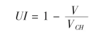
波动指数(undulation index,UI)反映动脉瘤瘤体凹陷程度的指标。其公式如下[33]:

式中VCH是在动脉瘤瘤体凸起部分的体积,V是动脉瘤瘤体总体积。UI随着VCH的减小而逐渐增大,即动脉瘤由饱满变扁平时,破裂风险增加。
椭圆度指数(elipticity index,EI)反映动脉瘤瘤体椭圆化程度的指标。其公式如下[33]:

式中VCH是在动脉瘤瘤体凸起部分的体积,SCH是动脉瘤瘤体凸起部分的表面积。EI随着VCH和SCH比值的减少而增大,动脉瘤破裂的风险也在增加。当EI为0时,即动脉瘤瘤体呈完全规则球形时,EI最小,破裂风险也最小。
非球形指数(nonsphericity index,NSI)是对EI和UI的概括总结,它是反映动脉瘤瘤体凹陷和椭圆化程度的指标。其公式为[34]:

式中,V为动脉瘤瘤体的体积;S为瘤体的表面积。NSI也是随着V和S的比值的减小而增大的,NSI越大,动脉瘤破裂的风险越大。
此外,体积瘤颈比(volume to neck ratio,VNR)也是三维形态学的重要参数,在预测动脉瘤破裂风险参数中,其重要性不亚于AR,实际上VNR和AR具有线性关系,因为它们都是反映瘤体大小与瘤颈大小程度的指标。研究发现动脉瘤体积越大,瘤颈越窄,流入动脉瘤内的血流越慢,而低速血流容易导致动脉瘤的生长和破裂[35]。
2.3 其他因素
颅内血管先天发育异常是导致动脉瘤的发生、发展、破裂的重要因素。因为血管变异会引起血流动力学的改变,而血流动力学的改变又可导致动脉瘤发生改变。以前交通动脉复合体为例,因为此处血管变异较多,大脑前动脉A1段常发生缺如,当A1段发生缺如时,同侧的颈内动脉只供应该侧的大脑中动脉,因此其血流量减少,而对侧的颈内动脉需要供应其侧的大脑中动脉及双侧的大脑前动脉,导致其血流量增加,从而引起血流动力学改变[36]。Alnaes等[37]发现,大脑前动脉A1段变异与前交通动脉瘤关系密切,以左侧A1段优势多见,而前交通动脉瘤也以偏左侧多见。种种迹象表明先天性血管发育异常是预测动脉瘤破裂的不可忽略的因素。
动脉瘤瘤内压力也是预测动脉瘤破裂的重要参数,对动脉瘤生长及破裂的影响是随着动脉瘤的生长而增加,这解释了临床上所观察到动脉瘤生长在初期较慢而到一定阶段加快并易于发生破裂的现象[38]。但是由于缺乏足够的病理状态下瘤壁的生物力学参数,目前对于特定的动脉瘤尚无法准确计算瘤壁内压力。
此外如性别、吸烟、高血压、相关的结缔组织病、动脉瘤病史和家族史[39],在判断动脉瘤破裂风险时,也是不可忽略的因素。
上述各种评估动脉瘤破裂的参数及因素较多且繁杂,目前只是将它们作为预测动脉瘤破裂风险的重要因素,为临床治疗提供参考,尚未形成统一的标准。需要我们进一步研究和探索。
[1]Etminan N,Beseoglu K,Barrow DL,et al.Multidiscip linary consensus on assessment of unruptured intracranial aneurysms: proposal of an international research group[J].Stroke,2014,45:1523-1530.
[2]Li MH,Chen SW,Li YD,et al.Prevalence of unruptured cerebral aneurysms in Chinese adults aged 35 to 75 years:a cross-sectional study[J].Ann Intern Med,2013,159:514-521.
[3]Amenta PS,Yadla S,Campbell PG,et al.Analysis of nonmodifiable risk factors for intracranial aneurysm rupture in a large,retrospective cohort[J].Neurosurgery,2012,70:693-699;discussion 699-701.
[4]Etminan N,Buchholz BA,Dreier R,et al.Cerebral aneurysms: formation,progression,and developmental chronology[J]. Transl Stroke Res,2014,5:167-173.
[5]Baek H,Jayaraman MV,Richardson PD,et al.Flow instability and wall shear stress variation in intracranial aneurysms[J].JR Soc Interface,2010,7:967-988.
[6]鲁刚,黄磊,张晓龙,等.颅内动脉瘤壁切应力与动脉瘤破裂关系的初步研究——基于患者的三维计算机模拟[J].介入放射学杂志,2009,18:568-571.
[7]Reneman RS,Arts T,Hoeks AP.Wall shear stress—an important determinant of endothelial cell function and structure—in the arterial system in vivo.Discrepancies with theory[J].JVasc Res,2006,43:251-269.
[8]Hassan T,Timofeev EV,Saito T,etal.Computational replicas: anatomic reconstructions of cerebral vessels as volume numerical grids at three-dimensional angiography[J].AJNR,2004,25: 1356-1365.
[9]Meng H,Wang Z,Hoi Y,et al.Complex hemodynamics at the apex of an arterial bifurcation induces vascular remodeling resembling cerebral aneurysm initiation[J].Stroke,2007,38: 1924-1931.
[10]Castro MA,Putman CM,Cebral JR.Computational fluid dynamics modeling of intracranial aneurysms:effects of parent artery segmentation on intra-aneurysmal hemodynamics[J]. AJNR,2006,27:1703-1709.
[11]Shojima M,Oshima M,Takagi K,et al.Magnitude and role of wall shear stress on cerebral aneurysm:computational fluid dynamic study of 20 middle cerebral artery aneurysms[J]. Stroke,2004,35:2500-2505.
[12]Omodaka S,Sugiyama S,Inoue T,et al.Local hemodynamics at the rupture point of cerebral aneurysms determined by computational fluid dynamics analysis[J].Cerebrovasc Dis,2012,34:121-129.
[13]Valencia A,Solis F.Blood flow dynamics and arterial wall interaction in a saccular aneurysm model of the basilar artery[J]. Comput Struct,2006,84:1326-1337.
[14]Hoi Y,Meng H,Woodward SH,et al.Effects of arterial geometry on aneurysm growth:three-dimensional computational fluid dynamics study[J].JNeurosurg,2004,101:676-681.
[15]Cebral JR,Castro MA,Appanaboyina S,etal.Efficient pipeline for image-based patient-specific analysis of cerebral aneurysm hemodynamics:technique and sensitivity[J].IEEE Trans Med Imaging,2005,24:457-467.
[16]Cebral JR,Sheridan M,Putman CM.Hemodynamics and bleb formation in intracranial aneurysms[J].AJNR,2010,31:304-310.
[17]Shojima M,Oshima M,Takagi K,etal.Role of the bloodstream impacting force and the local pressure elevation in the rupture of cerebral aneurysms[J].Stroke,2005,36:1933-1938.
[18]Ujiie H,Liepsch DW,Goetz M,et al.Hemodynamic study of the anterior communicating artery[J].Stroke,1996,27:2086-2093.
[19]Castro MA,Putman CM,Sheridan MJ,et al.Hemodynamic patterns of anterior communicating artery aneurysms:a possible association with rupture[J].AJNR,2009,30:297-302.
[20]UCAS Japan Investigators,Morita A,Kirino T,et al.The natural course of unruptured cerebral aneurysms in a Japanese cohort[J].N Engl JMed,2012,366:2474-2482.
[21]Burns JD,Huston J,Layton KF,et al.Intracranial aneurysm enlargement on serial magnetic resonance angiography: frequency and risk factors[J].Stroke,2009,40:406-411.
[22]Villablanca JP,Duckwiler GR,Jahan R,et al.Natural history of asymptomatic unruptured cerebral aneurysms evaluated at CT angiography:growth and rupture incidence and correlation with epidemiologic risk factors[J].Radiology,2013,269:258-265.
[23]Dhar S,TremmelM,Mocco J,etal.Morphology parameters for intracranial aneurysm rupture risk assessment[J].Neurosurgery,2008,63:185-196;discussion 196-7.
[24]Matsukawa H,Uemura A,Fujii M,et al.Morphological and clinical risk factors for the rupture of anterior communicating artery aneurysms[J].JNeurosurg,2013,118:978-983.
[25]Wermer MJ,Van Der Schaaf IC,Algra A,etal.Risk of rupture of unruptured intracranial aneurysms in relation to patient and aneurysm characteristics:an updated meta-analysis[J].Stroke,2007,38:1404-1410.
[26]Ujiie H,Tachibana H,Hiramatsu O,et al.Effects of size and shape(aspect ratio)on the hemodynamics of saccular aneurysms:a possible index for surgical treatment of intracranial aneurysms[J].Neurosurgery,1999,45:119-129;discussion 129-30.
[27]Tateshima S,Chien A,Sayre J,et al.The effect of aneurysm geometry on the intra-aneurysmal flow condition[J]. Neuroradiology,2010,52:1135-1141.
[28]Rahman M,Smietana J,Hauck E,et al.Size ratio correlates with intracranial aneurysm rupture status:a prospective study[J].Stroke,2010,41:916-920.
[29]Avolio A,Farnoush A,Morgan M,et al Hemodynamic models of cerebral aneurysms for assessment of effect of vessel geometry on risk of rupture[J].Conf Proc IEEE Eng Med Biol Soc,2009.
[30]Forget TR,Benitez R,Veznedaroglu E,et al.A review of size and location of ruptured intracranial aneurysms[J]. Neurosurgery,2001,49:1322-1325;discussion 1325-6.
[31]Baharoglu MI,Schirmer CM,Hoit DA,et al.Aneurysm inflowangle as a discriminant for rupture in sidewall cerebral aneurysms:morphometric and computational fluid dynamic analysis[J].Stroke,2010,41:1423-1430.
[32]Lin N,Ho A,Gross BA,et al.Differences in simple morphological variables in ruptured and unruptured middle cerebral artery aneurysms[J].JNeurosurg,2012,117:913-919.
[33]Raghavan ML,Ma B,Harbaugh RE.Quantified aneurysm shape and rupture risk[J].JNeurosurg,2005,102:355-362.
[34]Lauric A,Miller EL,Baharoglu MI,et al.3D shape analysis of intracranial aneurysms using thewrithe number as a discriminant for rupture[J].Ann Biomed Eng,2011,39:1457-1469.
[35]Ryu CW,Kwon OK,Koh JS,et al.Analysis of aneurysm rupture in relation to the geometric indices:aspect ratio,volume,and volume-to-neck ratio[J].Neuroradiology,2011,53:883-889.
[36]Hendrikse J,Van Raamt AF,Van Der Graaf Y,et al. Distribution of cerebral blood flow in the circle of W illis[J]. Radiology,2005,235:184-189.
[37]Alnaes MS,Isaksen J,Mardal KA,et al.Computation of hemodynamics in the circle of Willis[J].Stroke,2007,38: 2500-2505.
[38]Feng Y,Wada S,Tsubota K,etal.The application of computer simulation in the Genesis and development of intracranial aneurysms[J].Technol Health Care,2005,13:281-291.
[39]You SH,Kong DS,Kim JS,et al.Characteristic features of unruptured intracranial aneurysms:predictive risk factors for aneurysm rupture[J].JNeurol Neurosurg Psychiatry,2010,81: 479-484.
The effect of hem odynam ic and m orphological factors on the rupture of intracranial aneurysm s
JIANG Guo-quan,FANG Xing-gen,XU Shan-shui,ZHANG Lian-fu.Department of Neurosurgery,Affiliated Yijishan Hospital,Wannan Medical College,Wuhu,Anhui Province 241001,China
FANG Xing-gen,E-mail:fangxinggen@gmail.com
Along with the wide use of CT angiography and MR angiography in the clinical practice,more and more asymptomatic intracranial aneurysms have been detected.Clinically,it is of the first importance to identify these aneurysms that carry high risks to probably develop rupture as for these patients suitable treatment can be promptly adopted so as to ensure a satisfactory result.This paper aims tomake a brief review about hemodynamic and morphological risk factors that may cause the rupture of intracranial aneurysms.(JIntervent Radiol,2014,23:1109-1113)
intracranial aneurysm;hemodynamics;morphology
R578.1
A
1008-794X(2014)-12-1109-05
2014-06-04)
(本文编辑:李欣)
10.3969/j.issn.1008-794X.2014.12.023
弋矶山医院人才引进基金(YR201105)
241001芜湖皖南医学院附属弋矶山医院神经外科
方兴根E-mail:fangxinggen@gmail.com

