Dynamic reactive astrocytes after focal ischemia
Shinghua Ding
1 Dalton Cardiovascular Research Center, University of Missouri-Columbia, MO, USA
2 Department of Bioengineering, University of Missouri-Columbia, MO, USA
Dynamic reactive astrocytes after focal ischemia
Shinghua Ding1,2
1 Dalton Cardiovascular Research Center, University of Missouri-Columbia, MO, USA
2 Department of Bioengineering, University of Missouri-Columbia, MO, USA
Astrocytes are specialized and most numerous glial cell type in the central nervous system and play important roles in physiology. Astrocytes are also critically involved in many neural disorders including focal ischemic stroke, a leading cause of brain injury and human death. One of the prominent pathological features of focal ischemic stroke is reactive astrogliosis and glial scar formation associated with morphological changes and proliferation. This review paper discusses the recent advances in spatial and temporal dynamics of morphology and proliferation of reactive astrocytes after ischemic stroke based on results from experimental animal studies. As reactive astrocytes exhibit stem cell-like properties, knowledge of dynamics of reactive astrocytes and glial scar formation will provide important insights for astrocyte-based cell therapy in stroke.
ischemic stroke; reactive astrocytes; glial scar; morphology; cell proliferation; dynamics; cell therapy
Funding: This work was supported by the National Institutes of Health [Grant no. R01NS069726] and the American Heart Association Grant in Aid Grant [Grant no. 13GRNT17020004] to SD.
Ding S. Dynamic reactive astrocytes after focal ischemia. Neural Regen Res. 2014;9(23):2048-2052.
Introduction
Astrocytes are the most abundant glial cell type in the central nervous system (CNS). In a normal brain, there are generally two major types of astrocytes: Fibrous astrocytes in white matter found in the corpus callosum and protoplasmic astrocytes in grey matter found in the cortex. In addition to their morphologic differences, the processes of protoplasmic astrocytes completely wrap or ensheath synapses as well as blood vessels (Bushong et al., 2002; Wilhelmsson et al., 2006; Halassa et al., 2007). The spatial occupation and the intimate physical contact with both synapses and blood vessels render astrocytes as ideally situated to be involved in bidirectional interactions with neurons as well as with vasculature. Many studies also demonstrate that astrocytes are heterogeneous in morphology, molecular expression (Xie et al., 2010; Ding, 2013; Molofsky et al., 2014) and electrophysiological and Ca2+signaling properties (Zhou and Kimelberg, 2000; Takata and Hirase, 2008) (for review of this topic see Zhang and Barres, 2010). It has been thought that glial fi brillary acidic protein (GFAP) is a ‘pan-astrocyte’ marker, but its expression levels are different in fibrous and protoplasmic astrocytes. Aldh1L1 is the most widely and homogenously expressed astrocyte speci fi c protein (Cahoy et al., 2008).
Astrocytes have been found to play important roles in many diseases and respond to almost all forms of neural disorders ranging from severe brain injuries such as stroke and traumatic brain injury (TBI), and neurodegenerative diseases such as Alzheimer’s disease (AD), Parkinson’s disease (PD), and amyotrophic lateral sclerosis (ALS) through a process called astrogliosis (Sofroniew and Vinters, 2010; Verkhratsky et al., 2012). A hallmark of astrogliosis is the morphological changes and the increased expression of GFAP in astrocytes. Given the different causes and the onset of diseases, the temporal and spatial changes of these reactive astrocytes are different; thus, detailed studies on the dynamic changes of reactive astrocytes have been undertaken to provide information for potential therapeutic interventions. For extensive reviews of reactive astrocytes in various aspects in neural diseases, readers can consult reviews by Burda and Sofroniew (2014), Sofroniew and Vinters (2010), and Escartin and Bonvento (2008). This review article will focus on discussing the dynamics of reactive astrocytes in the peri-infarct region, i.e., the so called penumbra after focal ischemia in experimental animal models.
Spatial and temporal dynamics of reactive astrocytes in the penumbra after ischemia
Focal ischemic stroke, resulting from the blockage of cerebral blood vessels in a certain region of the brain, leads to cell death and brain damage and is a leading cause of human disability and death (Stapf and Mohr, 2002). Besides cell death in the ischemic core, ischemia induces a series of alterations at molecular and cellular levels in the penumbra over time, including Ca2+signaling, cellular proliferation, morphology changes and gene regulation (Panickar and Norenberg, 2005; Ding et al., 2009, 2013, 2014; Zamanian et al., 2012; Li et al., 2013). These alterations are temporal and spatial dependent with a common feature of high GFAP expression levels in reactive astrocytes and formation of glial scar in the penumbra that demarcates the ischemic core (infarction) from healthy tissue (Haupt et al.,2007; Hayakawa et al., 2010; Barreto et al., 2011; Shimada et al., 2011; Bao et al., 2012; Li et al., 2013). The clinical aim of stroke therapy is to salvage the cells in the penumbra; thus, in-depth studyon the dynamics of reactive astrocytes at molecular and cellular levels will provide insights for therapeutic strategy. Although the responses of astrocytes to ischemic stroke have been well documented in focal ischemic models, including photothrombosis (PT)-induced focal ischemia and middle cerebral artery occlusion (MCAO) models (Stoll et al., 1998; Schroeter et al., 2002; Haupt et al., 2007; Nowicka et al., 2008; Barreto et al., 2011; Shen et al., 2012; Li et al., 2013), detailed and quantitative studies on cell proliferation with a good temporal resolution are lacking. Our recent study presented a detailed evaluation of dynamic change of reactive astrocytes in the cortex after PT (Li et al., 2014). We used bromodeoxyuridine (BrdU) labeling and immunostaining to assess the spatial and temporal changes in cellular proliferation, morphology and glial scar formation. To precisely study the rate of cell proliferation of astrocytes and microglia at different times after ischemia, we designed a ‘time-block’BrdU labeling protocol to titrate proliferating cells in the penumbra. Mice were administered with BrdU at the beginning of days 1, 3, 4, 5, 9, 11, and 13 post PT for two consecutive days and sacrificed 1 day following the last injection. From this study, a few new results were obtained.
The spatial and temporal distribution of proliferating cells
Our results show that the densities of BrdU+cells in the region close to the ischemic core are higher than those regions further away from the ischemic core over time after PT (Figure 1B), suggesting spatial difference in cell proliferation rates (Li et al., 2014). On the other hand, BrdU+cells signi fi cantly increased from post ischemic day 1 to day 2 and reached a peak value during days 3 and 4 after PT, and then decreased over time and fi nally sustained their value for a prolonged time—until day 14, the longest time in the study (Figure 1B). These results demonstrate that the rate of proliferating cells generated in the penumbra after ischemia is highly spatiotemporal dependent, consitent with the report from Barreto et al. (2011).
Morphological changes of reactive astrocytes
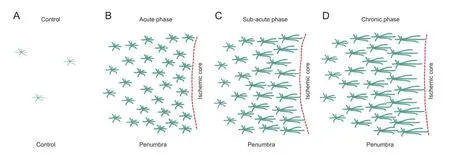
As glial fi brillary acidic protein (GFAP) is a prototypic marker for reactive astrocytes, we conducted immunostaining of GFAP to inspect the morphological change and proliferation of reactive astrocytes. There was little expression of GFAP in the cortex of control mice (also see previous studies (Zhang et al., 2010; Li et al., 2013)) (Figure 1A:A1). However, a signi fi cant increase of GFAP was observed at day 2 post PT (Figure 1A:A2). Up to day 4 post PT, astrocytes exhibited a stellate morphology and hypertrophy with highly upregulated GFAP expression (Figure 1A:A3). Starting from day 6, astrocytes in the penumbra were densely packed and formed a stream with their elongated (straight) processes pointing towards the ischemic core, i.e., a feature of astroglial scar formation (Figure 1A:A4). After day 10, the morphology of astrocytes at the scar border remained similar but with longer processes as compared with days 6—8, suggesting the maturation of astroglial scar tissue (Figure 1A:A5–A6). Signi fi cant increase in GFAP was also observed in the regions further away from the penumbra but with similar morphology to the astrocytes in the control condition. Thus, morphology of GFAP+astrocytes in the penumbra experienced dramatic changes over time after PT (Figure 1A), corroborating the results from other studies (Haupt et al., 2007; Nowicka et al., 2008). Mestriner et al. (2015) conducted a detailed study on the morphology of reactive astrocytes at 30 day after endotelin-1 induced ischemic stroke. Their results showed that rami fi cation and length of reactive astrocytes in the penumbra were different between sensorimotor cortex and dorsolateral striatum, indicating the regional heterogeneity inthe morphology of reactive astrocytes; however, morphological change in earlier stage might be more important in disease progress than in the chronic stage. The detailed study on morphology of reactive astrocytes was also conducted in rats at day 4 after MCAO (Wagner et al., 2012). Mean process volume, diameter and branching level in reactive astrocytes in the penumbra all increased compared with astrocytes in the remote region from ischemic core. However, the mean process length of reactive astrocytes in the penumbra is shorter than astrocytes in the remote region, confirming hypertrophic morphology of reactive astrocytes at this time point. Due to the heterogeneity of astrocytes in the brain even in the same region such as cortex (Takata and Hirase, 2008; Benesova et al., 2009), it is conceivable that astrocytes would respond to stroke in different manners. Thus detailed characterization of reactive astrocytes can only be done with lineage analysis and the availability of transgenic mice that express fl uorescent marker in different types of astrocytes.
Proliferating reactive astrocytes
It is known that reactive astrocytes are also characterized by progressive changes in proliferation and gene expression (Panickar and Norenberg, 2005; Haupt et al., 2007; Nowicka et al., 2008; Barreto et al., 2011; Zamanian et al., 2012). We further evaluated the rate of proliferating astrocytes using double staining of GFAP and BrdU. Although a large number of GFAP+astrocytes were emerged after PT, overall, the GFAP+BrdU+proliferating astrocytes only accounted for a small percentage of total BrdU+cells, which reached a peak value of about 6% from post ischemic days 3 to 4 and then decreased sharply over time (Figure 1D). On the other hand, the ratio of GFAP+BrdU+to GFAP+also reached the highest level within days 3 to 4 after PT (Li et al., 2014). These results demonstrated that stroke induces an increase in the number of proliferating reactive astrocytes in a highly time-dependent manner. The results indicate that the majority of GFAP+reactive astrocytes resulted from the upregulation of GFAP in existing astrocytes without proliferation. Nevertheless, this BrdU labeling protocol may underestimate the total number of BrdU+cells since a single daily injection will not label all proliferating astrocytes and other cells (Wanner et al., 2013).
Correlation of behavioral de fi cits with reactive astrogliosis
Our study demonstrated that focal ischemia-induced reactive astrocytes exhibit heterogeneity in morphology, GFAP expression levels and proliferating capability; furthermore, such heterogeneity is spatiotemporal dependent (Figure 2). After ischemia, the brain experiences spontaneous recovery process (Badan et al., 2003; Li et al., 2004; Clarkson et al., 2013). Since astrogliosis and glial scar formation is such an important pathological phenomenon, one is led to ask whether reactive astrogliosis is related to ischemia-induced behavioral de fi cits. To explore this, behavioral tests were conducted to study thetime courses of forelimb shift asymmetricity, strength, and sensory motor impairments (Li et al., 2014). The functional de fi cits have a similar time window to the infarct expansion, brain edema and swelling, and the highest rates of cell proliferation and reactive astrocyte generation. Functional de fi cits were recovered from day 6 after ischemia when glial scar tissue starts to form, suggesting that glial scarring might have a bene fi cial effect by stopping the expansion of the ischemic core. Thus, our study suggests that dynamic cellular proliferation and reactive astrogliosis correlate with the progress of brain and neuronal remodeling and functional recovery, and that targeting reactive astrocytes might be an important strategy to facilitate improvement of stroke outcomes.
Reactive astrogliosis and cell therapy in focal ischemia
Astrogliosis also occurs in chronic neurodegenerative diseases such as AD. Due to the slow reactivation processes associated with the disease progress and lack of glial scar tissue, reactive astrocytes are more evenly distributed in chronic diseases. Thus it is conceivable that the properties of reactive astrocytes in chronic neurodegenerative diseases are different from these in focal ischemia. Although profound progress has been made regarding the dynamics of reactive astrocytes in morphology and cell proliferation after strokes, studies on gene pro fi le of reactive astrocytes at different times after focal ischemia are needed to define the properties of reactive astrocyte at different stages. Our study suggests that the change of gene expression will likely be different at different times after ischemia as the morphology, the proliferating rate and the density of reactive astrocytes experience dynamic changes. Although single-point study of gene expression of reactive astrocyte after ischemia has been conducted (Zamanian et al., 2012), further studies in this area will likely elucidate the signaling pathways by which astrogliosis is induced after ischemia and derive new insights into the therapeutic potential of reactive astrocytes in ischemia.
On the other hand, growing evidence indicates that reactive astrocytes exhibit stem cell-like properties (Buffo et al., 2008; Robel et al., 2011; Shimada et al., 2012; Sirko et al., 2013; Dimou, 2014). They can express neural stem cell related proteins such as Nestin, Sox2 (Shimada et al., 2012), and DCX, an immature neural stem cell marker (Ohab et al., 2006). Moreover, it has been reported that astrocytes can be converted into neuroblasts and neurons by forced expression of single transcriptional factors such as Sox2 (Su et al., 2014), neurogenin-2 (Berninger et al., 2007; Heinrich et al., 2010), NeuroD1 (Guo et al., 2013), or a combination of multiple transcriptional factors such as ASCL1, LMX1B and NURR1 (Addis et al., 2011). Thus targeting reactive astrocytes and using local astrocytes are attractive strategies of cell therapy for stroke. Our study on dynamics of reactive astrocytes provides an important implication for the optimal timing for the pharmacological and genetic manipulations of reactive astrocytes to improve stroke outcomes in experimental and clinic studies of stroke therapy. To genetically manipulate reactive astrocytes in vivo, astrocyte-speci fi c approaches such as viral transduction (Xie et al., 2010) and Cre/loxP recombinase system with astrocyte-speci fi c Cre driver mouse lines (Mori et al., 2006) are required.
While growing evidence suggests that ischemic stroke dramatically increases neurogenesis in the subventricular zone (SVZ) and subgranular layer in dentate gyrus (Tobin et al., 2014), a recent study fi rst showed that ischemic stroke causes substantial reactive astrogliosis in SVZ (Young et al., 2013). The hypertrophic reactive astrocytes and their tortuous processes disrupt neuroblast migratory scaffold and thus might be the cause of SVZ reorganization after stroke. Future studies will be required to further explore whether SVZ astrocytes can function as neural stem cells and can be differentiated into neurons to contribute to the improvement of stroke outcomes.
Addis RC, Hsu FC, Wright RL, Dichter MA, Coulter DA, Gearhart JD (2011) Ef fi cient conversion of astrocytes to functional midbrain dopaminergic neurons using a single polycistronic vector. PLoS One 6:e28719.
Badan I, Buchhold B, Hamm A, Gratz M, Walker LC, Platt D, Kessler C, Popa-Wagner A (2003) Accelerated glial reactivity to stroke in aged rats correlates with reduced functional recovery. J Cereb Blood Flow Metab 23:845-854.
Bao Y, Qin L, Kim E, Bhosle S, Guo H, Febbraio M, Haskew-Layton RE, Ratan R, Cho S (2012) CD36 is involved in astrocyte activation and astroglial scar formation. J Cereb Blood Flow Metab 32:1567-1577.
Barreto GE, Sun X, Xu L, Giffard RG (2011) Astrocyte proliferation following stroke in the mouse depends on distance from the Infarct. PLoS One 6:e27881.
Benesova J, Hock M, Butenko O, Prajerova I, Anderova M, Chvatal A (2009) Quanti fi cation of astrocyte volume changes during ischemia in situ reveals two populations of astrocytes in the cortex of GFAP/EGFP mice. J Neurosci Res 87:96-111.
Berninger B, Costa MR, Koch U, Schroeder T, Sutor B, Grothe B, Götz M M (2007) Functional properties of neurons derived from in vitro reprogrammed postnatal astroglia. J Neurosci 27:8654-8664.
Buffo A, Rite I, Tripathi P, Lepier A, Colak D, Horn AP, Mori T, Gotz M (2008) Origin and progeny of reactive gliosis: A source of multipotent cells in the injured brain. Proc Natl Acad Sci U S A 105:3581-3586.
Burda J, Sofroniew M (2014) Reactive gliosis and the multicellular response to CNS damage and disease. Neuron 81:229-248.
Bushong EA, Martone ME, Jones YZ, Ellisman MH (2002) Protoplasmic astrocytes in CA1 stratum radiatum occupy separate anatomical domains. J Neurosci 22:183-192.
Cahoy JD, Emery B, Kaushal A, Foo LC, Zamanian JL, Christopherson KS, Xing Y, Lubischer JL, Krieg PA, Krupenko SA, Thompson WJ, Barres BA (2008) A transcriptome database for astrocytes, neurons, and oligodendrocytes: a new resource for understanding brain development and function. J Neurosci 28:264-278.
Clarkson AN, Lopez-Valdes HE, Overman JJ, Charles AC, Brennan KC, Thomas Carmichael S (2013) Multimodal examination of structural and functional remapping in the mouse photothrombotic stroke model. J Cereb Blood Flow Metab 33:716-723.
Dimou L, Götz M (2014) Glial cells as progenitors and stem cells: new roles in the healthy and diseased brain. Physiol Rev 94:709-737.
Ding S, Wang T, Cui W, Haydon PG (2009) Photothrombosis ischemia stimulates a sustained astrocytic Ca2+ signaling in vivo. Glia 57:767-776.
Ding S (2013) In vivo astrocytic Ca2+ signaling in health and brain disorders. Future Neurol 8:529-554.
Ding S (2014) Ca2+signaling in astrocytes and its role in ischemic stroke. Adv Neurobiol 11:189-211.
Escartin C, Bonvento G (2008) Targeted activation of astrocytes: a potential neuroprotective strategy. Mol Neurobiol 38:231-241.
Guo Z, Zhang L, Wu Z, Chen Y, Wang F, Chen G (2014) In vivo direct reprogramming of reactive glial cells into functional neurons after brain injury and in an Alzheimers disease model. Cell Stem Cell 14:188-202.
Halassa MM, Fellin T, Takano H, Dong JH, Haydon PG (2007) Synaptic islands de fi ned by the territory of a single astrocyte. J Neurosci 27:6473-6477.
Haupt C, Witte OW, Frahm C (2007) Up-regulation of Connexin43 in the glial scar following photothrombotic ischemic injury. Mol Cell Neurosci 35:89-99.
Hayakawa K, Nakano T, Irie K, Higuchi S, Fujioka M, Orito K, Iwasaki K, Jin G, Lo EH, Mishima K, Fujiwara M (2010) Inhibition of reactive astrocytes with fl uorocitrate retards neurovascular remodeling and recovery after focal cerebral ischemia in mice. J Cereb Blood Flow Metab 30:871-882.
Heinrich C, Blum R, Gascón S, Masserdotti G, Tripathi P, Sánchez R, Tiedt S, Schroeder T, Götz M, Berninger B (2010) Directing astroglia from the cerebral cortex into subtype speci fi c functional neurons. PLoS Biol 8:e1000373.

Figure 1 Time course of astrocyte proliferation and morphological changes after stroke.

Figure 2 Schematic representations of dynamic reactive astrocytes in the penumbra and glial scar formation at different stages after a focal ischemic stroke.
Li H, Zhang N, Lin H, Yu Y, Cai QM (2014) Histological, cellular and behavioral assessments of stroke outcomes after photothrombosis-induced ischemia in adult mice. BMC Neurosci 15:58.
Li H, Zhang N, Sun G, Ding S (2013) Inhibition of the group I mGluRs reduces acute brain damage and improves long-term histological outcomes after photothrombosis-induced ischaemia. ASN Neuro 5:195-207.
Li X, Blizzard KK, Zeng Z, DeVries AC, Hurn PD, McCullough LD (2004) Chronic behavioral testing after focal ischemia in the mouse: functional recovery and the effects of gender. Exp Neurol 187:94-104.
Mestriner RgG, Saur L, Bagatini PB, Baptista PP, Vaz SP, Ferreira K, Machado SA, Xavier Ld, Netto CA (2015) Astrocyte morphology after ischemic and hemorrhagic experimental stroke has no in fl uence on the different recovery patterns. Behav Brain Res 278:257-261.
Molofsky AV, Kelley KW, Tsai HH, Redmond SA, Chang SM, Madireddy L, Chan JR, Baranzini SE, Ullian EM, Rowitch DH (2014) Astrocyte-encoded positional cues maintain sensorimotor circuit integrity. Nature 509:189-194.
Mori T, Tanaka K, Buffo A, Wurst W, Kuhn R, Gotz M (2006) Inducible gene deletion in astroglia and radial glia is valuable tool for functional and lineage analysis. Glia 54:21-34.
Nowicka D, Rogozinska K, Aleksy M, Witte OW, Skangiel-Kramska J (2008) Spatiotemporal dynamics of astroglial and microglial responses after photothrombotic stroke in the rat brain. Acta Neurobiol Exp 68:155-68.
Ohab JJ, Fleming S, Blesch A, Carmichael ST (2006) A Neurovascular Niche for Neurogenesis after Stroke. J Neurosci 26:13007-13016.
Panickar KS, Norenberg MD (2005) Astrocytes in cerebral ischemic injury: morphological and general considerations. Glia 50:287-298.
Robel S, Berninger B, Gotz M (2011) The stem cell potential of glia: lessons from reactive gliosis. Nat Rev Neurosci 12:88-104.
Schroeter M, Jander S, Stoll G (2002) Non-invasive induction of focal cerebral ischemia in mice by photothrombosis of cortical microvessels: characterization of in fl ammatory responses. J Neurosci Methods 117:43-49.
Shen J, Ishii Y, Xu G, Dang TC, Hamashima T, Matsushima T, Yamamoto S, Hattori Y, Takatsuru Y, Nabekura J, Sasahara M (2012) PDGFR-β as a positive regulator of tissue repair in a mouse model of focal cerebral ischemia. J Cereb Blood Flow Metab 32:353-367.
Shimada IS, Borders A, Aronshtam A, Spees JL (2011) Proliferating reactive astrocytes are regulated by notch-1 in the peri-infarct area after stroke. Stroke 42:3231-3237.
Shimada IS, LeComte MD, Granger JC, Quinlan NJ, Spees JL (2012) Self-renewal and differentiation of reactive astrocyte-derived neural stem/ progenitor cells isolated from the cortical peri-infarct area after stroke. J Neurosci 32:7926-7940.
Sirko S, Behrendt G, Johansson PA, Tripathi P, Costa M, Bek S, Heinrich C, Tiedt S, Colak D, Dichgans M, Fischer IR, Plesnila N, Staufenbiel M, Haass C, Snapyan M, Saghatelyan A, Tsai LH, Fischer A, Grobe K, Dimou L, et al. (2013) Reactive glia in the injured brain acquire stem cell properties in response to sonic Hedgehog. Cell Stem Cell 12:426-439.
Sofroniew M, Vinters H (2010) Astrocytes: biology and pathology. Acta Neuropathol 119:7-35.
Stapf C, Mohr JP (2002) Ischemic stroke therapy. Annu Rev Med 53:453-475.
Stoll G, Jander S, Schroeter M (1998) In fl ammation and glial responses in ischemic brain lesions. Prog Neurobiol 56:149-171.
Su Z, Niu W, Liu ML, Zou Y, Zhang CL (2014) In vivo conversion of astrocytes to neurons in the injured adult spinal cord. Nat Commun 5:3338-3353.
Takata N, Hirase H (2008) Cortical layer 1 and layer 2/3 astrocytes exhibit distinct calcium dynamics in vivo. PLoS One 3:e2525.
Tobin MK, Bonds JA, Minshall RD, Pelligrino DA, Testai FD, Lazarov O (2014) Neurogenesis and in fl ammation after ischemic stroke: what is known and where we go from here. J Cereb Blood Flow Metab 10:1573-1584.
Verkhratsky A, Sofroniew MV, Messing A, deLanerolle NC, Rempe D, Rodroguez JJ, Nedergaard M (2012) Neurological diseases as primary gliopathies: a reassessment of neurocentrism. ASN Neuro 4.
Wagner DC, Scheibe J, Glocke I, Weise G, Deten A, Boltze J, Kranz A (2013) Object-based analysis of astroglial reaction and astrocyte subtype morphology after ischemic brain injury. Acta Neurobiol Exp (Wars) 73:79-87.
Wanner IB, Anderson MA, Song B, Levine J, Fernandez A, Gray-Thompson Z, Ao Y, Sofroniew MV (2013) Glial scar borders are formed by newly proliferated, elongated astrocytes that interact to corral inflammatory and fi brotic cells via STAT3-dependent mechanisms after spinal cord injury. J Neurosci 33:12870-12886.
Wilhelmsson U, Bushong EA, Price DL, Smarr BL, Phung V, Terada M, Ellisman MH, Pekny M (2006) Rede fi ning the concept of reactive astrocytes as cells that remain within their unique domains upon reaction to injury. Proc Natl Acad Sci U S A 103:17513-17518.
Xie Y, Wang T, Sun GY, Ding S (2010) Speci fi c disruption of astrocytic Ca2+signaling pathway in vivo by adeno-associated viral transduction. Neuroscience 170:992-1003.
Young CC, van der Harg JM, Lewis NJ, Brooks KJ, Buchan AM, Szele FG (2013) Ependymal ciliary dysfunction and reactive astrocytosis in a reorganized subventricular zone after stroke. Cereb Cortex 23:647-659.
Zamanian JL, Xu L, Foo LC, Nouri N, Zhou L, Giffard RG, Barres BA (2012) Genomic analysis of reactive astrogliosis. J Neurosci 32:6391-6410.
Zhang W, Xie Y, Wang T, Bi J, Li H, Zhang LQ, Ye SQ, Ding S (2010) Neuronal protective role of PBEF in a mouse model of cerebral ischemia. J Cereb Blood Flow Metab 30:1962-1971.
Zhang Y, Barres BA (2010) Astrocyte heterogeneity: an underappreciated topic in neurobiology. Curr Opin Neurobiol 20:588-594.
Zhou M, Kimelberg HK (2000) Freshly isolated astrocytes from rat hippocampus show two distinct current patterns and different [K+]ouptake capabilities. J Neurophysiol 84:2746-2757.
10.4103/1673-5374.147929
Shinghua Ding, Ph.D., Department of Bioengineering, University of Missouri-Columbia, Dalton Cardiovascular Aesearch Center, 134 Research Park Drive, Columbia, MO65211, USA, dings@missouri.edu.
http://www.nrronline.org/
Accepted: 2014-11-05
- 中国神经再生研究(英文版)的其它文章
- “Standby” EMT and “immune cell trapping” structure as novel mechanisms for limiting neuronal damage after CNS injury
- Angioplasty and stenting for severe vertebral artery ori fi ce stenosis: effects on cerebellar function remodeling veri fi ed by blood oxygen level-dependent functional magnetic resonance imaging
- Sulindac for stroke treatment: neuroprotective mechanism and therapy
- A novel functional electrical stimulation-control system for restoring motor function of post-stroke hemiplegic patients
- Neurotrophins and their receptors in satellite glial cells following nerve injury
- Restoration of function after cortical lesion: does it require an internal template?

