An expedited method for isolation of DNA for PCR from Magnaporthe oryzae stored on filter paper
Yulin Jia*,Yeshi A.Wamishe,Bo Zhou
aUSDA Agricultural Research Service,Dale Bumpers National Rice Research Service,Stuttgart,AR,USA
bUniversity of AR Cooperative Extension Service,Stuttgart AR72160,USA
cPlant Breeding,Genetics and Biotechnology,International Rice Research Institute,Los Baños,Philippines
1.Introduction
The filamentous ascomycete fungus Magnaporthe oryzae is the causal agent of a wide range of diseases including rice blast.It is destructive in several crops and is under intensive study worldwide[1].The classical method of fungal DNA preparation is multi-step and includes growing the fungus in liquid or solid medium,lyophilizing mycelia,disrupting cell walls,removing proteins with phenol and chloroform,and precipitating DNA with ethanol or isopropanol.This method is time-consuming and labor-intensive,and results in pollution from the phenol and chloroform compounds.Other procedures for extraction and purification of fungal DNA were modified from the CTAB method originally developed for plant tissue extraction[2]using organic solvents[3].The CTAB method was considered superior for removing carbohydrates.Although these techniques are available for extraction of fungal DNA,DNA isolation from some fungal mycelia and spores remains difficult.
A rapid and simple method for polymerase chain reaction(PCR)-based identification of fungal genotypes increases efficiency and enables the amplification of large numbers of samples in a relatively short time.Such a method would also be useful for screening for known genes and for studying the genetic identity of exotic pathogens under quarantine.Other rapid DNA extraction methods of M.oryzae for different purposes have been reported [4–8].However,PCR amplification of M.oryzae from desiccated filter papers has not.
The objective of this study was to develop a simple and fast method of direct amplification of a known gene in M.oryzae stored desiccated on filter paper for a relatively long time.Direct amplification of a gene of interest using PCR would save time and cost incurred by growing the fungus and then extracting DNA.
2.Materials and methods
2.1.Fungal materials
A total of 28 field isolates of blast fungus purified from a 2012–2013 Arkansas field collection were grown on Wattman filter paper as described by Jia[9].Specifically,an oatmeal agar piece(0.1 cm in diameter)containing the fungal structures was inoculated onto sterilized filter paper spread on an oatmeal plate[10].The fungus was allowed to grow for 1–2 weeks under continuous black and white fluorescent light at room temperature between 21 and 24 °C.Filter papers with fungal structures(mycelia and spores)were then dried in a desiccator.The filter papers were then cut aseptically into pieces of 0.5–1.0 cm diameter and transferred to sterile glass tubes.These were stored at –20 °C in a freezer.Isolate B1 was used as a positive control for AVR-Pi9 primers and isolate ZN61 as a positive control for AVR-Pita1 primers[11].
2.2.DNA preparation and PCR amplification
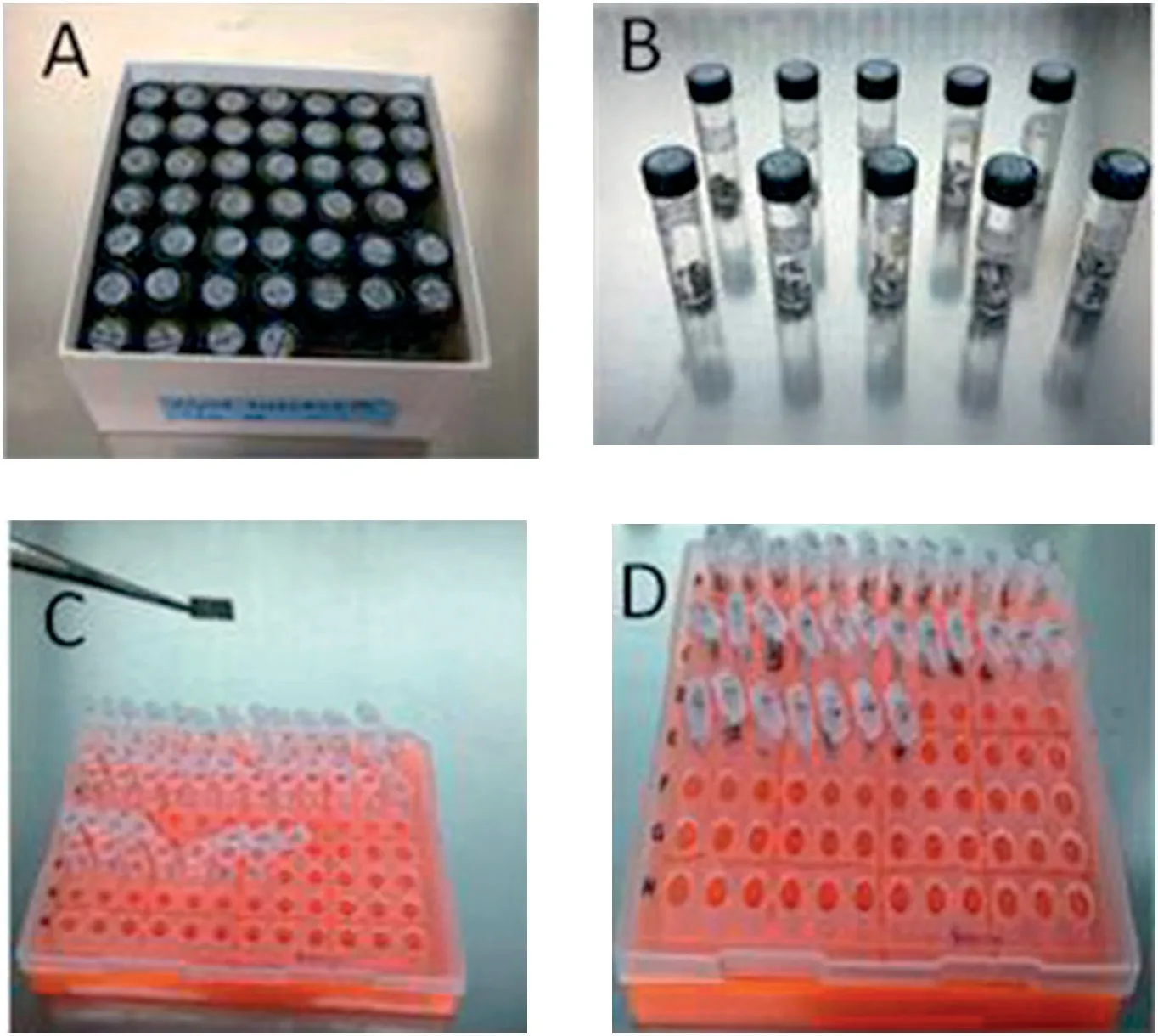
Under a class II type A/B3 flow hood,a filter paper piece from the stored tube containing 5-month-old mycelia and spores was removed and dipped in a 0.2 mL Eppendorf tube containing 100 μL 10× (Tris and EDTA,pH 7.5) (Fig.1).The tube was then heated at 95 °C for 10 min in a thermocycler(PTC-200,MJ Research,Waltham,MA,USA) and centrifuged at 3000 r min-1for 1 min.This DNA extracted for 11 min was stored at 4 °C in a refrigerator until use for PCR amplification.Two sets of primers were designed from the AVR-Pi9 gene(B.Zhou,unpublished data).One set was AVR9-BZ forward(5′-CTG CTC CAT CTT GTT TGG CC-3′),and AVR9-BZ reverse(5′-CAC TAG TAC AAG CAC TAA CC-3′) amplifying a 1 kb genomic fragment.The other set was AVR9-YJ-forward(5′-ATC CCC ATC CAC AGG ATT CC-3′) and AVR9-YJ-reverse (5′-GTG CTT ACT ACT TAG TAT AA-3′) amplifying a 660 bp genomic fragment.The latter were designed using PRIMER 3 (http://biotools.umassmed.edu/bioapps/primer3_www.cgi)based on a genomic sequence encompassing the AVR-Pi9 locus([10];Y.Jia and B.Zhou,unpublished data).These primers were known to amplify a fragment of about 660 bp of the AVR-Pi9 coding region.All PCR reactions were performed using Taq PCR Master Mix(Qiagen Inc.,Valencia,CA,USA).Each PCR consisted of the following components:10 μL of Taq PCR Master Mix(contains 5 U of Taq DNA polymerase,2 × Qiagen PCR buffer,3 mmol L-1MgCl2,and 400 μmol L-1of each dNTP),0.5 μL of each 100 μmol L-1primer,1 μL fungal genomic DNA solution,and 9 μL distilled water(provided by the Qiagen Kit)in a final reaction volume of 20 μL.Reactions were performed in a thermocycler(PTC-200,MJ Research,Waltham,MA,USA)with the following PCR program:1 cycle at 95 °C for 3 min for initial denaturation,29 cycles at 94 °C for 30 s,55 °C for 30 s,72 °C for 60 s,and a final extension at 72 °C for 8 min.
The PCR products were separated by 1.0%(w/v)agarose gel electrophoresis in 1 × TAE,and stained with SYBR Green Safe(Invitrogen Inc.,Grand Island,NY,USA).The gel was visualized and photographed using a Bio-Rad gel photographic system,Chemi Doc MP (Bio-Rad Laboratories,Inc.,Hercules,CA,USA).The size of the amplified fragment was estimated with a Bioline hyperladder 1 kb plus (Bioline USA Inc.,Taunton,MA,USA).
To evaluate the stability of the DNA extracted directly from inoculated filter paper pieces,PCRs were repeated on days 4,8,10,and 18 of refrigerated storage.The tests were performed independently using the same sets of samples following a similar amplification protocol.The same DNA samples were used to amplify AVR-Pita1 using primers YL149/YL169 on day 18 of storage using the protocol described by Dai et al.[11]For a positive control,DNA from ZN61 extracted conventionally was used[12].
3.Results
In an effort to genotype the AVR genes of M.oryzae from a 2012–2013 Arkansas collection,a fast and simple procedure was developed to prepare DNA for PCR amplification.The procedure included two steps: (1) M.oryzae-inoculated filter paper pieces were stored for a minimum of 5 months at –20 °C and transferred to 100 μL of TE (10×,pH 7.5,Tris and EDTA) in a 0.5-mL Eppendorf tube using a sterile loop(Fig.1).The tube was then incubated in a thermocycler at 95 °C for 10 min,and (2)after incubation,the tube was spun for 1 min at 3000 r min-1to prepare the DNA for PCR.
The PCR reaction was modified as follows.Instead of 1 μmol L-1of primer in the final PCR reaction,2.5 μmol L-1of primer was used to increase reproducibility and the success rate of PCR amplification.To evaluate the quality and stability of the extracted DNA,1 μL was repeatedly used throughout the PCR tests on the extraction day and on days 4,8,10,and 18 of refrigerated storage (Fig.2).Predicted PCR products were amplified from fungal structures maintained on filter paper,and from DNA prepared by a conventional procedure as a control (Fig.2).Isolates that did not yield predicted PCR products were confirmed by PCR amplification using another primer,AVR9-YJ that is specific to the coding region of the same gene (Fig.2-D).However,the presence of AVR-Pi9 in isolates 12,13,14,and 28 was undetermined (Fig.2-D).The same set of DNA was also tested using primers YL149/YL169,confirming the presence of AVR-Pita1 in 15 isolates.Again the four isolates in which AVR-Pi9 was not amplified showed no amplification of AVR-Pita1,suggesting problems with the fungal structures or their DNA quality for PCR(Fig.2-E).

Fig.1-Description of DNA preparation from desiccated filter papers.Glass bottles with samples of M.oryzae in a freezer box(A);glass bottles showing desiccated filter pieces carrying M.oryzae fungal mycelia and spores(B);A piece of filter paper containing the fungal structures removed from a glass bottle with a sterilized forceps to be transferred to a 0.2 mL Eppendorf tube containing 10 × TE for DNA extraction(C);and Eppendorf tubes with fungal DNA and filter pieces ready for PCR amplification(D).
4.Discussion
Gene detection using PCR is a common method of microbial identification and diagnosis.Although PCR amplification can be directly performed using various microbial cultures,prior isolation of DNA is often preferred.The DNA extraction process eliminates unknown interfering substances and appears largely to ensure consistent test results.Toward this end,considerable efforts have been made to improve DNA preparation from fungi[6–8,13,14].Many of these methods rely on using a grinder(with or without liquid nitrogen) to break up the mycelia.However,this is a time-consuming task when large number of samples are to be processed.In the present study,the whole procedure can be completed within 11 min at the cost only of TE buffer for sample preparation.It works by disrupting the cell wall and releasing DNA using a high temperature,95 °C,into a highly concentrated TE solution for 10 min.It is important to note that some samples failed to yield PCR products when only 1 μmol L-1of each primer was used(data not shown).However,2.5 μmol L-1of primer was able to ensure successful PCR amplification for most of the samples tested.Additionally,filter pieces used for conserving fungus were sterile and not expected to contribute foreign substances that inhibit the PCR process.As a result,filter paper pieces in 10 × TE may remain in the tube,and the DNA remains usable for repeated PCR amplification for as long as three weeks or more(data not shown).All procedures can be readily performed in a highly contained environment.Thus,this method will also be of great benefit to specialists who routinely perform PCR experiments on a variety of fungal pathogens.It will also be particularly useful when amplification of a fungus is prohibited or impossible owing to its potential toxicity or danger of escape during the pathogen quarantine process.Of the 28 samples tested for the presence of AVR-Pita1,15 showed amplified bands identical to that of the positive control ZN61[11](Fig.2-E).
The availability of a rapid,low-cost,and reliable DNA extraction procedure would considerably reduce not only the workload but also the test turnaround time.The same genomic DNA prepared following this procedure was repeatedly amplified by PCR with three primer pairs (Fig.2).Recently,DNAs prepared several months ago from filters have been successfully used to characterize the genetic diversity of M.oryzae,and filters stored for 1 to 9 years have been used to amplify AVR-Pita1,AVR-Pi9,and other fungal genes(X.Wang and Y Jia,unpublished data).Although this procedure is not suitable for producing large amounts of fungal DNA,we anticipate that it could be applied to the study of many other fungal cultures as a rapid,reliable,and low-cost alternative to the existing DNA extraction protocols for PCR used in research and clinical laboratories.Consequently,this method will be of great benefit for crop breeding and protection worldwide[13].

Fig.2-PCR products amplified using two sets of AVR-Pi9-and one set of AVR-Pita1-specific primers.PCR products of approximately 1 kb amplified with the AVR-Pi9-BZ primer set using freshly prepared DNA on the DNA extraction day(A),at 4 days(B),and at 8 days(C).PCR products of approximately 66 bp amplified using AVR-Pi9-YJ primer using DNA 10 days after extraction(D).PCR products at appropriately 1 kb amplified using primers YL149/YL169 using DNA 18 days after preparation(E).Isolates 1,5,6,8,9,15,16,17,18,19,20,22,25,26,and 27 carrying AVR-Pita1.Lane 1 is a KBM marker and lanes from 1 to 8 and from 9 to 29 were field blast isolates purified from Arkansas commercial rice fields in 2012 and 2013,respectively.
The authors thank Dr.Barbara Valent of Kansas State University and Guo-Liang Wang of Ohio State University for the technical support; Tracy Bianco and Michael Lin of USDA Agriculture Research Service for pathogen isolation,purification,storage,and other technical support; and Scott Belmar for reviewing the manuscript.This project was supported in part by Agriculture and Food Research Initiative Competitive Grant 2013-68004-20378 from the USDA National Institute of Food and Agriculture.USDA is an equal-opportunity provider and employer.
[1] Y.Jia,G.Liu,S.Costanzo,S.Lee,Y.Dai,Current progress on genetic interactions of rice with rice blast and sheath blight fungi,Front.Agric.China 3(2009) 231–239.
[2] M.A.Saghai-Maroof,K.M.Soliman,R.A.Jorgensen,R.W.Allard,Ribosomal DNA spacer-length polymorphisms in barley:Mendelian inheritance,chromosomal location and population dynamics,Proc.Natl.Acad.Sci.U.S.A.81(1984)8014–8018.
[3] N.Blin,D.W.Stafford,Isolation of high molecular weight DNA,Nucleic Acids Res.3(1976) 2303–2308.
[4] J.L.Cenis,Rapid extraction of fungal DNA for PCR amplification,Nucleic Acids Res.20(1992)2380.
[5] J.R.Xu,J.E.Hamer,Assessment of Magnaporthe grisea mating type by spore-PCR,Fungal Genet.Newsl.40 (1995) 80.
[6] D.W.Griffin,C.A.Kellogg,K.K.Peak,E.A.Shinn,A rapid and efficient assay for extracting DNA from fungi,Lett.Appl.Microbiol.34(2002) 210–214.
[7] K.Saitoh,K.Togashi,T.A.T.Teraoka,A simple method for a mini-preparation of fungal DNA,J.Gen.Plant Pathol.72(2006)348–350.
[8] G.C.Graham,P.Mayers,R.J.Henry,A simplified method for the preparation of fungal genomic DNA for PCR and RAPD analysis,BioTechniques 16(1994) 48–50.
[9] Y.Jia,A user-friendly method to isolate and single spore the fungi Magnaporthe oryzae and Magnaporthe grisea obtained from diseased field samples.On line,Plant Health Prog.(2009),http://dx.doi.org/10.1094/PHP-2009-1215-01-BR.
[10] B.Valent,M.S.Crawford,C.G.Weaver,F.G.Chumley,Genetic studies of pathogenicity and fertility of Magnaporthe grisea,Iowa State J.Res.60(1986) 569–594.
[11] Y.Dai,E.Winston,J.C.Correll,Y.Jia,Inducation of avirulence by AVR-Pita1 in virulent U.S.field isolates of Magnaporthe oryzae,Crop J.2(2014) 1–9.
[12] E.Zhou,Y.Jia,P.Singh,J.Correll,F.N.Lee,Instability of the Magnaporthe oryzae avirulence gene AVR-Pita alters virulence,Fungal Genet.Biol.44(2007) 1024–1034.
[13] S.Rozen,H.Skaletsky,Primer3 on the WWW for general users and for biologist programmers,Methods Mol.Biol.132(2000) 365–386.
[14] D.Liu,S.Coloe,R.Baird,J.Pedersen,Molecular determination of dermatophyte fungi using the arbitrarily primed polymerase chain reaction,Br.J.Dermatol.137(1997)351–355.
- The Crop Journal的其它文章
- Morphological,cytological and molecular analyses of a synthetic hexaploid derived from an interspecific hybrid between Gossypium hirsutum and Gossypium anomalum
- Genetic dissection of tetraploid cotton resistant to Verticillium wilt using interspecific chromosome segment introgression lines
- The impacts of conservation agriculture on crop yield in China depend on specific practices,crops and cropping regions
- Exp2 polymorphisms associated with variation for fiber quality properties in cotton(Gossypium spp.)
- Isolation and characterization of a novel wall-associated kinase gene TaWAK5 in wheat(Triticum aestivum)
- Effect of subsoil tillage depth on nutrient accumulation,root distribution,and grain yield in spring maize

