脉搏血氧波形振幅变异率在急诊感染性休克患者容量复苏中的作用
刘业成 徐军 朱华栋 王仲 于学忠


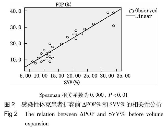
【摘要】目的 脉搏血氧波形振幅变异率(respiratory variations in the pulse oximetry plethysmographic waveform amplitude,ΔPOP)作为容量反应的动态评估指标正在被广泛研究。本研究旨在探讨ΔPOP在急诊感染性休克患者的容量反应评估中的作用。方法 本实验前瞻性地研究2010年10月到2011年10月北京协和医院急诊科抢救室和EICU收治的28例感染性休克患者,记录所有患者在扩容(羟乙基淀粉万汶500 mL)前后的各项血流动力学参数,如心输出量、每搏输出量变异率(stroke volume variation, SVV)和ΔPOP等,容量反应阳性定义为扩容后患者每搏输出量较输液前增加15%以上。结果 容量反应阳性患者扩容前的ΔPOP较容量反应阴性患者高(P<0,01)。扩容前的SVV和ΔPOP两指标之间存在显著的正相关关系,Spearman相关系数为0,900(P<0,01)。结论 ΔPOP对急诊感染性休克患者的容量反应预测准确性较高,和SVV有很好的相关性,优于CI、SVRI、CVP等静态指标,值得进一步研究。
【关键词】容量反应;感染性休克;急诊科
The performance of ΔPOP in the assessment of fluid responsiveness in septic shock patients in emergency department Liu Yecheng, Xu Jun, Zhu Huadong, Wang Zhong, Yu Xuezhong. Department of Emergency Medicine, Peking Union Medical College Hospital, Beijing 100730, China
Corresponding author: Yu Xuezhong, Email: dryxz@sina,com
【Abstract】Objective Respiratory variations in the pulse oximetry plethysmographic waveform amplitude (ΔPOP) have been popularly studied as a dynamic indicators for fluid responsiveness assessment. The authors hypothesized that ΔPOP can indicate fluid responsiveness in septic shock patient in emergency department.Methods A prospective study of 28 patients with septic shock was carried out in Emergency Room and Emergency Intensive Care Unit from 1 October, 2010 to 30 September, 2011. Hemodynamic data including cardiac index, stroke volume variation (SVV) and ΔPOP were recorded before and after volume expansion treatment. Fluid responsiveness was defined as an increase in cardiac index of 15% or greater.Results Changes in ΔPOP after volume expansion were greater in responders than that in non-responders (P<0,01). There was a significant relation between ΔPOP and SVV before volume expansion (r=0,900, P<0,0001). Conclusions ΔPOPcan indicate fluid responsiveness non-invasively in septic shock patient in emergency department. This marker has potential clinical application with high sensitivity and reliability.
【Key words】Fluid responsiveness; Septic shock; Emergency department
容量复苏在感染性休克的早期目标治疗中占据极为重要的地位[1]。足够的血管内容量是使用血管活性药物的前提条件,但过多的容量又会造成肺水肿等副作用,因此对患者容量反应的评估尤为重要。常规的静态评估指标如中性静脉压(CVP)、肺动脉楔压(PCWP)目前被认为不能很好地反映患者的容量状态;而动态评估指标利用了患者每搏量随呼吸的变化,已被证实能较静态指标更好地预测容量反应[2]。但是,动态指标的获取常常有一定困难:每搏输出量变异率(SVV)和脉压变异率(PPV)是有创的,相关的并发症不可回避[3];下腔静脉直径变异率、主动脉流速的脉冲多普勒异率等则在技术上有一定要求,难以广泛开展。目前对于上呼吸的机患者,脉搏血氧波形振幅变异率(respiratory variations in the pulse oximetry plethysmographic waveform amplitude,ΔPOP) 正在被广泛研究[4]。研究发现,ΔPOP对心室的前负荷变化很敏感[5],能在手术室中很好地预测患者的容量反应[6]。本研究旨在探讨ΔPOP在急诊感染性休克患者的容量反应评估中的作用。
1 资料与方法
1,1 一般资料
本实验前瞻性地研究2010年10月到2011年10月北京协和医院急诊科抢救室和EICU收治的感染性休克患者,所有患者满足《2004严重感染和感染性休克治疗指南》的感染性休克诊断标准。入组患者同时满足以下条件:(1)控制性机械通气;(2)有氧饱和度监测;(3)有脉搏波形指示的连续心输出量(pulse indicated continuous cardiac output, PiCCO)監测。患者的入组排除标准:(1)有自主呼吸;(2)有持续的心律失常;(3)已有明确血容量过多的临床表现。
所有患者置入中心静脉导管(颈内静脉或锁骨下静脉)和外周动脉导管(股动脉)进行PiCCO监测,采用热稀释方法测定心输出量,自中心静脉注射15 mL低于8 ℃的生理盐水,描记热稀释曲线,计算得到心输出量指数(CI)、外周血管阻力指数(SVRI)、经胸血管内容量指数(ITBVI)、SVV等。采集数据时对患者充分镇静、镇痛,采用容量控制通气,潮气量8~10 mL/kg,呼吸频率12~15 次/min,呼气末正压设置为0~5 cmH2
另一方面,从感染性休克PiCCO监测的各项指标看,笔者的研究发现常规血流动力学参数心率、平均动脉压、中心静脉压、心输出量指数乃至外周血管阻力对于预测患者的容量反应效果不佳,再一次说明静态指标的局限性,每个患者的心输出量指数、中心静脉压、外周血管阻力等的绝对数并不能提示患者的血管内容量是否足够。在所有的静态指标中,只有PiCCO所特有的容量指标ITBVI在笔者的研究中和患者的容量反应有一定关系。但在实际应用上,笔者也发现,对于一些特殊患者,如本身有心脏问题:心肌肥厚、心腔较小,或心脏扩张、心腔较大的患者,ITBVI的结果也无法用正常人的参考值进行衡量,此时用它预测容量反应并不可靠。而动态容量指标SVV和笔者关注的ΔPOP在感染性休克患者的容量负荷试验中,阳性组和阴性组之前有很显著地差异,说明它们预测患者的容量反应都很可靠。
当然,ΔPOP也有其局限性。首先是动态容量指标的普遍问题,为了保证每次呼吸对循环的影响一致,需要患者在严格的控制性机械通气下才能进行,对非插管上机的患者不适用[15];其次,ΔPOP的应用前提是心律齐,在心律不齐如房颤患者ΔPOP是不适用的;再次,脉搏血氧波形会受患者局部因素的干扰。虽然本研究中没有出现,但皮肤过厚、肤色过深或手指循环太差、皮温过低等因素,都会导致脉搏血氧波形无法显示。
综上所述,笔者的研究认为ΔPOP对急诊感染性休克的患者的容量反应预测准确性较高,和SVV有很好地相关性,优于CI、SVRI、CVP等静态指标,其价值有待于进一步前瞻性研究的证实。
参考文献
[1]姚咏明,刘峰, 盛志勇.多器官功能障碍综合征与脏器功能支持策略[J].中华急诊医学杂志,2006,15(4):293-294.
[2] Pinsky MR, Teboul JL. Assessment of indices of preload and volume responsiveness[J].Curr Opin Crit Care, 2005, 11(3):235-239.
[3] Dong ZZ, Fang Q, Zheng X, et al. Passive leg raising as an indicator of fluid responsiveness in patients with severe sepsis[J]. World J Emerg Med, 2012, 3(3): 191-196.
[4]Shelley KH, Awad AA, Stout RG, et al. The use of joint time frequency analysis to quantify the effect of ventilation on the pulse oximeter waveform[J]. J Clin Monit Comput, 2006, 20(2):81-87.
[5]Han Y, Song ZJ, Tong CY, et al. Effects of hypothermia on the liver in a swine model of cardiopulmonary resuscitation[J]. World J Emerg Med, 2013,4(4): 298-303.
[6] Solus-Biguenet H, Fleyfel M, Tavernier B, et al. Non-invasive prediction of fluid responsiveness during major hepatic surgery[J]. Br J Anaesth, 2006, 97(6):808-816.
[7]Della Rocca G, Costa MG, Coccia C, et al. Preload and haemodynamic assessment during liver transplantation:a comparison between the pulmonary artery catheter and transpulmonary indicator dilution techniques[J]. Eur J Anaesthesiol, 2002,19(12):868-875.
[8]李家琼,李茂琴,许继元,等.脉搏轮廓法在感染性休克早期液体复苏中的运用[J].中华急诊医学杂志,2011,20(1) :30-34.
[9] Melot J, Sebbane M, Dingemans G, et al. Use of indicators of fluid responsiveness in septic shock: a survey in public emergency departments[J]. Ann Fr Anesth Reanim,2012,31(7):583-590.
[10]Iijima T, Iwao Y, Sankawa H. Circulating blood volume measured by pulse dye-densitometry: comparison with (131)I-HSA analysis[J]. Anesthesiology,1998, 89(6):1329-1335.
[11]Shamir M, Eidelaman LA, Floman Y, et al. Pulse oximetry plethysmographic wave form during changes in blood volume[J]. Br J Anaesth, 1999, 82(2): 178-181.
[12]Desebbe O, Cannesson M. Using ventilation induced plethysmographic variations to optimize patient fluid status[J]. Curr Opin Anaesthesiol, 2008, 21(6):772-778.
[13]Natalini G, Rosano A, Franschetti ME, et al. Variations in arterial blood pressure and photoplethysmography during mechanical ventilation[J]. Anesth Analg, 2006, 103(5):1182-1188.
[14] Dorlas JC, Nijboer JA. Photo-electric plethysmography as a monitoring device in anaesthesia[J]. Br J Anaesth, 1985, 57(5): 524-530.
[15]Perner A, Faber T.Stroke volume variation does not predict fluid responsiveness in patients with septic shock on pressure support ventilation[J]. Acta Anaesthesiol Scand, 2006,50(9):1068-1073.
(收稿日期:2013-06-13)
(本文编辑:邵菊芳)
DOI:10,3760/cma,j,issn,1671-0282,2014,01,005
作者单位:100730 北京,北京协和医院急诊科
通信作者:于学忠,Email:dryxz@sina,com
P15-18

