血清非羧化MGP蛋白在强直性脊柱炎中的表达及意义
黄汉清,朱立新,李留洋,钱俊,王坤
血清非羧化MGP蛋白在强直性脊柱炎中的表达及意义
黄汉清,朱立新,李留洋,钱俊,王坤
目的探讨强直性脊柱炎(AS)患者血清非羧化MGP蛋白(ucMGP)的水平及其诊断价值,了解ucMGP与AS炎症及骨化病程的关系。方法选择2010年1月-2012年7月珠江医院收治的82例AS患者作为AS组,同期75例健康体检者作为对照组。评估AS患者各项临床指标(年龄、性别、病程、疾病活动度)、影像学进展以及反映骨代谢或炎症的指标[红细胞沉降率(ESR)、C反应蛋白(CRP)、骨钙素(OC)、骨碱性磷酸酶(BALP)]。疾病活动度依据BASDAI评分标准,影像学进展以mSASSS评分标准为依据。采用竞争性ELISA法检测AS患者血清ucMGP浓度。利用ROC曲线分析ucMGP诊断AS的价值。运用相关分析法分析ucMGP与疾病活动度、影像进展、骨代谢及炎症生物标志的关系。结果AS组ESR及血清CRP水平高于对照组,但ucMGP浓度(2958±654nmol/L)低于对照组(4551±1036nmol/L,P<0.01)。ROC曲线分析显示,ucMGP=3859nmol/L(截断点)时的诊断特异性及敏感性分别为69.7%、91.5%,Youden指数为0.612,曲线下面积(AUC)为0.825,其诊断效力为中级。相关分析显示,血清ucMGP水平与ESR、CRP、BALP、OC以及BASDAI评分无明显相关性。影像学mSASSS评分与ucMGP水平呈显著负相关(r=-0.715,P<0.01),当mSASSS>0、1、10时,r分别为-0.715、-0.741、-0.776(P<0.01),当mSASSS<10时,r=-0.297(P=0.028),相关性不明显。结论AS患者血清ucMGP可作为一种新的血清诊断指标及反映骨化进展的生物标志,尤其是在AS骨化病程后期。
Gla蛋白,基质,非羧化;脊柱炎,强直性;骨化
强直性脊柱炎(ankylosing spondylitis,AS)是一种以附着点炎及韧带骨赘形成为主要病理特征的慢性进展性炎性疾病,最终导致进展性脊柱强直及活动受限[1]。尽管韧带骨赘形成代表骨质增生,但目前研究显示大部分AS患者尤其是晚期患者表现为骨密度下降[2]。因此,X线片以及能够反映骨及软骨代谢的指标对于检测AS的疾病活动情况、骨质疏松或者骨质改变具有重要意义。为量化AS的病理进程,可对传统X线片进行影像评分以评价骨组织的改变情况[3]。在目前常用的X线片影像评分系统中,mSASSS(modified Stoke Ankylosing Spondylitis Spine Score)被认为具有较高的临床应用价值[4]。基质Gla蛋白(matrix Gla protein,MGP)是一种分子量为14kD的低分子胞外基质蛋白,因含有5个可羧化γ-谷氨酸残基而得名[5],另外还带有多个可磷酸化的丝氨酸残基[6]。MGP主要由软骨细胞[7]及血管平滑肌细胞分泌[8],可以抑制血管钙化[9]及软骨成骨分化。MGP活性决定于羧化的γ-谷氨酸残基,其羧化过程具有维生素K依赖性[10]。MGP的羧化或者磷酸化使血液中存在多种MGP分子形式,其中非羧化MGP蛋白被称为ucMGP。目前尚未见关于AS患者血清或局部MGP蛋白水平的大样本临床研究。Silaghi等[11]研究发现,关节炎患者群体(包括5例AS患者)总体上表现出关节液与血液中ucMGP的分布异常。AS骨赘形成主要以软骨化骨的方式进行,推测MGP可能在AS的骨化过程中发挥负性调节作用。为此本研究观察AS患者的血清ucMGP水平,及其与AS相关的临床指标如BASDAI(bath AS disease activity index),影像评分(mSASSS),血清中炎症指标如红细胞沉降率(ESR)、C反应蛋白(CRP)水平等,骨代谢指标如骨碱性磷酸酶(BALP)、骨钙素(OC)等的关系,以期为AS的临床诊断提供新的思路。
1 资料与方法
1.1病例资料 选择2010年1月-2012年7月珠江医院收治的82例AS患者及同期75例健康体检者加入随机对照研究。AS患者均符合修改的纽约诊断标准(1984年)[12],尽量避免因病情活动造成的选择偏倚。排除具有心血管疾病、慢性肾脏疾病、感染、其他风湿性疾病,以及正使用抗肿瘤坏死因子生物制剂,维生素K及其拮抗剂如华法林,维生素D等的患者。对照组来自健康体检人群,无心血管疾病、肝肾疾病、风湿性疾病、炎症及感染,未服用维生素K及其拮抗剂、维生素D。纳入研究的AS患者及健康对照均能提供研究所涉及的各项临床资料及检测结果,否则予以排除。
1.2临床评估 收集AS患者及健康对照的临床资料,包括一般临床资料[年龄、性别、病程、体重指数(BMI)],HLA-B27表型,影像学资料(颈椎及腰椎侧位X线片),ESR(Westergren 法),CRP浓度(免疫比浊法测定)。参照BASDAI评分标准[13]设计随访问卷(0-10分),获得每例患者的BASDAI评分。由3位具有影像专业知识背景的医师对患者脊柱双侧侧位X线片进行独立阅片,按照mSASSS标准[4]进行评分,最终分值为3位医师评分的平均值。
1.3血清保存及检测 抽取AS患者及健康对照的静脉血,置于真空非抗凝管,30min内迅速保存于-20℃,并在1个月内通过离心(2400×g,10min)获取上层血清后转移到-80℃长期保存。BALP、OC浓度检测采用ELISA法,具体操作按照试剂盒(NovaTeinBio,USA)说明书进行。ucMGP浓度检测采用竞争ELISA法。以带有生物素标记的ucMGP多肽序列(ucMGP35-54)的人工合成类似物(Pepscan,Leylystad,Netherlands)作为标准品,该多肽序列带生物素标记后(biotinylated ucMGP 35–54,b-ucMGP35–54)则作为竞争抗原。具体检测过程如下:多克隆兔抗鼠IgG抗体以0.1mol/L碳酸盐溶液(pH9.6)稀释2000倍后,铺板(100μl/孔),37℃孵育1.5h,10%小牛血清封闭(200μl/L),室温下孵育2h。微孔板洗板5次,加入100μl针对ucMGP Gla结构域(35-54序列)的单克隆抗体(mAb-ucMGP;VitaK BV,Maastricht,Netherlands),37℃孵育过夜,洗板5次。取5μl标准品或血清样品加入70μl PBS-Tween 20缓冲液稀释,取20μl稀释后的标准品或血清样品与100μl b-ucMGP35–54 (0.7μg/ml)混合。取100μl上述混合液加入微孔板,4℃孵育过夜。洗板3次后,加入稀释10 000倍的链霉素抗生物素蛋白-过氧化酶(streptavidin-peroxide)100μl,37℃孵育0.5~1h,洗板5次,加入100μl TMP显色,加入50μl 1mol/L H2SO4,450nm光谱下读板。通过标准品曲线计算ucMGP浓度。
1.4统计学处理 采用SPSS 16.0软件进行统计分析。计量资料以±s表示,组间比较采用Mann-Whitney U检验,计数资料比较采用χ2检验。通过ROC曲线(receiver operating characteristic curve)分析确定ucMGP作为AS诊断指标的截断点,并确定其在该点的诊断特异性、敏感性、曲线下面积(AUC)、Youden指数。诊断效力评级:AUC≤0.7为低级,0.7<AUC<0.9为中级,AUC≥0.9为高级。ucMGP水平与临床及血清指标的关系分析采用Spearman秩相关法。P<0.01为差异有统计学意义。
2 结 果
2.1AS患者与健康对照临床资料、血清炎症及骨代谢相关指标的比较 AS组年龄、性别、BMI、BALP、OC等指标与对照组比较差异无统计学意义,但ESR及CRP水平明显高于对照组,而血清ucMGP浓度明显低于对照组,差异有统计学意义(P<0.01,表1)。
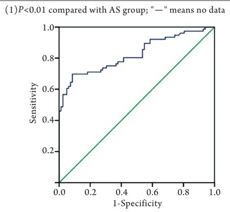
2.2血清ucMGP作为AS诊断指标的最佳截断点及诊断价值 ROC曲线分析显示,ucMGP诊断AS的界值(最佳截断点)为3859nmol/L,此时可获得最优化的诊断特异性(69.7%)及敏感性(91.5%)组合,Youden指数为0.612,AUC为0.825(图1),诊断效力可评为中级。

表1 临床指标和血清生物学标记的比较Tab. 1 Comparison of clinical parameters and serum

图1 血清ucMGP浓度水平作为诊断指标的ROC曲线分析Fig. 1 ROC curve analysis based on ucMGP level as diadynamic criteria
2.3AS患者血清ucMGP浓度与各指标的相关性Spearman相关分析显示,AS患者血清ucMGP浓度与ESR、CRP、BALP、OC、BASDAI评分均无明显相关性(P>0.05),但与颈椎及腰椎侧位X线片mSASSS评分呈显著负性相关(r=-0.715,P<0.01)。绘制ucMGP-mSASSS散点图(图2)并进行分区间分析显示,分别取mSASSS>0、1、10时,Spearman相关系数r分别为-0.715、-0.741、-0.776,P值均小于

图2 ucMGP血清浓度值与mSASSS 评分关联性特征散点图Fig. 2 Relevance between mSASSS score and serum ucMGP concentration
3 讨 论
本研究通过前瞻性对照发现AS患者血清ucMGP水平明显低于健康对照人群,其均值仅为对照组的65%,而Silaghi等[11]的研究报道5例AS患者血清ucMGP低于对照组,但差异无统计学意义。为了解ucMGP对AS的诊断价值,本研究利用ROC曲线分析,结果显示,以ucMGP=3859nmol/L作为界值诊断AS的特异性及敏感性分别为69.7%、91.5%,Youden指数为0.612,AUC为0.825,诊断价值为中级。而ucMGP诊断AS的特异性较低,考虑与多种疾病如心血管疾病、慢性肾衰竭、类风湿等有关,维生素K、D等的摄入也可能影响其浓度,即使在研究设计时将上述疾病纳入排除标准,但由于疾病早期临床常难以鉴别,因此可能对实验结果造成一定影响。ucMGP通过ELISA试剂盒即可检测,相对于HLA-B27需采用流式细胞术进行测定,成本更低,检测速度更快,如果与HLA-B27结合将有望提高AS的诊断率。
为了解MGP蛋白在AS病理过程中的作用,本研究对其与AS炎症及骨代谢指标的关系进行了分析,结果显示AS患者CRP、ESR明显偏高,而其他指标与对照组比较无明显差异,且CRP、ESR、BALP、OC、BASDAI等与ucMGP无明显相关性。既往研究显示,AS患者的CRP、ESR、BASDAI与其活动期炎症过程密切相关[14-15],但反映AS的病理骨化进程不敏感[15]。本研究显示ucMGP与CRP、ESR、BASDAI无明显相关,亦提示其与AS活动期炎症的联系并不紧密。虽然BALP、OC与ucMGP无显著相关,但不能因此确定ucMGP未参与AS的骨化过程,因为BALP、OC虽然可很好地反映成骨代谢水平[16],但对AS的病理性骨化过程并不敏感[15,17]。
本研究采用mSASSS评分系统对AS患者的影像学进展进行量化评分,结果显示血清ucMGP浓度与mSASSS评分呈显著负相关,散点图观察及分区间分析显示,随着mSASSS评分增加,二者的关联性增强。mSASSS是目前评价AS脊柱骨化进展最敏感的影像指标,其值越高表明AS骨化程度越严重,因此本研究结果提示血清ucMGP浓度可作为反映AS骨化程度的指标,尤其是在AS病程后期。
既往研究显示MGP蛋白对于异位骨化[18]及软骨化骨[19]具有负性调节作用,其抑制钙化及骨化的机制包括:①与钙离子结合清除组织中富余的钙离子[20],或与钙盐晶体结合阻止羟基磷灰石晶体形成[21];②抑制BMP-2的成骨活性,以调节软骨细胞成熟及成骨分化[12]。因此,对于AS患者血清ucMGP浓度降低,我们认为是由于ucMGP易聚集于AS骨化活跃的病灶处,并可能在骨化病程中扮演负性调节角色,骨化越严重,ucMGP在局部的聚集越明显,从而循环ucMGP的浓度越低。但该观点尚需要病理学研究支持。
综上所述,本研究发现,AS患者血清ucMGP可作为一种新的诊断指标及反映骨化进展的生物学标志物,尤其是在AS骨化病程后期。
[1]van der Linden S, van der Heijde DM. Clinical and epidemiologic aspects of ankylosing spondylitis and spondyloarthropathies[J]. Curr Opin Rheumatol, 1996, 8(4): 269-274.
[2]Karberg K, Zochling J, Sieper J, et al. Bone loss is detected more frequently in patients with ankylosing spondylitis with syndesmophytes[J]. J Rheumatol, 2005, 32(7): 1290-1298.
[3]Braun J, Golder W, Bollow M, et al. Imaging and scoring in ankylosing spondylitis[J]. Clin Exp Rheumatol, 2002, 20(6 Suppl 28): S178-S184.
[4]Wanders AJ, Landewe RB, Spoorenberg A, et al. What is the most appropriate radiologic scoring method for ankylosing spondylitis? A comparison of the available methods based on the Outcome Measures in Rheumatology Clinical Trials filter[J]. Arthritis Rheum, 2004, 50(8): 2622-2632.
[5]Price PA, Urist MR, Otawara Y. Matrix Gla protein, a new gamma-carboxyglutamic acid-containing protein which is associated with the organic matrix of bone[J]. Biochem Biophys Res Commun, 1983, 117(3): 765-771.
[6]Price PA, Rice JS, Williamson MK. Conserved phosphorylation of serines in the Ser-X-Glu/Ser(P) sequences of the vitamin K-dependent matrix Gla protein from shark, lamb, rat, cow, and human[J]. Protein Sci, 1994, 3(5): 822-830.
[7]Luo G, D'Souza R, Hogue D, et al. The matrix Gla protein gene is a marker of the chondrogenesis cell lineage during mouse development[J]. J Bone Miner Res, 1995, 10(2): 325-334.
[8]El-Maadawy S, Kaartinen MT, Schinke T, et al. Cartilage formation and calcification in arteries of mice lacking matrix Gla protein[J]. Connect Tissue Res, 2003, 44(Suppl 1): 272-278.
[9]Luo G, Ducy P, Mckee MD, et al. Spontaneous calcification of arteries and cartilage in mice lacking matrix GLA protein[J]. Nature, 1997, 386(6620): 78-81.
[10]Vermeer C. Gamma-carboxyglutamate-containing proteins and the vitamin K-dependent carboxylase[J]. Biochem J, 1990, 266(3): 625-636.
[11]Silaghi CN, Fodor D, Cristea V, et al. Synovial and serum levels of uncarboxylated matrix Gla-protein (ucMGP) in patients with arthritis[J]. Clin Chem Lab Med, 2011, 50(1): 125-128.
[12]van der Linden S, Valkenburg HA, Cats A. Evaluation of diagnostic criteria for ankylosing spondylitis. A proposal for modification of the New York criteria[J]. Arthritis Rheum, 1984, 27(4): 361-368.
[13]Garrett S, Jenkinson T, Kennedy LG, et al. A new approach to defining disease status in ankylosing spondylitis: the Bath Ankylosing Spondylitis Disease Activity Index[J]. J Rheumatol, 1994, 21(12): 2286-2291.
[14]Spoorenberg A, van der Heijde D, de Klerk E, et al. Relative value of erythrocyte sedimentation rate and C-reactive protein in assessment of disease activity in ankylosing spondylitis[J]. J Rheumatol, 1999, 26(4): 980-984.
[15]Park MC, Chung SJ, Park YB, et al. Bone and cartilage turnover markers, bone mineral density, and radiographic damage in men with ankylosing spondylitis[J]. Yonsei Med J, 2008, 49(2): 288-294.
[16]Christenson RH. Biochemical markers of bone metabolism: an overview[J]. Clin Biochem, 1997, 30(8): 573-593.
[17]Korczowska I, Przepiera-Bedzak H, Brzosko M, et al. Bone tissue metabolism in men with ankylosing spondylitis[J]. Adv Med Sci, 2011, 56(2): 264-269.
[18]Murshed M, Schinke T, Mckee MD, et al. Extracellular matrix mineralization is regulated locally; different roles of two glacontaining proteins[J]. J Cell Biol, 2004, 165(5): 625-630.
[19]Yagami K, Suh JY, Enomoto-Iwamoto M, et al. Matrix GLA protein is a developmental regulator of chondrocyte mineralization and, when constitutively expressed, blocks endochondral and intramembranous ossification in the limb[J]. J Cell Biol, 1999, 147(5): 1097-1108.
[20]Hackeng TM, Rosing J, Spronk HM, et al. Total chemical synthesis of human matrix Gla protein[J]. Protein Sci, 2001, 10(4): 864-870.
[21]O'Young J, Liao Y, Xiao Y, et al. Matrix Gla protein inhibits ectopic calcification by a direct interaction with hydroxyapatite crystals[J]. J Am Chem Soc, 2011, 133(45): 18406-18412.
Expression of uncarboxylated matrix Gla protein in ankylosing spondylitis and its significance
HUANG Han-qing1, ZHU Li-xin1*, LI Liu-yang2, QIAN Jun2, WANG Kun1
1Department of Orthopedics,
2Department of Organ Transplantation, Zhujiang Hospital, South Medical University, Guangzhou 510280, China
*
, E-mail: zhulixin1966@163.com
ObjectiveTo investigate the serum level of uncarboxylated matrix Gla protein (ucMGP) in ankylosing spondylitis (AS) patients, and to evaluate its diagnostic value and the relation of ucMGP to inflammation and ossification process in AS.MethodsEight-two AS patients and 76 healthy controls were enrolled in this randomized controlled study. The clinical indices (age, gender, course of disease, disease activity), changes in radiographic studies, and indices of bone metabolism or inflammation, including erythrocyte sedimentation rate (ESR), C-reactive protein (CRP), osteocalcin (OC), and bone-specific alkaline phosphatase (BALP) were evaluated or measured. The disease activity was assessed by Bath Ankylosing Spondylitis Disease Activity Index (BASDAI), and changes in radiographic pictures were evaluated according to the modified Stoke AS Spine Score (mSASSS), and serum level of ucMGP was measured by a competitive ELISA. The relationship between ucMGP and clinical indexes, radiographic scoring, indices in bone metabolism or inflammation was estimated by SPSS software, and the diagnostic value of ucMGP was analyzed by receiver operator characteristic (ROC) curve.ResultsThe levels of ESR and CRP in AS patients were higher than those in healthy controls, but the serum ucMGP was lower (2958±654nmol/L) compared with healthy controls (4551±1036nmol/L,P<0.01). ROC analysis revealed that the diagnostic specificity and sensitivity of ucMGP was 91.5% and 69.7% with a cut-off of 3859nmol/L (Youden index 0.612, AUC 0.825, 95% CI 0.758-0.891), and the diagnostic validity was moderate. There was no significant correlation between ucMGP and ESR, CRP, BALP, OC, BASDAI score. There was a significant negative correlation between the radiographic progression of the lesion and serum ucMGP (r=-0.715,P<0.01). This correlation was stronger when radiographic scores rose (mSASSS>0, r=-0.715,P<0.01; mSASSS >1, r=-0.741,P<0.01; mSASSS >10, r=-0.776,P<0.01; mSASSS <10, r=-0.297, P=0.028).ConclusionSerum ucMGP may serve as a diagnostic biomarker of AS and progression index of ossification, especially in late stage of AS.
Gla protein, matrix, uncarboxylated; spondylitis, ankylosing; ossification
R593.23
A
0577-7402(2013)06-0489-04
2012-12-20;
2013-04-22)
(责任编辑:胡全兵)
黄汉清,医学硕士。主要从事骨科的基础与临床研究
510280 广州 南方医科大学附属珠江医院骨科(黄汉清、朱立新、王坤),器官移植科(李留洋、钱俊)
朱立新,E-mail:zhulixin1966@163.com

