Surgical treatment of hepatocellular carcinoma with inferior vena cava tumor thrombus: a new cIassif i cation for surgicaI guidance
Shanghai, China
Surgical treatment of hepatocellular carcinoma with inferior vena cava tumor thrombus: a new cIassif i cation for surgicaI guidance
Ai-Jun Li, Wei-Ping Zhou, Chuan Lin, Xi-Long Lang, Zhen-Guang Wang, Xiao-Yu Yang, Qing-He Tang, Ran Tao and Meng-Chao Wu
Shanghai, China
BACKGROUND:Hepatic resection is the main treatment modality for hepatic tumors. Advances in diagnostic technique, preoperative preparation, surgical technique, and postoperative management increased the success rate. The present study aimed to evaluate hepatectomy and resection of inferior vena cava tumor thrombus (IVCTT) in patients with hepatocellular carcinoma, and the relationship between IVCTT classif i cation and selection of surgical technique.
METHODS:We retrospectively reviewed 13 patients with hepatocellular carcinoma who had undergone hepatectomy with IVCTT resection between May 1997 and August 2009. Age, gender, diagnosis, fi ndings of physical examination, results of preoperative laboratory investigations, radiological examination, criteria for resection, postoperative pathological results, incisions, operative technique, intraoperative transfusion, drains, and intraoperative and postoperative complications were evaluated for all patients.
RESULTS:Type I IVCTT (10 patients) was posterior to the liver and below the diaphragm; type II IVCTT (2 patients) was above the diaphragm but still outside the atrium; and type III IVCTT (1 patient) was above the diaphragm and in the right atrium. Type I was treated by radical hepatectomy and removal of IVCTT with total hepatic vascular exclusion. Type II was treated by radical hepatectomy and removal of IVCTT by incision of thediaphragm. Type III was treated by hepatectomy and resection of the thrombus from the right atrium under cardiopulmonary bypass. There were no surgical complications and one patient has been survived for 4 years with cancer-free status. The median survival time was 18.2 months, and the 1- and 2-year survival rates were 53.8% and 15.4%, respectively.
CONCLUSION:Surgical treatment is safe and feasible for treatment of IVCTT in patients with hepatocellular carcinoma, and surgical resectability can be judged according to the classif i cation of tumor thrombus.
(Hepatobiliary Pancreat Dis Int 2013;12:263-269)
liver tumor;inferior vena cava; hepatectomy; tumor thrombus; total hepatic vascular exclusion
Introduction
Hepatocellular carcinoma (HCC), a common malignant tumor,[1,2]is seen in 11%-23% of cases associated with venous thrombus,[3]3.8% with inferior vena cava tumor thrombus (IVCTT),[1]and 2% with tumor thrombus extending to the right atrium.[2]HCC with IVCTT is very rare and only approximately 300 cases have been reported.[4-19]HCC with IVCTT may be complicated by lung metastasis, pulmonary infarction, secondary Budd-Chiari syndrome, ball-valve thrombus syndrome, and heart failure.[3,4]Furthermore, it is associated with an increased risk of sudden death due to heart failure or pulmonary embolism and urgent management is needed. The prognosis of such patients is extremely poor, and the median survival is 1.9 months,[5]which is worse than that of patients with portal vein tumor thrombus.
The inferior vena cava (IVC) is removed when it wasinvaded by a liver tumor. Only a few investigators[2,3,6-13]have reported surgery for HCC associated with IVCTT, partly because it carries a high risk and is technically diff i cult. Technical developments such as total hepatic vascular exclusion (THVE) have made this type of operation both feasible and prevalent. Many reports on IVC resection for the treatment of liver tumors were documented following the inauguration of THVE. It is generally accepted that the prognosis is good for radical hepatectomy plus the resection of tumor thrombus in the hepatic vein and the removal of IVCTT, as long as there is no local or distant metastasis. However, a standard treatment modality has not been established. Any treatment that controls liver cancer potentially could prolong the survival of patients. Treatment options include surgery, local ablation, chemotherapy, immunochemotherapy, isolated hepatic perfusion therapy, radiotherapy and transarterial chemoembolization (TACE).[1,4,9,14,15]The current literature does not provide evidence of survival benef i t from any treatment for patients with HCC associated with IVCTT.
The present study reviewed 13 liver cancer patients with IVCTT who underwent surgical resection between May 1997 and August 2009 in terms of clinical, IVCTT classif i cation, and surgical perspectives. The eff i cacy of hepatectomy combined with IVCTT resection was evaluated. The aim of this report was to describe the classif i cation of IVCTT and its guidance to surgery. The different types of IVCTT require different surgical methods.
Methods
Clinical data
All patients who had been operated upon for hepatic tumors with IVCTT between May 1997 and August 2009 were included in this study. The choice of treatment was made by the patient after a full discussion with our multidisciplinary treatment team including surgeons, hepatologists and oncologists. Patient details, treatment procedure, postoperative course, and disease progress were collected. The outcomes included overall survival rate, procedure-related complications and hospital mortality. Patient survival was calculated from the date of treatment to the last follow-up or death. Accumulative survival rate was calculated using the Kaplan-Meier method.
IVCTT invasion was assessed by computed tomography (CT), ultrasonography, IVC venography, and magnetic resonance imaging (MRI) in all patients, and fi nally by surgery. The follow-up included a complete history, physical examination, abdominal ultrasonography or contrast CT and MRI, and laboratory evaluation of the liver, renal function tests and tumor markers. The followup was carried out every month for the fi rst 3 years, and then the interval of the follow-up was increased gradually.
Classif i cation of IVCTT
IVCTT is usually disseminated from the hepatic veins or short hepatic veins. In 13 patients of this series, 8 had IVCTT extended from the right hepatic vein, 1 from the middle hepatic vein, 1 from the left hepatic vein, and 3 from the right posterior branch of the short hepatic vein. Clinically, IVCTT was classif i ed into three types according to its anatomic location close to the heart: (1) posterior hepatic type (type I), where the tumor thrombus was in the IVC posterior to the liver and below the diaphragm; (2) superior hepatic type (type II), where the tumor thrombus was in the IVC above the diaphragm but still outside the atrium; and (3) intracardiac type (type III), where the tumor thrombus was above the diaphragm and entered the right atrium.
Surgical techniques
The fi rst procedure was the assessment of resectability. After endotracheal induction of general anesthesia, the patient was positioned in the recumbent position with a pad under the lower thoracic and upper lumbar region to facilitate exposure. Bilateral subcostal incisions were made and extended superiorly in the midline to the xiphoid. The location and size of the tumor and the type of IVCTT were explored. This improved visualization of the conf l uence of the hepatic veins. The liver was then mobilized by dividing the falciform, right and left triangular ligaments, and coronary ligaments. The hepatoduodenal ligament was identif i ed and surrounded with a vascular tape to expedite a subsequent Pringle's maneuver. The suprahepatic and infra-hepatic IVCs were exposed, vascular occlusion bands were placed around the IVC. Two vascular clamps were placed across the suprahepatic and infrahepatic IVC. The hepatic parenchyma was fi rst dissected after Pringle's occlusion, leaving only one hepatic vein in connection with the tumor on the liver cut surface. Partial division of the hepatic parenchyma was required before the division into the hepatic vein and the IVC could be seen. This resulted in a clear demarcation of the hepatic vein and the IVC. The fully fi lled hepatic vein was dissected without being removed and ligated temporarily.
For type I tumor thrombus, Pringle's maneuver was performed fi rst, followed by occlusion of the suprahepatic and infrahepatic IVC with vascular tape or occlusion forceps and then the hepatic vein and theconnecting IVC wall were incised under direct vision (by opening the infrahepatic IVC longitudinally). The tumor and thrombus were removed together under direct vision. The IVC was washed, and the wall was sewn up. The head of the thrombus was round, smooth and red-white with a bullet shape. If the tumor thrombus/ embolus was ruptured, it was removed with an oval clamp. The IVC was washed repeatedly. Among the 10 type I tumor thrombi in our study, the tumor and IVCTT were removed under direct vision in 7 patients, the tumor was dissected from the IVC wall in 2, and the IVC was partially resected and repaired in 1 (Figs. 1, 2 and 3).

Fig. 1.Enhanced CT scan of the abdomen: (type I) tumor thrombus of the IVC.
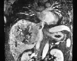
Fig. 2.MRI of the abdomen shows a (type I) tumor thrombus causing intraluminal invasion of the IVC.

Fig. 3.Surgical specimen: the cut surface of the tumor invading into the right hepatic vein and IVC. Tumor thromb in the right hepatic vein (arrow), and IVC (right).
As blood occlusion at the infradiaphragmatic IVC is prohibited for type II tumor thrombus, we fi rst dissected the infrahepatic IVC; pulled the hepatic round ligament downward; dissected the falciform and bilateral triangular ligaments; dissected the posterior hepatic IVC under the diaphragm by about 4 cm; incised the diaphragm muscle anterior to the suprahepatic IVC to enter the diaphragm and expose the right atrial appendage; performed Pringle's maneuver; occluded the infrahepatic IVC with vascular tape; and then occluded the suprahepatic IVC above the tumor thrombus with the vascular tape or occluded the right atrial appendage with a Satinsky clamp. This technique ensures that tumor thrombus is not slipping or falling-off, and therefore, avoids acute pulmonary embolism. We incised the posterohepatic IVC longitudinally, resectedthe tumor and thrombus under direct vision, and fi nally sewed up the pericardium, diaphragm muscle and IVC wall. The Satinsky clamp or the suprahepatic IVC over the tumor thrombus with the vascular tape was released fi rst, and then the infrahepatic IVC occlusion, and last, the Pringle's maneuver (Fig. 4).
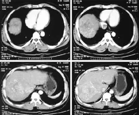
Fig. 4.CT-scan of the abdomen shows a solid mass in the right lobe of the liver. (Type II) tumor thrombus in the right hepatic vein and suprahepatic IVC above the diaphragm.
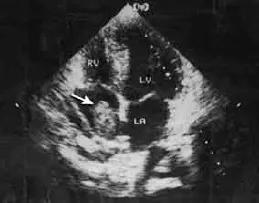
Fig. 5.Color Doppler imaging confirms the diagnosis (arrow). (Type III) tumor thrombus extending into the atrium.
For type III, tumor thrombus extended into the right atrium, hepatectomy plus resection of IVCTT and right atrium TT were performed by liver and cardiothoracic surgeons together. The liver tumor and thrombus were removed together under direct vision, and cardiothoracic surgery was performed under cardiopulmonary bypass (CPB) to remove the IVCTT growing into the right atrium (Fig. 5).
Results
General information
The median age of the patients was 49.7 years (range 35-72). There were 11 males and 2 females, and all 13 patients were classif i ed as Child-Pugh class A. The main clinical manifestations were upper abdominal pain and/ or discomfort (all patients). One patient had a history of edema of the lower extremities, which disappeared on admission. There was no ascites or blood stasis of the lower body in any of the patients. One patient had received radiotherapy preoperatively. The tumor size was 4-18 cm in diameter (mean 10.2).
Ten patients with type I underwent radical hepatectomy plus resection of the tumor thrombus; 2 patients with type II underwent radical hepatectomy plus resection of the tumor thrombus by opening longitudinally the infrahepatic IVC; and 1 patient with type III underwent right hepatectomy plus resection of the tumor thrombus from the right atrium by CPB opening of the right atrium to remove the tumor thrombus. Tumor was located in the right lobe in 10 patients, in the left lobe in 1 patient, and segment IV+V+VIII (of Couinaud) and segment V+VI+VII+ paracaval portion (PCP) in 1 patient each. No distant metastasis or portal vein tumor thrombus was found in all of the patients during the preoperative examination (Table). There were 10 type I cases, 2 type II cases, and 1 type III case. Table summarizes the patient characteristics, treatment details and outcomes.
Using intermittent Pringle's maneuver at room temperature, surgery was successful in all 13 patients, with no surgical mortality or intraoperative pulmonary embolism. At the same time, total hepatic vascular occlusion was performed within 6 to 25 minutes in 12 patients. There was no signif i cant change in pulse, bloodpressure and central venous pressure before and after occlusion for type I IVCTT. However, the blood pressure in one of the type II patients was increased from 40 to 60 mmHg after occlusion. The length of IVCTT was 2-6 cm, which was consistent with the preoperative MRI fi ndings. There was no signif i cant adhesion between the tumor thrombus and the IVC wall in 10 patients, in whom the tumor thrombus was removed completely. In 2 of the 10 patients, close adhesion was found between the tumor thrombus and the IVC wall. The IVC was partially resected and repaired in one patient. In all 13 patients, blood loss during operation was 300-8000 mL (mean 2276.9). Postoperative pathology conf i rmed the diagnosis of HCC in all 13 patients.
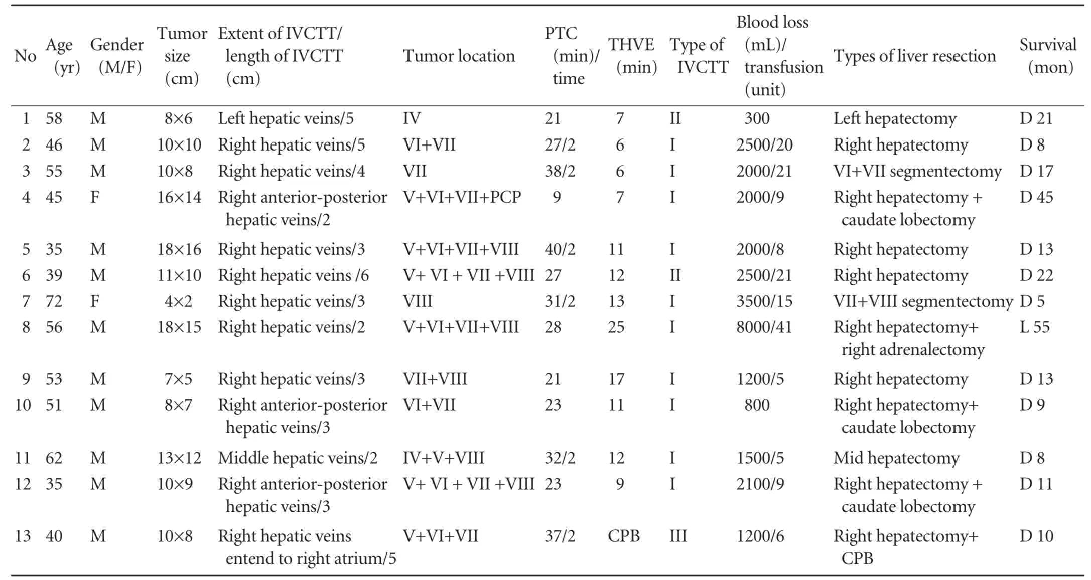
Table.Patients' characteristics
Complications
No perioperative mortality or postoperative hemorrhage was observed in addition to biliary leakage, stricture, or jaundice, and long-term complication. Liver enzyme, total protein and albumin returned to normal within 6-8 days. Postoperative bilirubin and aspartate aminotransferase levels and prothrombin time were also reversed to normal.
Follow-up
The patients were followed up postoperatively for 1-6 years. during this period, 9 patients received TACE, and 3 received radiotherapy, microwave therapy and radiofrequency therapy, respectively. One patient developed jaundice and another patient had multiple intrahepatic metastases 5 months later after surgery. One patient received a second operation for tumor recurrence during the following year. In the fourth year after surgery, one patient died from cerebral metastasis and another from pulmonary metastasis. Nine of the 13 patients died from HCC recurrence. One patient survived for 4 years. The 1- and 2-year survival rates were 53.8% and 15.4%, respectively (Fig. 6).

Fig. 6.Overall survival curve of 13 patients after surgery. The 1-and 2-year survival rates were 53.8% and 15.4%, respectively.
Discussion
HCC invades the hepatic vein and causes venous thrombus. The thrombus further extends to the IVC to form IVCTT, or even to the right atrium.[3]Only a few studies[2,3,6-13]have shown that surgical removal of tumor thrombus may be feasible, but surgery is rarely performed owing to the limited hepatic reserve of patients. However, no single treatment has yet been established as the gold standard.
Yamanaka and colleagues[7]have reported longterm prognosis of liver resection for HCC with tumor thrombus in the hepatic vein, vena cava, and atrium. It was previously believed that the prognosis of liver cancer with IVCTT was poor[5]and surgical treatment was not preferable. However, recently, Liu et al[9]reported that hepatectomy plus thrombectomy with THVE was practical for patients with HCC and portal vein tumor thrombus and IVCTT, and that the long-term survival and recurrence-free survival were superior to those of chemotherapy alone. However, the authors did not def i ne the classif i cation of IVCTT in detail.
For the classif i cation of IVCTT, the location of type I tumor thrombus is below the diaphragm. For type II IVCTT, if subdiaphragmatic occlusion of the IVC, thrombus may cause rupture of the IVC. Thrombus may fall-off, migrate to the heart, and result in cardiac tamponade, acute pulmonary embolism and other severe complications. For type III IVCTT, tumor thrombus may extend into the right atrium, surgery is performed under CPB to remove the IVCTT. So, the different types of IVCTT require different surgical methods.
Preoperatively, Doppler ultrasound, CT and MRI, magnetic resonance angiography are able to detect the size, location, length and degree of tumor thrombus directly and clearly, which guide the selection of surgical modalities, assessment of the degree of surgical diff i culty and risks, and preparation of countermeasures. It has been recognized in recent years that, as long as there is no local or distant metastasis, the prognosis is good for radical hepatectomy plus resection of tumor thrombus in the hepatic vein and IVC.
Koo et al[4]reported that the survival was signif i cantly higher in those patients who underwent TACE and three-dimensional conformal radiotherapy for HCC with IVCTT than in those treated with TACE alone, with a median survival time of 11.7 and 4.7 months, respectively. In our study, the median survival time was 18.2 months with the 1- and 2-year survival rates of 53.8% and 15.4%, respectively, which are similar to those reported by Yamanaka et al,[7]and better than those by Lee et al[1]and Koo et al.[4]
There are some important issues that warrant further discussion. First is the characteristic of IVCTT. Most hepatic venous blood fl ows into the IVC via the left, middle and right hepatic veins, and the rest via various short hepatic veins. ICVTT is developed when the tumor invades the IVC, the hepatic veins or short hepatic veins, blood fl ows were blocked. Most IVCTTs extend along the vena cava without involving the venous wall.[1]When the IVC is obstructed completely, Budd-Chiari syndrome may develop;[3]however, if the obstruction is incomplete, there may be no signif i cant symptoms and signs.
The second important issue is surgical methods. About 11%-23% of patients who have undergone liver cancer resection may develop hepatic venous tumor thrombus and treatment is relatively easy for them. Thrombus can be removed from the liver cut surface, or by extrahepatic occlusion of the hepatic veins, or THVE. However, when the IVC is involved, surgery carries high risks, such as massive intraoperative hemorrhage, air embolism, or ectopic tumor embolism. Treatment for liver cancer with IVCTT includes surgery, TACE[1]and radiotherapy.[1,15]The selection of surgical methods depends on the anatomic level of tumor thrombus (the proximal end of tumor thrombus) extending to the IVC and whether the venous wall is involved. We classif i ed IVCTT into three types according to the location. Based on these types, different surgical protocols were designed.
As the location of type I tumor thrombus is below the diaphragm, it can be removed without much diff i culty by standard radical hepatectomy and whole liver blood occlusion. If the venous wall is involved and the IVC is completely obstructed, the involved segment of the vena cava can be removed. If the IVCTT is adhered to the vessel wall, thrombus can be removed by incision of the IVC. If the IVCTT cannot be removed completely, IVC replacement can be considered. In three of our patients, the tumor thrombus was closely adhered to the venous wall, and the upper level of the thrombus was at the junction between the hepatic vein and IVC, below the level of the diaphragm.
In type II tumor thrombus, the proximal end of the thrombus surpasses the diaphragm, the tumor thrombus must be removed by incision of the mediastinum and pericardium.
In type III tumor thrombus, the thrombus extends to the right atrium, the thrombus must be removed by incision of the atrium under CPB.[2,7]Georgen et al[2]have removed tumor thrombus extending to the right atrium without extracorporeal bypass, using fi ngerassisted palpation of the tumor through the invaginated wall of the right atrium, with concomitant retraction of the liver. The thrombus is below the suprahepatic IVC tourniquet. We think that this operation is dangerous when the fi ngers in this maneuver easily breaks the thrombus and causes acute pulmonary embolism.
The third important issue is the prevention of the fall-off of the tumor thrombus. The patient is placed in the Trendelenburg position to lower the caval and hepatic venous pressure and reduce the risk of air embolism. However, tumor movement and IVC compression are unavoidable when dissecting a large tumor, which often detaches the thrombus. This problem can be avoided by placing a device in the IVC proximal to the thrombus before dissecting the tumor.[16]Control of intraoperative hemorrhage is another issue that needs special attention. Many lumbar venous collateral vessels are fully opened during whole liver blood occlusion, therefore, intraoperative hemorrhage is possible. In our patients, the maximum intraoperative hemorrhage was 8 L, and the longest THVE occlusion lasted 25 minutes. Postoperative blood pressure was stable. Serum creatinine, blood urea nitrogen and urine output were normal. There was no signif i cant inf l uence on postoperative liver function.
In conclusion, surgical treatment is safe and feasible for the treatment of IVCTT in patients with HCC. Aggressive treatment may result in prolonged survival.
Contributors:LAJ proposed the study, performed the research and wrote the fi rst draft. WZG, YXY and TQH collected and analyzed the data. All authors contributed to the design and interpretation of the study and to further drafts. WMC is the guarantor.
Funding:This study was supported by a grant from the Chinese Key Project for Infectious Diseases (2008ZX10002-025).
Ethical approval:Not needed.
Competing interest:No benef i ts in any form have been received or will be received from a commercial party related directly or indirectly to the subject of this article.
1 Lee IJ, Chung JW, Kim HC, Yin YH, So YH, Jeon UB, et al. Extrahepatic collateral artery supply to the tumor thrombi of hepatocellular carcinoma invading inferior vena cava: the prevalence and determinant factors. J Vasc Interv Radiol 2009;20:22-29.
2 Georgen M, Regimbeau JM, Kianmanesh R, Marty J, Farges O, Belghiti J. Removal of hepatocellular carcinoma extending in the right atrium without extracorporal bypass. J Am Coll Surg 2002;195:892-894.
3 Sung AD, Cheng S, Moslehi J, Scully EP, Prior JM, Loscalzo J. Hepatocellular carcinoma with intracavitary cardiac involvement: a case report and review of the literature. Am J Cardiol 2008;102:643-645.
4 Koo JE, Kim JH, Lim YS, Park SJ, Won HJ, Sung KB, et al.Combination of transarterial chemoembolization and threedimensional conformal radiotherapy for hepatocellular carcinoma with inferior vena cava tumor thrombus. Int J Radiat Oncol Biol Phys 2010;78:180-187.
5 Okuda K, Ohtsuki T, Obata H, Tomimatsu M, Okazaki N, Hasegawa H, et al. Natural history of hepatocellular carcinoma and prognosis in relation to treatment. Study of 850 patients. Cancer 1985;56:918-928.
6 Fukuda S, Okuda K, Imamura M, Imamura I, Eriguchi N, Aoyagi S. Surgical resection combined with chemotherapy for advanced hepatocellular carcinoma with tumor thrombus: report of 19 cases. Surgery 2002;131:300-310.
7 Yamanaka J, Iimuro Y, Kanno H, Kuroda N, Hirano T, Okada T, et al. Liver resection for hepatocellular carcinoma with tumor thrombus in hepatic vein, vena cava, and atrium: long-term prognosis. Gastroenterol 2003;124:A695
8 Li AJ, Wu MC, Zhou WP, Yang JM. Surgical treatment of hepatic cancer invading inferior vena cava. Zhonghua Yi Xue Za Zhi 2006;86:1671-1674.
9 Liu J, Wang Y, Zhang D, Liu B, Ou Q. Comparison of survival and quality of life of hepatectomy and thrombectomy using total hepatic vascular exclusion and chemotherapy alone in patients with hepatocellular carcinoma and tumor thrombi in the inferior vena cava and hepatic vein. Eur J Gastroenterol Hepatol 2012;24:186-194.
10 Kuehnl A, Schmidt M, Hornung HM, Graser A, Jauch KW, Kopp R. Resection of malignant tumors invading the vena cava: perioperative complications and long-term follow-up. J Vasc Surg 2007;46:533-540.
11 Ohwada S, Ogawa T, Kawashima Y, Ohya T, Kobayashi I, Tomizawa N, et al. Concomitant major hepatectomy and inferior vena cava reconstruction. J Am Coll Surg 1999;188: 63-71.
12 Le Treut YP, Hardwigsen J, Ananian P, Saésse J, Grégoire E, Richa H, et al. Resection of hepatocellular carcinoma with tumor thrombus in the major vasculature. A European casecontrol series. J Gastrointest Surg 2006;10:855-862.
13 Okada S. How to manage hepatic vein tumour thrombus in hepatocellular carcinoma. J Gastroenterol Hepatol 2000;15: 346-348.
14 Woodall CE, Scoggins CR, Ellis SF, Tatum CM, Hahl MJ, Ravindra KV, et al. Is selective internal radioembolization safe and effective for patients with inoperable hepatocellular carcinoma and venous thrombosis? J Am Coll Surg 2009;208: 375-382.
15 Fleming CJ, Andrews JC, Wiseman GA, Gansen DN, Roberts LR. Hepatic vein tumor thrombus as a risk factor for excessive pulmonary deposition of microspheres during TheraSphere therapy for unresectable hepatocellular carcinoma. J Vasc Interv Radiol 2009;20:1460-1463.
16 Stambo GW, Leto J, Van Epps K, Woeste T, George C. Endovascular treatment of intrahepatic inferior vena cava obstruction from malignant hepatocellular tumor thrombus utilizing Luminexx self-expanding nitinol stents. South Med J 2006;99:1148-1149.
17 Hashimoto T, Minagawa M, Aoki T, Hasegawa K, Sano K, Imamura H, et al. Caval invasion by liver tumor is limited. J Am Coll Surg 2008;207:383-392.
18 Hung KC, Hsieh PM, Yeh SA, Sun PL, Huang LC, Chen YS. Successful resection of huge hepatocellulav carcinoma with inferior uena cava thrombus after downsizing by threedimensional conformed radiation therapy and transaoterial chemoembolization. Formosam J Surg 2011;44:156-159.
19 Yoshidome H, Takeuchi D, Kimura F, Shimizu H, Ohtsuka M, Kato A, et al. Treatment strategy for hepatocellular carcinoma with major portal vein or inferior vena cava invasion: a single institution experience. J Am Coll Surg 2011;212:796-803.
Received April 12, 2012
Accepted after revision November 5, 2012
AuthorAff i liations:Division of Special Treatment II (Li AJ, Yang XY, Tang QH and Wu MC) and Division of Hepatic Surgery Third (Zhou WP, Lin C and Wang ZG), Eastern Hepatobiliary Surgery Hospital, Second Military Medical University, Shanghai 200438, China; Department of Cardiothoracic Surgery, Changhai Hospital, Second Military Medical University, Shanghai 200433, China (Lang XL); Center for Organ Transplantation and Department of General Surgery, Ruijin Hospital, Shanghai 200020, China (Tao R)
Meng-Chao Wu, MD, Division of Special Treatment II, Eastern Hepatobiliary Surgery Hospital, Second Military Medical University, Shanghai 200438, China (Tel: 86-21-81875531; Fax: 86-21-65562400; Email: qiufujian@sina.com)
© 2013, Hepatobiliary Pancreat Dis Int. All rights reserved.
10.1016/S1499-3872(13)60043-0
 Hepatobiliary & Pancreatic Diseases International2013年3期
- Hepatobiliary & Pancreatic Diseases International的其它文章
- Clinical features and outcomes of patients with severe acute pancreatitis complicated with gangrenous cholecystitis
- Survival outcomes of right-lobe living donor liver transplantation for patients with high Model for End-stage Liver Disease scores
- Propofol inhibits the adhesion of hepatocellular carcinoma cells by upregulating microRNA-199a and downregulating MMP-9 expression
- Double-blind randomized sham controlled trial of intraperitoneal bupivacaine during emergency laparoscopic cholecystectomy
- Pancreatic Castleman disease treated with laparoscopic distal pancreatectomy
- Pancreatic duct disruption and nonoperative management: the SEALANTS approach
