温和高温下胰腺癌细胞株PANC1对吉西他滨耐药性的影响
刘明浩 李淑德 李兆申 高军 吴洪玉 龚艳芳
·论著·
温和高温下胰腺癌细胞株PANC1对吉西他滨耐药性的影响
刘明浩 李淑德 李兆申 高军 吴洪玉 龚艳芳
目的观察温和高温下胰腺癌细胞株PANC1对吉西他滨敏感性的影响。方法应用1 μmol/L吉西他滨处理胰腺癌PANC1细胞1 h后,分别置37、42、45℃水浴锅孵育1 h,再继续培养0、24、48 h。采用CCK-8法检测细胞增殖,AV/PI染色后上流式细胞仪检测细胞凋亡率。结果42℃及45℃孵育细胞24、48 h后的细胞增殖均较37℃孵育的细胞显著降低(0.96±0.05、0.88±0.03比1.05±0.02;1.28±0.04、0.94±0.04比1.49±0.09;t值分别为4.367、25.120、3.510、12.101,P值均<0.05),且45℃孵育的细胞增殖显著低于42℃孵育的细胞(t值分别为3.348、11.732,P值均<0.05)。37、42、45℃孵育的吉西他滨处理的PANC1细胞48 h后的细胞凋亡率分别为(7.125±0.064)%、(9.985±0.615)%、(14.845±1.987)%,3组间的差异均具有统计学意义(t值分别为10.320、9.832、4.575,P值均<0.05)。结论温和高温可以增加PANC1细胞对吉西他滨的敏感性。
胰腺肿瘤; 温和高温; 吉西他滨; 药物疗法
胰腺癌患者手术切除率低,对放、化疗不敏感,生存期短[1]。文献报道,温和高温(mild temperature hyperthermia)(40~45℃,1~2 h)处理肿瘤细胞可提高其对化疗药物的敏感性[2-3]。近期文献报道,局部高温处理(42℃,30 min)肿瘤联合化疗治疗的胰腺癌患者平均生存期长于仅化疗的患者[4]。为此,本研究观察温和高温联合吉西他滨对胰腺癌细胞耐药性的影响,为温和高温的临床应用提供实验依据。
材料和方法
一、温和高温联合吉西他滨处理PANC1细胞
PANC1胰腺癌细胞株购于中国科学院细胞库,常规培养传代。取对数生长期细胞接种于96孔板,每孔加入5×10⁴个细胞(100 μl),应用1 μmol/L吉西他滨(法国礼来公司生产,每支200 mg,批号:A674393A)处理细胞。将处理后细胞分别置于37、42、45℃水浴锅孵育1 h,再继续常规培养0、24、48 h。每组每时间点设6个复孔。
二、细胞增殖检测
将上述各组各时间点细胞去培养液,每孔加入10 μl CCK-8(Dojindo公司)溶液,再加入90 μl DMEM培养液继续孵育4 h,应用自动酶标仪测各孔450 nm处吸光度值(A450)。
三、细胞凋亡检测
将温和高温联合吉西他滨处理的细胞继续培养48 h,收集细胞,调整细胞数为5×10⁶/ml。应用70%乙醇固定后置-20℃过夜,用PBS洗涤2次后应用Annexin V-fluorescein isothiocyanate/propidium iodide(eBioscienc)试剂处理细胞,上流式细胞仪分析细胞凋亡率。
四、统计学分析
结 果
一、胰腺癌PANC1细胞增殖的改变
37℃孵育组PANC1细胞在0、24、48 h时的A450值分别为0.68±0.01、1.05±0.02、1.49±0.09;42℃孵育组分别为0.68±0.02、0.96±0.05、1.28±0.04;45℃孵育组分别为0.68±0.02、0.88±0.03、0.94±0.04。42℃及45℃孵育24 h后的细胞增殖较37℃显著降低(t=4.367,P<0.05;t=25.120,P<0.01);孵育48 h的细胞增殖亦较37℃显著降低(t=3.510,P<0.05;t=12.101,P<0.01)。同时,45℃孵育的细胞增殖显著低于42℃孵育的细胞(t=3.348,P<0.05;t=11.732,P<0.01)。
二、胰腺癌PANC1细胞凋亡的变化
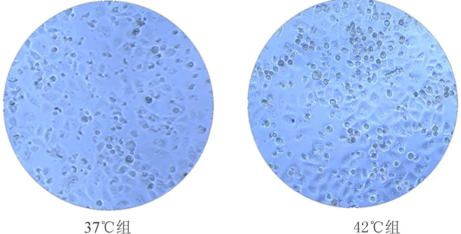
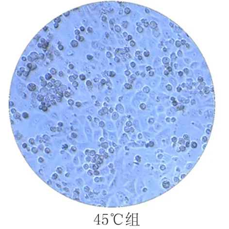
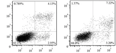
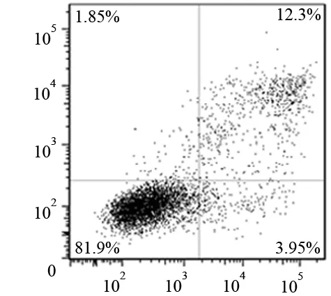
显微镜下见42、45℃孵育组的悬浮细胞明显多于37℃组(图1)。37、42、45℃孵育48 h后的PANC1细胞的凋亡率分别为(7.125±0.064)%、(9.985±0.615)%、(14.845±1.987)%(图2)。37℃孵育组的细胞凋亡率显著低于42℃组(t=10.320,P<0.05)及45℃组(t=9.832,P<0.05);42℃组又显著低于45℃组(t=4.575,P<0.05)。


图1不同温度孵育吉西他滨处理的PANC1细胞48 h的细胞形态(×100)


图237(a)、42(b)、45℃(c)孵育吉西他滨处理的PANC1细胞48 h的细胞凋亡 (流式细胞仪)
讨 论
温度对肿瘤影响的研究可以追溯到1970年。当时发现当达到一定温度时细胞会被阻滞在不同细胞周期,进而死亡。导致细胞死亡的温度是有特异性的,不同细胞可以耐受不同的温度。之后发现细胞的死亡主要原因是高温促使蛋白变性。如果使肿瘤组织蛋白变性坏死,温度至少要达到或超过60℃,但临床上使肿瘤组织达到或超过该温度并不容易,且该温度作用于人体的安全性较差。为此,有学者对温和高温处理肿瘤组织是否可以提高化疗的敏感性进行了研究,结果显示温和高温联合化疗处理软组织肉瘤可以提高患者平均生存期。将发生转移的软组织肉瘤患者整体温度提高到41.8℃维持1 h可以使患者对化疗药物的敏感性提高24%[5-6],维持机体39~40℃ 4~6 h可以达到抗肿瘤的效果[7]。Adachi等[8]发现,高温(43℃ 1 h)可以诱导胰腺癌细胞热休克蛋白70表达,从而提高肿瘤细胞对化疗药物的敏感性。有学者报道,提高机体温度可以增加肿瘤组织血流及血管通透性,使化疗药物更容易作用于肿瘤细胞,从而达到增加其对化疗药物的敏感性。但有学者认为,高温对微血管的损伤及增加细胞间质的压力可能会抵消这种血流增加的作用[9]。因而温度对提高化疗药物敏感性的机制尚未十分明确。
本研究结果显示,温和高温联合吉西他滨处理能明显抑制胰腺癌细胞的增殖,增加细胞的凋亡率,表明温和高温联合吉西他滨能提高胰腺癌PANC1细胞对吉西他滨的敏感性。虽然,45℃较42℃的效果更好,但临床将患者局部温度加热到45℃时的安全性较差。42℃高温相比传统高温(>45℃)应用于临床有其明显的优势。首先,42℃高温可以作用患者全身,不会出现传统高温治疗肿瘤所发生的遗漏肿瘤细胞,导致肿瘤复发;其次,42℃高温操作简便,可以使用水浴及红外设备直接加热患者,不必进行有创操作;最后,42℃高温可以提高肿瘤患者对化疗药物的敏感性,延长患者的预后,有效地平衡了治疗的有效性及安全性。
[1] Jemal A, Siegel R, Ward E, et al. Cancer statistics, 2008. CA Cancer J Clin, 2008,58:71-96.
[2] Fiegl M, Schlemmer M, Wendtner CM, et al. Ifosfamide, carboplatin and etoposide (ICE) as second-line regimen alone and in combination with regional hyperthermia is active in chemo-pre-treated advanced soft tissue sarcoma of adults. Int J Hyperthermia,2004,20:661-670.
[3] Westermann AM, Wiedemann GJ, Jager E, et al. A Systemic Hyperthermia Oncologic Working Group trial. Ifosfamide, carboplatin, and etoposide combined with 41.8 degrees C whole-body hyperthermia for metastatic soft tissue sarcoma. Oncology,2003,64:312-321.
[4] Maluta S, Schaffer M, Pioli F, et al. Regional hyperthermia combined with chemoradiotherapy in primary or recurrent locally advanced pancreatic cancer: an open-label comparative cohort trial. Strahlenther Onkol, 2011,187:619-625.
[5] Fiegl M, Schlemmer M, Wendtner CM, et al. Ifosfamide, carboplatin and etoposide (ICE) as second-line regimen alone and in combination with regional hyperthermia is active in chemo-pre-treated advanced soft tissue sarcoma of adults. Int J Hyperthermia,2004,20:661-670.
[6] Westermann AM, Wiedemann GJ, Jager E, et al. A Systemic Hyperthermia Oncologic Working Group trial. Ifosfamide, carboplatin, and etoposide combined with 41.8 degrees C whole-body hyperthermia for metastatic soft tissue sarcoma. Oncology,2003,64:312-321.
[7] Kraybill WG, Olenki T, Evans SS, et al. A phase I study of fever-range whole body hyperthermia (FR-WBH) in patients with advanced solid tumours: correlation with mouse models. Int J Hyperthermia,2002,18:253-266.
[8] Adachi S, Kokura S, Okayama T, et al. Effect of hyperthermia combined with gemcitabine on apoptotic cell death in cultured human pancreatic cancer cell lines. Int J Hyperthermia,2009,25:210-219.
[9] Jain RK. Normalization of tumor vasculature: an emerging concept in antiangiogenic therapy. Science,2005,307:58-62.
MildhyperthermiaaffectschemoresistanceofgemcitabineinpancreaticcancerPANC1cellline
LIUMing-hao,LIShu-de,LIZhao-shen,GAOJun,WUHong-yu,GONGYan-fang.
DepartmentofGastroenterology,ChanghaiHospital,SecondMilitaryMedicalUniversity,Shanghai200433,China
LIZhao-shen,Email:zhaoshenli@hotmail.com;LIShu-de,Email:lishude57@126.com
ObjectiveTo investigate the effect of mild hyperthermia on chemoresistance of gemcitabine in the pancreatic cancer cell line PANC1.MethodsThe PANC1 cell was treated with gemcitabine (1 μmol/L) for 1 h, then was heated at 37℃, 42℃ and 45℃ for 1 hour, and was cultured for 0 h, 24 h, 48 h. Cell growth was analyzed by CCK-8 assay. Cell apoptosis was analyzed by Annexin V-fluorescein isothiocyanate (FITC)/propidium iodide (PI) staining.ResultsThe proliferation of cells at 42℃ and 45℃ for 24 h and 48 h were significantly lower than that at 37℃(0.96±0.05,0.88±0.03vs1.05±0.02;1.28±0.04,0.94±0.04vs1.49±0.09;t=4.367, 25.120,P<0.05). The proliferation of cells at 45℃ was significantly lower than that at 42℃(t=3.348, 11.732,P<0.05).The cell apoptosis rates at 37℃, 42℃, 45℃ after 48 h were (7.125±0.064)%, (9.985±0.615)%, (14.845±1.987)%, the difference among the 3 groups was statistically significant (t=10.320, 9.832, 4.575,P<0.05).ConclusionsMild hyperthermia can reduce chemoresistance of gencitabine in pancreatic cancer cell PANC1.
Pancreatic neoplasms; Mild hyperthermia; Gemcitabine; Drug therapy
10.3760/cma.j.issn.1674-1935.2012.03.014
上海卫生局科研基金
200433 上海,第二军医大学长海医院消化内科
李兆申,Email:zhaoshenli@hotmail.com;李淑德,Email:lishude57@126.com
2011-12-19)
(本文编辑:屠振兴)

