
Acupuncture activates signal transduction pathways related to brain-tissue restoration after ischemic injury**☆
2012-09-12 06:19:32HaomeiTianHongZhangJunbaoZhuJuanZhangHeningCaiYuchenZhangChutaoChen
中国神经再生研究(英文版) 2012年24期
Haomei Tian, Hong Zhang, Junbao Zhu, Juan Zhang, Hening Cai, Yuchen Zhang, Chutao Chen
1 Key Laboratory of Meridians and Viscera, Hunan University of Traditional Chinese Medicine, Changsha 410007, Hunan Province, China
2 Liuzhou Hospital of Traditional Chinese Medicine, Liuzhou 545001, Guangxi Zhuang Autonomous Region, China
3 Ningxiang Hospital of Traditional Chinese Medicine, Ningxiang 410600, Hunan Province, China
4 Postgraduate College, Hunan University of Traditional Chinese Medicine, Changsha 410007, Hunan Province, China
5 Laboratory of Acupuncture Bioinformatics, Hunan University of Traditional Chinese Medicine, Changsha 410007, Hunan Province, China
Acupuncture activates signal transduction pathways related to brain-tissue restoration after ischemic injury**☆
Haomei Tian1, Hong Zhang1, Junbao Zhu2, Juan Zhang3, Hening Cai4, Yuchen Zhang4, Chutao Chen5
1 Key Laboratory of Meridians and Viscera, Hunan University of Traditional Chinese Medicine, Changsha 410007, Hunan Province, China
2 Liuzhou Hospital of Traditional Chinese Medicine, Liuzhou 545001, Guangxi Zhuang Autonomous Region, China
3 Ningxiang Hospital of Traditional Chinese Medicine, Ningxiang 410600, Hunan Province, China
4 Postgraduate College, Hunan University of Traditional Chinese Medicine, Changsha 410007, Hunan Province, China
5 Laboratory of Acupuncture Bioinformatics, Hunan University of Traditional Chinese Medicine, Changsha 410007, Hunan Province, China
Abstract
A middle cerebral artery occlusion-model was established in rats using the improved thread embolism method. Rats were treated with acupuncture at either Dazhui (DU14), Renzhong (DU26),Baihui (DU20), or a non-meridian point. Detection with protein-chip technology showed that the level of protein phosphorylation in both groups was upregulated or downregulated depending on the signaling pathway compared with the model group that did not receive acupuncture. Analysis of proteins showing downregulated phosphorylation revealed that five signaling pathways were activated in the acupuncture-treatment group, while only two were activated in the acupuncturecontrol group. In contrast, analysis of proteins showing upregulated phosphorylation revealed only one pathway was activated in the acupuncture-treatment group, whereas four pathways were activated in the acupuncture-control group. Furthermore, the number of activated proteins in the acupuncture-treatment group was not only higher than the acupuncture-control group, but unlike the acupuncture-control group, the majority of activated proteins were key proteins in the signaling pathways. Our findings indicate that acupuncture at specific points can activate multiple signaling pathways to promote the restoration of brain tissue following ischemic injury, and that this is based on a combination of effects resulting from multiple pathways, targets, and means.
Key Words
acupuncture; cerebral ischemia; Dazhui; Baihui; Renzhong; protein-chip technology;signal-transduction pathways; nerve injury; brain injury; ischemic injury; neural regeneration
Research Highlights
(1) A protein chip was used to understand the changes in brain-tissue proteins after acupuncture at Dazhui (DU14), Baihui (DU20), and Renzhong (DU26).
(2) The number of activated proteins increased after acupuncture at Dazhui, Baihui, and Renzhong,and they were mostly key proteins that contributed substantially to signaling pathways.
(3) Acupuncture can activate multiple signaling pathways to promote the restoration of brain tissue following ischemic injury, and the effects of this process are based on the combination of multiple pathways, targets, and means.
Haomei Tian☆, M.D.,Lecturer, Key Laboratory of Meridians and Viscera,Hunan University of Traditional Chinese Medicine, Changsha 410007,Hunan Province, China
Corresponding author:Chutao Chen, M.D., Lecturer,Laboratory of Acupuncture Bioinformatics, Hunan University of Traditional Chinese Medicine, Changsha 410007, Hunan Province,China
doctor_cct@163.com
Received: 2012-05-11 Accepted: 2012-07-26(N20111216001/YJ)
Tian HM, Zhang H, Zhu JB,Zhang J, Cai HN, Zhang YC,Chen CT. Acupuncture
activates signal transduction pathways related to
brain-tissue restoration after ischemic injury. Neural
Regen Res.
2012;7(24):1866-1872.
www.crter.cn
www.nrronline.org
INTRODUCTION
The pathogenesis of cerebral ischemia is a dynamically evolving process that appears as a series of neurotoxic and neuroprotective interactions and associated reactions. The proteins underlying these reactions exist as part of signaling pathways, and typically undergo phosphorylation in response to external stimuli.Acupuncture is known to be an effective treatment for cerebral ischemia, and current research on its mechanism of action focuses primarily on protein expression and single channels[1-3]. However, because the evidence regarding protein interactions and regulation of multiple signal transduction pathways is not large, comprehensive analysis of signal molecules is necessary to fully explore the mechanism by which acupuncture modulates complex network signals in the process of repairing cerebral ischemic injury.Antibody protein-chip technology is used to determine protein phosphorylation, protein-expression profiling,protein interactions[4-7], and to screen signal pathways[8].Advances in proteomic technology have led to a wide interest in the mechanisms underlying successful acupuncture treatment[9-12]. For example, Yi et al[13]used protein-chip technology to investigate the influence of acupuncture on protein phosphorylation when applied to the Stomach Channel of Foot-Yangming during repair of rat gastric mucosa, and found that electroacupuncture works through multiple channels, multiple targets, and multiple pathways to promote gastric mucosa repair.This experiment was aimed to verify whether acupuncture at Du meridian points can promote signal transduction pathways of phosphorylated proteins related to brain tissue after ischemic injury, and to provide experimental evidence that acupuncture and moxibustion are effective treatments of brain-tissue ischemic disease and meridian organ (Du meridian and brain) associated research.
RESULTS
Quantitative analysis of experimental animals
Forty Sprague-Dawley rats were used, 10 of which were randomly chosen as a sham-operation group. The remaining 30 rats were used to establish the middle cerebral artery occlusion model using the improved thread-embolism method, and then divided into three 10-rat groups: the model group, acupuncture-control group, and acupuncture-treatment group.Acupuncture-treatment group rats were subjected to acupuncture at Dazhui (DU14), Renzhong (DU26), and Baihui (DU20) and acupuncture-control group rats were given acupuncture at a non-meridian point 0.3 cm lateral.All rats were included in the resulting analyses.
Influence of acupuncture on signal-transduction protein phosphorylation during repair of rat brain ischemic injury
The expression profiles of the antibody microarray in each group are shown in Figure 1. Results of the calibration and comparison of antibody microarrays between groups showed that protein phosphorylation levels changed, with varying degrees of upregulation and downregulation depending on the signaling pathways involved. This experiment only included proteins whose phosphorylation level changed by at least 1.5 times(compared with the model group), and that could be attributed to known signal-transduction pathways.
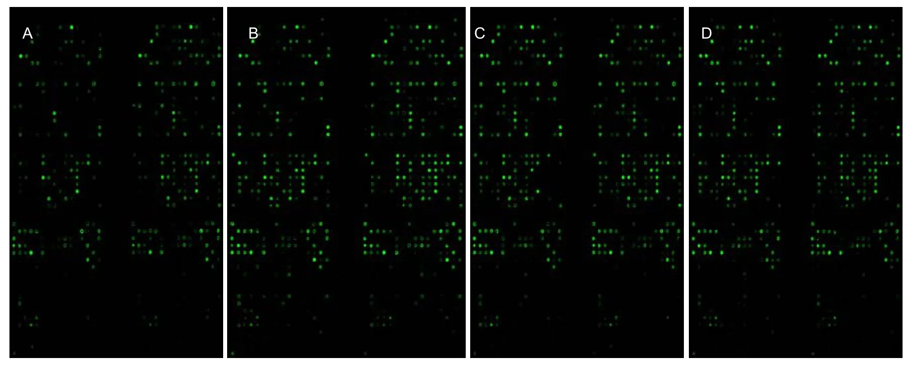
Figure 1 Antibody microarray expression profiling in rat brain tissue.(A) Sham-operation group; (B) model group; (C) acupuncture-control group; (D) acupuncture-treatment group. Green dots represent protein detection-points.
Signal transduction pathways of proteins whose phosphorylation levels were downregulated after acupuncture
Compared with the model group, acupuncture after cerebral ischemia at Dazhui, Renzhong, and Baihui induced reduction in phosphorylation levels by at least 1.5 times in the cell-apoptosis, mitogen activated protein kinases, cell-cycle regulated, adhesion-molecule, and receptor tyrosine kinase signaling pathways. Specifically,downregulated phosphorylation was observed in c-fos,TRADD, Cytochrome C, bcl-X, DFF45/ICAD, Bim (BOD),Bak and AIF-proteins known to be part of the cell-apoptosis pathway[14-15], with AIF, Bim, bcl-X, Bak and Cytochrome C being key channel proteins. Sinilr downregulation was observed in Raf-1, Mekk-1, Mek2,and Stat-1, key proteins from the mitogen activated protein kinase signaling pathway[16]. In addition,phosphorylation levels were reduced in Cdk8, CDC37,p73 and CDC34 from the cell-cycle regulated pathway,MHC II (HLA-DP) from the adhesion-molecule signal pathway[17], and platelet-derived growth factor-alpha and CD115/c-fms/CSF-1R/M-CSFR from the receptor tyrosine kinase signaling pathway[18]. In contrast,downregulated phosphorylation was observed in only three proteins from two pathways in the acupuncture-control group: Cytochrome C and DR3cell from the apoptosis signaling pathways and Paxillin from adhesion-molecule signaling pathways (Table 1).Acupuncture at Dazhui, Baihui and Renzhong promotes brain-tissue repair through multiple signal transduction pathways, including mitogen activated protein kinases,cell-apoptosis, and the cell-cycle signaling pathways.The specific proteins affected are AIF, Bim, bcl-X, Bak(BOD), Cytochrome C in the cell-apoptosis signaling pathways, Cdk8, CDC37, p73, CDC34 in the cell-cycle regulation signaling- transduction pathway, and Raf-1,Mekk-1, Mek2, Stat-1, and other key proteins in mitogen activated protein kinase signaling pathways. Together these lead to effective signal transduction. In other pathways, only a few of the associated proteins are activated. However, non- receptor tyrosine kinase signaling pathways and adhesion-molecule signal transduction pathways can activate mitogen activated protein kinase signaling pathways either directly or indirectly. Because key proteins are activated in three signal transduction pathways, this may constitute effective signal transduction, thereby protecting and repairing brain tissue against ischemic injury. Although some proteins involved in the signal transduction pathways were observed in the acupuncture-control group, they were not key proteins, and the signal transduction cannot be deemed effective. These findings indicate acupuncture at Dazhui, Baihui and Renzhong can promote the restoration of damaged brain tissue in part by downregulating phosphorylation in multiple signal transduction pathways.

Table 1 Signal transduction pathway of the proteins with down-regulated phosphorylation levels (≥1.5 times) in acupuncture group and acupuncture control group compared with model group
Signal transduction pathways of proteins whose phosphorylation levels were upregulated after acupuncture
Compared with the model group, an increase in phosphorylation levels by at least 1.5 times was found in the cell-cycle signal transduction pathway of the acupuncture-treatment group (Table 2).In contrast, the acupuncture-control group showed upregulated phosphorylation in multiple pathways.These included proteins related to cell-cycle regulation,cell apoptosis, mitogen activated protein kinases, and adhesion-molecule signal transduction pathways (Cdk7,p27Kip1 and Cdk1/p34cdc2). Specifically, upregulated phosphorylation was observed in the adenovirus-E2 promoter 5 and p19ARF from the cell-cycle regulation pathway, Bcl-2 associated athanogene 1, Bax, and Fas-ligand from the cell-apoptosis signaling pathway,Raf-1, Mekk-1, and Stat-1 from the mitogen activated protein kinase signaling pathway, and CD138 from the adhesion-molecule signaling pathway. However,although these four signal pathways in the acupuncture-control group were activated, the number of proteins in each of these pathways was too small to form an effective signal-transduction pathway,indicating a lack of physiological significance. This further supports the notion that acupuncture only activates upregulation in cell-cycle signal-transduction pathways.
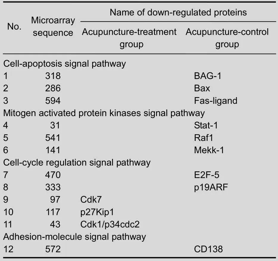
Table 2 Signal transduction pathway of the proteins with up-regulated phosphorylation levels (≥1.5 times) in acupuncture-treatment group and acupuncture-control group compared with model group
The ischemic tissues in the acupuncture group and acupuncture-control group were compared. For proteins that underwent upregulated phosphorylation (≥1.5 times), only the cell-cycle signal pathway was activated in the acupuncture-treatment group, which may prevent cell proliferation through inhibition of signal transduction.Although activation of cell-apoptosis, mitogen activated protein kinases, cell-cycle, and adhesion-molecule signaling pathways were visible in the acupuncture-control group, with the exception of the mitogen activated protein kinase signaling pathway,there were no key proteins activated. This makes it likely that effective signal transduction did not occur,and was of little significance in the repair of ischemic brain tissue. Our results show that acupuncture at Dazhui, Baihui and Renzhong promotes the restoration of damaged brain tissue through activation of multiple signal transduction pathways, in particular mitogen activated protein kinases, cell apoptosis, and cell-cycle signaling pathways.
DISCUSSION
Clinical and experimental studies have verified the contribution of acupuncture towards treatment of cerebral ischemia[19-21]. However, research into the mechanisms underlying this treatment has primarily concentrated on a few proteins or a single signal pathway, and no research has reported a change in brain tissue proteins by different interventions.Conducting more complete studies is therefore urgently needed to understand how acupuncture works to assist the repair of cerebral ischemia. Song et al[22]found that acupuncture can inhibit the proliferation of glial cells in a rat model of middle cerebral artery occlusion and reperfusion injury, thus providing a beneficial environment for neural regeneration and functional reconstruction. The underlying mechanism might depend on the regulation of the cell cycle. Bim reports that activation of the c-Jun NH2-terminal kinase pathway following cerebral ischemia reperfusion injury can induce neuronal apoptosis[23-24]. In this experiment,protein microarray-analysis showed that acupuncture at Dazhui, Baihui, and Renzhong can alter protein phosphorylation in the damaged brain tissue, primarily through downregulation in multiple signal transduction pathways. Our results support this notion and identified mitogen activated protein kinases, cell cycle, and apoptosis signaling pathways as being particularly affected. However, the specific mechanism of action of each pathway in the repair process of ischemic injury in brain tissue remains unknown, and must be further explored. Significant differences between the acupuncture-treatment group and the acupuncture-control group regarding the number of activated proteins and their pathways were observed,indicating that acupuncture promotes the repair of brain tissue damage through multiple channels, multiple targets, and multiple level processes. Additionally, a certain acupoint specificity exists that provides reliable justification for continued in-depth research of acupuncture and moxibustion for treatment of cerebral ischemia.
MATERIALS AND METHODS
Design
A proteomic experiment.
Time and setting
Experiments were performed from July 2010 to November 2011 in the Experimental Animal Center of Hunan University of Traditional Chinese Medicine,Level-Three Laboratory of the State Administration of Traditional Chinese Medicine of China, Key Laboratory of Meridians and Viscera, Hunan University of Traditional Chinese Medicine, China.
Materials
A total of 40 pathogen-free Sprague-Dawley rats, aged 5 weeks, weighing 230-250 g, were provided by Hunan SLAC Jingda Experimental Animal Co. Ltd., China with license No. SCXK (Xiang) 2009-0004. All animals were fed in the Experimental Animal Center of Hunan University of Traditional Chinese Medicine, at 20-22°C in 65-70% relative humidity. Experimental disposal of animals complied with the Guidance Suggestions for the Care and Use of Laboratory Animals, issued by the Ministry of Science and Technology of China[25].
Methods Establishment of the rat brain ischemic injury model through middle cerebral artery occlusion using the suture method
The middle cerebral artery was occluded one week prior to the experiment to allow rats time to adapt to the new environment and food schedule. All experimental animals were allowed water, but no food for 12 hours prior to surgery. Under intraperitoneal anesthesia with 10% chloral hydrate (30 mg/100 g), rats were fixed in a supine position on the working table, a 1-cm longitudinal incision was made 0.3 cm left of the median line, and then, to fully expose the common carotid, branch internal carotid, and external carotid arteries, subcutaneous fascia, muscle, and thyroid were stripped using hemostatic forceps. A blunt dissection was performed to dissect the common carotid arteries and vagus nerve,exposing blood vessels and nerves, and the suture lines were placed at the proximal end of the common carotid,internal carotid, and external carotid arteries. The internal carotid artery was temporarily blocked with a micro-artery clamp, followed by the proximal end of the common carotid and external carotid arteries. Then a small incision was cut 3 mm away from the common carotid artery bifurcation, the suture was inserted into the internal carotid artery and tightly fastened to the distal end of the artery at a depth of 19 mm (from blood vessel bifurcation). Afterward, the artery clamp was loosened,followed by wound suture and single-cage rearing. The sham operation group was only subjected to separation of the left common carotid artery and ligation of the left common carotid and external carotid arteries, while the internal carotid artery was not occluded[26]. After animals’vital signs were confirmed to be stable postoperatively,their neurological function was assessed with the Longa scale[26]. Only rats with a score ranging from 1 to 3,indicating successful modeling, were used in the experiment. Whole-brain anatomy revealed that infarcts occurred in temporal cortex, a region that is innervated by the middle cerebral artery, further indicating the success of experimental models. When the brain tissue was removed and inspected, rats with significant subarachnoid hemorrhages were excluded from the experiment. Rats were housed at 20°C, and allowed food and water ad libitum.
Acupuncture at Baihui, Dazhui and Renzhong
After breathing and heartbeat stabilized for 4.82 ±0.84 hours, acupuncture treatment using 0.3 mm ×25 mm acupuncture needle (Suzhou Medical Supplies Factory, Suzhou, China) was directly administered at the Dazhui point (between the seventh cervical vertebra and the first thoracic vertebra, the median back) 5 mm,horizontally at the Baihui point (the median parietal bone)10 mm, and at the Renzhong point (1 mm away from the median-cleft lip nose) 2 mm towards the nasal septum,according to Experimental Acupuncture[27]and the Rat Acupuncture Points Map[28]. The inserted needle was rotated for 1 minute at each point, and again during maintenance every 12 hours, for a total of six times. In the acupuncture-control group, acupuncture was administered to a non-meridian point 0.3 cm lateral. The sham-operation and ischemic-injury model groups underwent the occlusion procedure only, and did not receive any acupuncture.
Harvesting specimens
Two days after acupuncture treatment, rats from each group were decapitated under 10% chloral hydrate anesthesia, and the left-sided ischemic brain tissue was harvested, preserved in liquid nitrogen for 10 seconds,and stored at -80°C.
Antibody protein-chip detection
To extract protein, protein-extraction reagent was added to brain tissue at a concentration of 1 mL/250 mg tissue and supplemented with 5 μL protease-inhibitor mixed liquid, 5 μL phenylmethylsulfonyl fluoride and 5 μL phosphatase mixture. The mixture was homogenated for 30 seconds at 1-minute intervals in an ice bath until tissues were completely lysed, centrifuged at 14 000 × g for 15 minutes, and the supernatant was collected.Proteins were quantified using the BCA method, and prepared into electrophoresis samples containing 6 μg/μL protein. The protein was labeled with biotin (with a biotin:protein ratio of 1:7) and then purified (Changsha Chemical Reagent Factory, Changsha, China). The biotin-labeled protein samples were hybridized with 720 phosphorylation protein chips (Springbio, Preston,CA, USA) for microarray detection. Image scanning was performed with Genepix4000B (Axon, Rowley, NC, USA)and a fluorescent scanner (Taiwan MSI Technology Company, New Taipei, China) at 532 nm excitation,images in TIF format were analyzed with Genepix Pro 6(Axon Instruments, Union City, CA, USA)[29], and the phosphorylation level was determined according to the fluorescence intensity.
Protein microarray data analysis
Phosphorylation levels of proteins in the acupuncturetreatment group and the acupuncture-control group were compared with the model group. If upregulated or downregulated levels changed by more than 1.5 times,the protein signal transduction pathways were explored and the correlation with brain tissue damage and protection were analyzed.
Funding: This study was supported by the National Natural Science Foundation of China for Youth, No. 201130901901 and a grant by Youth Fund Project of the Ministry of Education of China, No. 11B092.
Author contributions: Chutao Chen designed the study.
Haomei Tian was responsible for data analysis and drafted the manuscript. Hong Zhang revised the experiment. Junbao Zhu,Juan Zhang, Hening Cai and Yuchen Zhang performed animal experiments and specimen processing.
Conflicts of interest: None declared.
Ethical approval: Experimental disposals were approved by the Animal Ethics Committee of Hunan University of Traditional Chinese Medicine in China.
REFERENCES
[1] Liu R, Yi W, Huang KB, et al. Regulation of the JAK-STAT signal transduction system in cerebral ischemia and acupuncture intervention. Zhongguo Laonian Xue Zazhi.2011;31(4):717-718.
[2] Yang Zh, Xu NG, Yi W, et al. Research of intervention with electroacupuncture through regulating MAPK/ERK pathway for cerebral ischemia rats. Guangzhou Zhongyiyao Daxue Xuebao. 2010;27(1):23-30.
[3] Chen DF, Lai Z, Zhang SJ, et al. Effects of electroacupuncture on PI3-K/Akt path in reperfused local cerebral ischemia rats. Zhongguo Kangfu Yixue Zazhi.2009;24(6):502-504.
[4] Celis JE, Moreira JM, Cabezón T, et al. Identification of extracellular and intracellular signaling components of the mammary adipose tissue and its interstitial fluid in high risk breast cancer patients: toward dissecting the molecular circuitry of epithelial-adipocyte stromal cell interactions. Mol Cell Proteomics. 2005;4(4):492-522.
[5] Lim YB, Pyun BJ, Lee HJ, et al. Proteomic identification of radiation response markers in mouse intestine and brain.Proteomics. 2011;11(7):1254-1263.
[6] Xu FF, Liu XH. Proteomic technology: its progress and application in microcirculation research. Sheng Li Ke Xue Jin Zhan. 2010;41(6):429-434.
[7] Lin X, Wang Q, Cheng Y, et al. Changes of protein expression profiles in the amygdala during the process of morphine-induced conditioned place preference in rats.Behav Brain Res. 2011;221(1):197-206.
[8] He HJ, Zong Y, Bernier M, et al. Sensing the insulin signaling pathway with an antibody array. Proteomics Clin Appl. 2009;3(12):1440-1450.
[9] Yan J, Zhang YJ, Tian HM, et al. Effect of electroacupuncture on gastric mucosal damage related signal molecules in rats. Zhongyi Zazhi. 2009;50(11):1002-1005.
[10] Kim HK, Thu VT, Heo HJ, et al. Cardiac proteomic responses to ischemia-reperfusion injury and ischemic preconditioning. Expert Rev Proteomics. 2011;8(2):241-261.
[11] Jiang L, He K, Chen CT, et al. Electroacupuncture intervention on the expression of mitogen activated protein kinase phosphatase-1 and phosphorylated extracellular signal-regulated kinase 1/2 in aorta of rats with spontaneous hypertension. Zhongguo Zuzhi Gongcheng Yanjiu yu Linchuang Kangfu. 2010;14(20):3686-3690.
[12] Koh PO. Identification of proteins differentially expressed in cerebral cortexes of Ginkgo biloba extract (EGb761)-treated rats in a middle cerebral artery occlusion model-a proteomics approach. Am J Chin Med. 2011;39(2):315-324.
[13] Yi SX, Tian HM, Yan J, et al. Changes of multiple protein phosphorylation signal transduction pathways in the process of repairing rat gastric mucosa through stimulation of stomach meridian with electro-acupuncture.Zhongguo Zuzhi Gongcheng Yanjiu yu Linchuang Kangfu.2009;13(41):8075-8079.
[14] Yu XJ, SHao XM, Fang JQ. Apoptosis pathway research progress. Shiyong Zhongyi Zazhi. 2011;27(1):71-72.
[15] Sun YY, Wang J. Apoptosis pathway research progress.ZHongguo Wuzhen Xue Zazhi. 2011;31:7579-7580.
[16] Sun X, Liu C, Qian M, et al. Ceramide from sphingomyelin hydrolysis differentially mediates mitogen-activated protein kinases (MAPKs) activation following cerebral ischemia in rat hippocampal CA1 subregion. Shengwu Yixue Yanjiu Zazhi. 2010;24(2):132-137.
[17] Fang YX, Feng HY, Mo SK, et al. Cytokine fusion protein technology and its application prospects. Xibao yu Fenzi Mianyi Xue Zazhi. 2009;9:856-859.
[18] Li X, Gao JH, Chen GL. Progress on the research of the protein tyrosine kinase inhibitors. Shenyang Yaoke Daxue Xuebao. 2011;28(12):1005-1012.
[19] Xiao L, Wang LX, Cui Q, et al. Clinical effects of acupuncture combined with herbal medicine in treating constipation of phlegm-heat and bowel-repletion in patients with acute cerebral infarction. Jiefangjun Yiyao Zazhi. 2011;23(1):15-17.
[20] Song HY, Fang W, Zhou LZ, et al. Therapeutic effect observation of Yin-yang meridians acupuncture on acute ischemic stroke. Hebei Zhongyi. 2010;32(5):719-721.
[21] Li SJ, Li J. 67 cases with cerebral infarction treated by acupuncture combined drug. Zhongguo Shiyong Shenjing Jibing Zazhi. 2010;13(7):38-39.
[22] Song TS, Zhou MF. The effects of acupuncture on astrocyte proliferation after cerebral ischemia-reperfusion injury in rats. Zhonghua Wuli Yixue yu Kangfu Zazhi. 2008;30(4):244-247.
[23] Bouillet P, Strasser A. BH3-only proteins-evolutionarily conserved proapoptotic Bcl-2 family members essential for initiating programmed cell death. J Cell Sci. 2002;115(Pt 8):1567-1574.
[24] Weston CR, Davis RJ. The JNK signal transduction pathway. Curr Opin Cell Biol. 2007;19(2):142-149.
[25] The Ministry of Science and Technology of the People’s Republic of China. Guidance Suggestions for the Care and Use of Laboratory Animals. 2006-09-30.
[26] Longa EZ, Weinstein PR, Carlson S, et al. Reversible middle cerebral artery occlusion without craniectomy in rats. Stroke. 1989;20(1):84-91.
[27] Guo Y. Experimental Acupuncture. Beijing: China Press of Traditional Chinese Medicine, 2008.
[28] Hua XB, Li CR, Zhou HL. The development of the rat acupuncture points map. Shiyan Dongwu yu Dongwu Shiyan. 1991(1):1.
[29] Yang L, Guo S, Li Y, et al. Protein microarrays for systems biology. Acta Biochim Biophys Sin (Shanghai). 2011;43(3):161-171.
(Edited by Huang LA, Sun XJ/Yang Y/Wang L)
10.3969/j.issn.1673-5374.2012.24.004
- 中国神经再生研究(英文版)的其它文章
- Effects of wind-dispelling drugs and deficiency-nourishing drugs of Houshiheisan compound prescription on astrocyte activation and inflammatory factor expression in the corpus striatum of cerebral ischemia rats****☆
- Acupuncture and moxibustion for visceral pain
- Therapeutic effect of nerve growth factor on cerebral infarction in dogs using the hemisphere anomalous volume ratio of diffusion-weighted magnetic resonance imaging*★
- Pre-ischemia electro-acupuncture potentiates the expression of Bcl-2 and transforming growth factor-beta 1 in rat brains*☆△◇
- Yizhijiannao Granule and a combination of its effective monomers, icariin and Panax notoginseng saponins, inhibit early PC12 cell apoptosis induced by beta-amyloid (25-35)☆
- No association between a polymorphism of the adenylate cyclase type IX gene and major depressive disorder in the Chinese Han population*☆