
Outcomes of loco-regional therapy for down-staging of hepatocellular carcinoma prior to liver transplantation
2011-07-03 12:40:18
Beijing, China
Outcomes of loco-regional therapy for down-staging of hepatocellular carcinoma prior to liver transplantation
Xian-Jie Shi, Xin Jin, Mao-Qiang Wang, Li-Xin Wei, Hui-Yi Ye, Yu-Rong Liang, Ying Luo and Jia-Hong Dong
Beijing, China
BACKGROUND: The number of loco-regional therapies (LRTs) for hepatocellular carcinoma (HCC) has increased dramatically during the past decade. Many patients with HCC who were beyond the Milan criteria were allowed to receive a liver transplantation (LT) once the HCC was successfully down-staged. This retrospective study aimed to analyze the outcomes of LRTs prior to LT in patients with HCC beyond the Milan criteria.
METHODS: We analyzed 56 patients treated from June 2006 to March 2010: 22 met the Milan criteria (T1+T2, 39.3%), 16 had T3 tumors (28.6%), and 11 had T4a tumors (19.6%), while 7 were suspected of tumor vascular invasion (T4b, 12.5%). All patients underwent preoperative LRTs, including transcatheter arterial chemoembolization, radiofrequency ablation, percutaneous ethanol injection, liver resection, and/or microwave coagulation therapy. The number of the patients who were successfully down-staged before LT, the types of LRTs used before LT, and their outcomes after LT were recorded.
RESULTS: Eleven patients had necrotic tumors (pT0, 19.6%); 6 had pT1 tumors (10.7%), 22 had pT2 tumors (39.3%), 6 had pT3 tumors (10.7%), 5 had pT4a tumors (8.9%), and 6 had pT4b tumors (10.7%). The histopathologic tumors of 39 patients (69.6%) were down-staged and met the established Milan criteria (pT0-2). Imaging-proven under-staging was present in 5 HCC patients (8.9%) who had tumors involving the intrahepatic venous system. Twenty-three patients (41.1%) had stable HCC and 10 (17.9%) died. The 1-, 3- and 4-year survival rates were 96%, 73% and 61%, respectively, with a mean survival time of 22.29±1.63 months. Six patients died of tumor recurrence. The 1-, 3- and 4-year recurrence-free survival (RFS) rates were 88%, 75% and 66%, respectively. The 3-year RFS of patients with pT0-2 tumors was 82%, which was markedly greater than that of patients with pT3 tumors (63%,P=0.018) or pT4 tumors (17%,P=0.000). Although the 3-year RFS of patients with pT3 tumors was greater than that of patients with pT4 tumors, the difference was not significant.
CONCLUSIONS: Successful down-staging of HCCs can be achieved in the majority of carefully selected patients by LRTs. Importantly, patients who are successfully down-staged and undergo LT may have a higher RFS rate.
(Hepatobiliary Pancreat Dis Int 2011; 10: 143-150)
hepatocellular carcinoma; tumor down-staging; loco-regional therapy; liver transplantation
Introduction
Hepatocellular carcinoma (HCC) is the third leading cause of cancer-related death and accounts for as many as 500 000 deaths annually.[1]A variety of therapeutic modalities can be used in the treatment of HCC, but orthotopic liver transplantation (OLT) is considered the only curative treatment option because OLT is claimed to simultaneously cure the malignant disease and replace the premalignant cirrhotic liver.[2]In the late 1980s, the outcomes of patients undergoing OLT for treatment of HCC were disappointing, with high levels of early recurrence and 5-year survival rates ranging between 18% and 40%.[3]These discouraging outcomes and a shortage of liver donors compelled the transplant community to establish stringent selection criteria for predicting the post-transplant survival rates of patients with HCC. In 1996, Mazzaferro et al,[4]a group from Milan, Italy, published a prospective study involving48 patients who underwent OLT for treatment of HCC. These patients met a restrictive set of criteria, which was based on tumor size and number (a single lesion ≤5 cm or two or three lesions ≤3 cm) according to pre-operative imaging. The 4-year overall patient survival rate after OLT was 75% and the recurrence-free survival rate (RFS) was 83%. Since the publication of this landmark study, these criteria, now termed the Milan criteria, have been validated by several other groups, who showed 5-year survival rates to be consistently 70% or higher.[5,6]
The risk of HCC progression correlates with time on the transplantation waiting list; therefore, strategies such as transcatheter arterial chemoembolization (TACE), percutaneous ethanol injection (PEI), and radiofrequency ablation (RFA) have been used to prevent the disease from progressing beyond the Milan criteria while these patients await a donor for OLT.[7]Such strategies are also used to decrease the recurrence of HCC after LT and for being able to down-stage the severity of the tumors so that they fall within the Milan criteria, which is more controversial. Transplant units face difficult situations, which include: 1) more and more patients with HCC, and 2) an organ shortage leading to longer waiting periods and increased tumor development. An alternative approach is to apply loco-regional therapies (LRTs) to reduce the size of these tumors, which allows for down-staging of the tumor and meeting the established Milan criteria for receiving an LT.[8]This study aimed to review the efficacy and survival benefit of various treatments of HCC prior to LT.
Methods
Clinical data
We retrospectively analyzed the clinical data of 56 HCC patients who had undergone LT following down-staging treatments from June 2006 to March 2010. Patients who underwent LT and gave informed consent entered this series of studies, which was approved by the Investigation and Ethics Committee of the General Hospital of the PLA. There were 47 men and 9 women. Their median age was 55 years (range 31-68 years), and follow-up time was 22 months (range 3-44 months). A majority of the patients had virus-related HCC [hepatitis B virus (n=55), hepatitis C virus (n=1)]. The Child-Pugh classification was A and B in 46 patients and C in 10. The level of alphafetoprotein (AFP) in 29 patients was below 400 ng/ml. All patients received routine serological AFP measurement, abdominal ultrasonic examination, CT and abdominal cavity blood vessel reconstruction examination. Some patients with tumor diameters >5 cm underwent positron emission tomography to exclude extrahepatic metastasis and intrahepatic vascular invasion. Baseline characteristics of the 56 patients are summarized in Tables 1 and 2. Staging of the HCCs was performed before and after OLT using the American Liver Tumor Study Group modified TNM classification.[9]Twenty-two patients met the Milan criteria (T1+T2, 39.3%), 16 had T3 tumors (28.6%), and 11 had T4a tumors (19.6%) at the time of diagnosis, while the other 7 were suspected of having tumors that had invaded the vascular system (T4b, 12.5%). All patients underwent preoperative LRT, using TACE, PEI, RFA, and/or microwave coagulation therapy (MCT) (Table 1). Forty-nine patients (87.5%) underwent deceased donor LT, and 7 underwent living donor LT. All explanted specimens underwent pathologic examination. The pathologic stage of the tumor, the histological grade of the tumor (well-, moderately-, or poorlydifferentiated), and the presence or absence of vascular invasion were recorded. The total tumor diameter for patients with multiple tumor nodules was calculated as the sum of the maximal diameter of each lesion in centimeters. The pathologic tumor stage was initially reported using the pathological tumor-node-metastasis (pTNM) classification.

Table 1. Baseline characteristics of patients
Down-staging
Successful down-staging was defined as a reduction in the size and number of viable tumors so that they fell within the UNOS T2 criteria (Milan criteria). Patients who were or were not successfully down-staged to fall within the UNOS T2 criteria had their response to treatment evaluated by measuring their AFP levels and analyzing their radiological and histological findings. The radiological response was evaluated according to tumor size, which was based on the RECIST and necrosis criteria. A complete response was defined as complete tumor necrosis (lack of tissue enhancement on arterial phase images or histological evidence of complete tumor necrosis after LT) and normalization of AFP levels.[10]
Treatment after LT
Immunosuppressive therapy after LT consisted of a triple-drug regimen of cyclosporine or tacrolimus combined with corticosteroids and/or mycophenolate mofetil. Patients were weaned off corticosteroids after three months. Then, all patients were followed up regularly with serum AFP quantification and ultrasonography performed once every month for the first six months. Suspected recurrences were confirmed with CT or MRI. Chest CTs or bone scans were used when distant metastases were suspected. All recurrences were properly evaluated for new treatment options. The choice of treatment was related to the number and size of the tumors, the presence of extrahepatic disease, liver function, and the general health of the patient.
Statistical analysis
Statistical analyses were performed using SPSS 17.0 for Windows (SPSS, Chicago, IL). The Kaplan-Meier product-limit method with the log-rank test was used to evaluate the survival rates. All causes of mortality were analyzed instead of using the mortality rates based solely on HCC. This allowed for the avoidance of underestimating the impact of HCC on mortality. Overall survival (OS) was defined as the interval between LT and death or the last observation. The data were censored for patients who were still alive at the last observation and for those who underwent secondary LT. RFS was calculated considering patients whether they were alive or dead at the time of analysis. The data were censored for living patients without signs of recurrence. APvalue less than 0.05 was considered statistically significant.
Results
LRT for HCC down-stagingLRTs used for HCC down-staging in this study are summarized in Table 1. The majority of patients (n=41) received only a single-treatment modality, while 15 received combined treatments. Of the 15 patients, 2 had combined TACE and RFA, 3 had combined TACE and MCT, and 8 received liver resection prior to TACE. These patients developed tumor recurrence after primary resection, which was followed by TACE. One patient underwent open biopsy of an 8.0 cm lesion in segment IV of the left hepatic lobe followed by TACE. TACE and RFA were the primary treatment modalities for the purpose of HCC down-staging (38× TACE, 3× RFA).

Table 2. HCC characteristics of 56 patients
Histopathologic studies
Of the 56 patients treated, 11 (19.6%) had completely necrotic tumors with no viable malignant cells and were staged pathologic T0 (pT0). Six patients had pT1 tumors (10.7%), 22 had pT2 tumors (39.6%), 6 had pT3 tumors (10.7%), 5 had pT4a tumors (8.9%), and 6 had pT4b tumors (10.7%) (Table 2).
Three patients suspected of having portal vein tumor thrombus (T4b) by preoperative imaging were treated by TACE; 2 of them were not found to have vascular invasion pathologically after TACE, and one was diagnosed with complete tumor thrombus necrosis after TACE. Three patients with tumor recurrence after resection underwent LT, and one of these underwent one course of TACE treatment prior to LT. The pathological reports of the 3 patients undergoing LT showed liver cirrhosis and moderate to severe dysplasia. No tumor recurrence occurred in these patients after LT.
The histopathologically positive tumors of 39patients (69.6%) were down-staged and met the Milan criteria (pT0-2). Tumors in 5 patients (8.9%) were under-staged according to imaging and the involvement of the intrahepatic venous system (Table 3). Twentythree patients (41.1%) were stable, and one had preoperative portal vein thrombosis (T4b), which was found to be completely necrotic after TACE; however, specimen examination showed micro-vascular invasion after he died from lung metastasis 9 months after LT.

Table 3. Correlation of pre-orthotopic LT radiologic staging with explant pathology
Overall survival rate and RFS after LT
There was no peri-operative mortality in the 56 OLT recipients. The 1-, 3- and 4-year overall survival rates were 96%, 73% and 61%, respectively, with a mean survival time of 22.29±1.63 months. Ten of the 56 patients died during the median follow-up of 22 months (range 3-44 months). Another 2 patients with complete tumor necrosis (pT0) died from ischemic-type biliary lesion (ITBL) after LT (6 and 34 months). One patient received a second LT due to chronic rejection, but died from multi-organ failure 14 days after surgery. Another patient died of graft-versus-host disease (GVHD) after 6 months. HCC recurrence was observed in 9 patients, including all 6 with pT4b tumors, 2 with pT4a tumors, and 1 with a pT3 tumor, with a mean recurrence time of 12.3 ± 6.4 months (range 3-23 months). Six patients died from tumor recurrence and metastasis. The 1-, 3-and 4-year RFSs were 88%, 75% and 66%, respectively. All tumor recurrences were observed within 2 years and occurred in the lung (4/9), liver (3/9), bone (1/9), and brain (1/9). The 3-year RFS of patients with pT0-2 tumors was 82%, which was markedly better than that of patients with pT3 tumors (63%, χ2=5.608,P=0.018) or pT4 tumors (17%, χ2=25.598,P=0.000). The 3-year RFS of patients with pT3 tumors was still better than that of patients with pT4 tumors (χ2=1.264,P=0.261), but this difference was not significant (Fig. A).
The 3-year overall survival rate was 73% after LT. According to the pathological results, 2 of 11 patients with pT0 tumors died of ITBL and GVHD. Therefore, the 3-year OS was just 60%, which was lower than 100% of the pT1 group (P=0.335), 86% of the pT2 group (P=0.134), and 63% of the pT3 group (P=0.350), but only better than that of the pT4 group (26%,P=0.057) (Fig. B). Hence, all patients were divided into the following groups: pT0-2 (n=39), pT3 (n=6), and pT4 (n=11), and the 3-year OS was 82%, 63% and 26%, respectively. The pT0-2 group was better than the pT3 group (χ2=5.313,P=0.021) and pT4 group (χ2=7.490,P=0.006). The difference between the pT3 and the pT4 groups was not significant (χ2=1.688,P=0.421) (Fig. C).
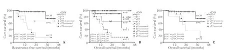
Fig. Cumulative proportion of recurrence-free (A) and overall (B, C) survival (Kaplan-Meier curves) by pathologic TNM group.
Discussion
The possibility of HCC down-staging
Experience using various loco-regional procedures to treat malignant liver tumors has encouraged some researchers to investigate whether these procedures are also useful in down-staging advanced HCCs so that patients may meet the criteria for LT. The concept of using LRTs, such as TACE, to reduce the size of HCCs, thereby facilitating resection or OLT, was first tested on patients with one or more lesions >3 cm.[11]HCC downstaging prior to transplantation has three potential goals: 1) control of tumor growth and vascular invasion while waiting for an organ; 2) using neoadjuvant therapy to improve the post-transplant outcome by reducing the risk of post-operative recurrence; and in some cases 3) using down-staging as a selection tool to more accurately predict whether patients have a low risk of tumor recurrence.[12]The fundamental principle behind downstaging is to select a subset of patients with tumors that are more likely to respond to treatment and do well after OLT.[8]
Among all the patients, 34 (60.7%) were beyond the established Milan criteria (T2) initially, based on preoperative image staging. Seven patients (12.5%) were diagnosed as having tumor vascular involvement (T4b). After HCC down-staging, 28 patients (50.0%) showed pathological tumor staging less than the pre-operative imaging, and 27 met the Milan criteria (pT0-2). Eleven patients (19.6%) showed complete tumor necrosis, while the other 23 (41.1%) of the same stage had controlled tumor progression. Only five patients (9.0%) were under-staged, far lower than that the literature reports (20%-30%).[13-15]This may explain why pre-operative LRT can control tumor growth, while significantly reducing the tumor burden and sustaining the patient's opportunity for LT.[12]
Chapman et al[16]reported that 18 of 76 (23.7%) patients with stage pT3-4 HCC had adequate downstaging to qualify for OLT under the Milan criteria. At a median of 19.6 months (range 3.6-104.7 months), 16/17 (94.1%) patients who had undergone OLT were still alive. Therefore, they concluded that patients who had been successfully down-staged and transplanted had excellent mid-term recurrence-free and overall survival rates similar to those of patients with stage T2 tumors. Bharat et al[17]reported that patients in the LRT group had a better than 5-year survival rate (82.4% vs. 51.8%;P=0.01), while the patients in the LRT group with stage pT2-4 tumors were better than those in the non-LRT group (77.6 vs. 37.4%;P=0.016). Sixteen LRT patients revealed complete tumor necrosis with no viable tumor cells upon explant pathology (pT0). These patients did not experience any long-term recurrence, in contrast to those with similar pre-OLT tumors. Yao et al[18]found favorable long-term RFS associated with LRT in patients with pT2 and pT3 HCC. Regalia et al[19]and Majno et al[11]also reported a trend toward improved survival associated with pre-OLT TACE. These studies suggested that some patients whose tumor size and number were within a certain upper limit may benefit from LRT-induced complete tumor necrosis, allowing for downstaging of the tumor to meet the criteria for LT. In this study, we found the 3-year OS and RFS patients, who met the Milan criteria (pT0-2) were markedly better than patients with pT3 (OS:P=0.021; RFS:P=0.018) and pT4 tumors (OS:P=0.006, RFS:P=0.000), but there was no significant difference between patients with pT3 and pT4 tumors (OS:P=0.421; RFS:P=0.247). Therefore, acceptable outcomes after OLT rely on the Milan criteria and offer a wider "safety margin'' for tumor under-staging by pre-operative imaging.[8]
Liver resection
Resection can be used as a treatment for HCC prior to LT in three different settings. First, resection can be used as a primary therapy, while LT can be reserved as a salvage therapy for patients who develop recurrence or liver failure. Second, resection can be used as an initial therapy to select patients who might benefit from LT based on pathological examinations of the tumor and the surrounding liver parenchyma. Third, resection can be used as a bridge therapy for patients who are alreadyenlisted for LT.[12]
A total of 8 patients (14.3%) had undergone liver resection pre-OLT while developing tumor recurrence after resection, combined with TACE (1-4 courses) to control tumor progression. One patient with HCC stage T1 showed complete tumor necrosis. Another patient with a moderately-differentiated HCC after resection had tumor recurrence. That patient then underwent three courses of TACE and received an LT. Histopathologically the explanted tumor showed microvascular invasion (a pT4b tumor), and the patient died of lung metastases within 9 months after transplant surgery.
It is widely accepted that liver resection as an adjuvant therapy pre-OLT increases the difficulty of a later LT.[20]Therefore, few centers in Europe and the United States use liver resection as a preoperative method for reducing the burden of HCCs, but LT is reserved as a salvage therapy for patients who had one liver resection, yet still showed tumor recurrence or liver failure. We suggest that this strategy will significantly increase the survival rate of HCC patients. Majino et al[11]also thought that the outcome of secondary liver transplant was similar to that of one-stage liver resection, assuming at least 60% tumor recurrence within the Milan criteria[21,22]
TACE
TACE consists of the selective embolization of the artery feeding a tumor via injection of chemotherapeutic agents. The use of a combination of highly concentrated chemotherapy drugs, such as cisplatin or doxorubicin, and some degree of ischemia in the tumor, is postulated to work synergistically to cause tumor necrosis.[23]The last two randomized clinical trials showed that patients with unresectable HCC and preserved liver function had a clear benefit from TACE treatment.[24-26]The rationale for using TACE as a neoadjuvant therapy prior to LT is to control tumor growth by inducing tumor necrosis while the patient awaits an organ. In patients with tumors slightly exceeding the Milan criteria, TACE can be used to gain time and learn more about the progression of the particular tumor prior to LT.[23]
Fifty-three of the 56 (94.6%) patients had received TACE, as the most common method of LRT. Thirtyeight patients (67.9%) received only TACE (1-8 courses), 7 (12.5%) with tumor recurrence received TACE after liver resection, and 6 (10.7%) had TACE combined with other methods for controlling tumor progression. Twenty-seven patients (48.2%) had tumor down-staging, 23 (41.1%) had stable tumors, and 5 (8.9%) were understaged. A total of 10 patients exhibited complete tumor necrosis (pT0) after 1.3 courses of TACE (range from 1 to 3 courses), accounting for 19.2% of the TACE-treated group. Eight of the 10 patients received TACE treatment only, while the other 2 patients with tumor recurrence received TACE treatment after liver resection. Pathologic examination showed that 43 patients (81.1%) exhibited complete tumor necrosis for more than 50% of the tumor area. This was attributed to repeated TACE treatment in the short-term to achieve maximal tumor necrosis. Three-year OS and RFS of the TACE treatment were 77% and 72%. Yao et al[18]further reported that 168 patients with pathologic T2 and T3 tumors received preoperative LRT, while 88 underwent preoperative TACE. Among the patients meeting the Milan criteria, the 5-year OS of the TACE group and the non-TACE group was 96% and 87% (P=0.12), respectively. However, in patients who did not meet the Milan criteria, the 5-year OS in the TACE group and the control group was significantly different (86% vs. 51%,P=0.005). Therefore, TACE can be used for screening the biological behaviors of tumors less aggressively.
Local ablation therapy
Local ablation therapy is a less-invasive method of killing tumor cells than use of drugs or ablation. Local ablation is commonly used in the treatment of small HCCs, in patients with multiple tumors, and for slowing tumor growth while patients are waiting for an organ donor. The most common treatments are PEI, RFA, and MCT. PEI induces tumor necrosis by cellular dehydration, protein denaturation, and thrombosis of small vessels. HCC is softer than the surrounding cirrhotic liver and is often encapsulated, thus allowing selective diffusion of ethanol within the tumor mass.[20]It is generally agreed that patients with HCCs 3 cm in size or smaller and 3 or fewer in number are the best candidates for PEI. Histopathologic studies have shown that PEI induces complete tumor necrosis in about 70% of patients with HCCs smaller than 3 cm.[27]One advantage of PEI is that it is easy to repeat, which allows further treatment of both local and distant intrahepatic recurrences. In fact, PEI is widely used for treating recurrence in the liver remnant after resection of HCC. RFA can directly cause tumor necrosis by thermal ablation. Thus, RFA is commonly used for the treatment of patients with HCCs less than 3 cm in diameter. In recent years, RFA has gradually replaced PEI as the main method of local ablation.[28,29]Among the 15 patients, 3 received RFA treatment (2 received combined RFA and TACE), and one exhibited complete tumor necrosis.
Local ablation and TACE have been used as alternative methods for the treatment of HCC. Manytransplant centers often combine several modalities of LRT to bridge the treatment with eventual LT. The common combination of treatments are TACE+RFA, TACE+PEI, and TACE+liver resection. In this study, the 3-year OS of the combined treatment group was better than that of the single treatment group (92% vs. 64%;P=0.233). TACE and RFA as primary treatments are used alone or in combination in HCC patients.[30]TACE combined with RFA is used for residual or satellite tumor lesions (≤4 cm). TACE was first performed to limit heat loss by convection (heat-sink effect) during RFA; on the other hand, TACE after RFA was mainly used to clear up the residual viable tumor after ablation of a large HCC.[17,30]
Over the past 10 years, encouraging long-term survival of carefully selected patients with small HCC has been achieved by OLT.[12-15,30]However, the grim reality of scarce liver sources will not change in the near future. There are differences in etiology of HCC between Eastern and Western populations. For Eastern populations, HBV infection is the major etiology, and patients with HBV infection-associated HCC have good liver function reserve before OLT and show a better response to local tumor treatment.[21]This study enrolled 60.7% (34/56) of patients beyond the Milan criteria. The tumors in 27 out of the 56 patients were down-staged after LRT, and 19.6% of the 56 patients exhibited complete tumor necrosis, and especially the long-term prognosis of patients with pathological stage T3 and T4a was better. LRTs are an effective strategy in slowing tumor progression and reducing the rate of recurrence. Yao et al[8]recommended a "protocol of UCSF down-staging" defining a clear upper limit of tumor load as an inclusion and exclusion criterion. The use of a variety of therapies for tumor down-staging brings curative hope to patients with HCC who do not meet the established Milan criteria for LT. This optimistic strategy still requires more clinical investigations to improve the outcome of such patients.
Funding: None.
Ethical approval: This study was approved by the Institutional Ethics Committee of the hospital.
Contributors: SXJ proposed the study. SXJ and JX wrote the first draft. JX, WLX and YHY analyzed the data. All authors contributed to the design and interpretation of the study and to further drafts. SXJ is the guarantor.
Competing interest: No benefits in any form have been received or will be received from a commercial party related directly or indirectly to the subject of this article.
1 Parkin DM, Bray F, Ferlay J, Pisani P. Estimating the world cancer burden: Globocan 2000. Int J Cancer 2001;94:153-156.
2 Rampone B, Schiavone B, Martino A, Viviano C, Confuorto G. Current management strategy of hepatocellular carcinoma. World J Gastroenterol 2009;15:3210-3216.
3 Ringe B, Pichlmayr R, Wittekind C, Tusch G. Surgical treatment of hepatocellular carcinoma: experience with liver resection and transplantation in 198 patients. World J Surg 1991;15:270-285.
4 Mazzaferro V, Regalia E, Doci R, Andreola S, Pulvirenti A, Bozzetti F, et al. Liver transplantation for the treatment of small hepatocellular carcinomas in patients with cirrhosis. N Engl J Med 1996;334:693-699.
5 Llovet JM, Bruix J, Fuster J, Castells A, Garcia-Valdecasas JC, Grande L, et al. Liver transplantation for small hepatocellular carcinoma: the tumor-node-metastasis classification does not have prognostic power. Hepatology 1998;27:1572-1577.
6 Hayashi PH, Trotter JF, Forman L, Kugelmas M, Steinberg T, Russ P, et al. Impact of pretransplant diagnosis of hepatocellular carcinoma on cadveric liver allocation in the era of MELD. Liver Transpl 2004;10:42-48.
7 Schwartz M, Roayaie S, Uva P. Treatment of HCC in patients awaiting liver transplantation. Am J Transplant 2007;7:1875-1881.
8 Yao FY. Expanded criteria for hepatocellular carcinoma: downstaging with a view to liver transplantation--yes. Semin Liver Dis 2006;26:239-247.
9 Marsh JW, Dvorchik I, Bonham CA, Iwatsuki S. Is the pathologic TNM staging system for patients with hepatoma predictive of outcome? Cancer 2000;88:538-543.
10 Jang JW, You CR, Kim CW, Bae SH, Yoon SK, Yoo YK, et al. Benefit of downsizing hepatocellular carcinoma in a liver transplant population. Aliment Pharmacol Ther 2010;31:415-423.
11 Majno PE, Adam R, Bismuth H, Castaing D, Ariche A, Krissat J, et al. Influence of preoperative transarterial lipiodol chemoembolization on resection and transplantation for hepatocellular carcinoma in patients with cirrhosis. Ann Surg 1997;226:688-703.
12 Belghiti J, Carr BI, Greig PD, Lencioni R, Poon RT. Treatment before liver transplantation for HCC. Ann Surg Oncol 2008; 15:993-1000.
13 Mazzaferro V, Chun YS, Poon RT, Schwartz ME, Yao FY, Marsh JW, et al. Liver transplantation for hepatocellular carcinoma. Ann Surg Oncol 2008;15:1001-1007.
14 Bruix J, Fuster J, Llovet JM. Liver transplantation for hepatocellular carcinoma: Foucault pendulum versus evidence-based decision. Liver Transpl 2003;9:700-702.
15 Duffy JP, Vardanian A, Benjamin E, Watson M, Farmer DG, Ghobrial RM, et al. Liver transplantation criteria for hepatocellular carcinoma should be expanded: a 22-year experience with 467 patients at UCLA. Ann Surg 2007;246: 502-511.
16 Chapman WC, Majella Doyle MB, Stuart JE, Vachharajani N, Crippin JS, Anderson CD, et al. Outcomes of neoadjuvant transarterial chemoembolization to downstage hepatocellular carcinoma before liver transplantation. Ann Surg 2008;248:617-625.
17 Bharat A, Brown DB, Crippin JS, Gould JE, Lowell JA, Shenoy S, et al. Pre-liver transplantation locoregional adjuvant therapy for hepatocellular carcinoma as a strategy to improve longterm survival. J Am Coll Surg 2006;203:411-420.
18 Yao FY, Kinkhabwala M, LaBerge JM, Bass NM, Brown R Jr, Kerlan R, et al. The impact of pre-operative locoregional therapy on outcome after liver transplantation for hepatocellular carcinoma. Am J Transplant 2005;5:795-804.
19 Regalia E, Coppa J, Pulvirenti A, Romito R, Schiavo M, Burgoa L, et al. Liver transplantation for small hepatocellular carcinoma in cirrhosis: analysis of our experience. Transplant Proc 2001;33:1442-1444.
20 Belghiti J, Cortes A, Abdalla EK, Régimbeau JM, Prakash K, Durand F, et al. Resection prior to liver transplantation for hepatocellular carcinoma. Ann Surg 2003;238:885-893.
21 Adam R, Azoulay D, Castaing D, Eshkenazy R, Pascal G, Hashizume K, et al. Liver resection as a bridge to transplantation for hepatocellular carcinoma on cirrhosis: a reasonable strategy? Ann Surg 2003;238:508-519.
22 Hu RH, Ho MC, Wu YM, Yu SC, Lee PH. Feasibility of salvage liver transplantation for patients with recurrent hepatocellular carcinoma. Clin Transplant 2005;19:175-180.
23 Poon RT, Fan ST, Tsang FH, Wong J. Locoregional therapies for hepatocellular carcinoma: a critical review from the surgeon's perspective. Ann Surg 2002;235:466-486.
24 Lo CM, Ngan H, Tso WK, Liu CL, Lam CM, Poon RT, et al. Randomized controlled trial of transarterial lipiodol chemoembolization for unresectable hepatocellular carcinoma. Hepatology 2002;35:1164-1171.
25 Cammà C, Schepis F, Orlando A, Albanese M, Shahied L, Trevisani F, et al. Transarterial chemoembolization for unresectable hepatocellular carcinoma: meta-analysis of randomized controlled trials. Radiology 2002;224:47-54.
26 Llovet JM, Bruix J. Systematic review of randomized trials for unresectable hepatocellular carcinoma: Chemoembolization improves survival. Hepatology 2003;37:429-442.
27 Matsumata T, Okamoto M, Ishikawa H, Nozoe T, Suehiro T, Funahashi S, et al. Necrosis of the hepatocellular carcinoma nodule can aggravate metastasis. Nippon Geka Gakkai Zasshi 1998;99:197-200.
28 Mazzaferro V, Battiston C, Perrone S, Pulvirenti A, Regalia E, Romito R, et al. Radiofrequency ablation of small hepatocellular carcinoma in cirrhotic patients awaiting liver transplantation: a prospective study. Ann Surg 2004;240:900-909.
29 Lu DS, Yu NC, Raman SS, Lassman C, Tong MJ, Britten C, et al. Percutaneous radiofrequency ablation of hepatocellular carcinoma as a bridge to liver transplantation. Hepatology 2005;41:1130-1137.
30 Yao FY, Kerlan RK Jr, Hirose R, Davern TJ 3rd, Bass NM, Feng S, et al. Excellent outcome following down-staging of hepatocellular carcinoma prior to liver transplantation: an intention-to-treat analysis. Hepatology 2008;48:819-827.
Received September 25, 2010
Accepted after revision January 19, 2011
Author Affiliations: Department of Hepatobiliary Surgery (Shi XJ, Jin X, Liang YR, Luo Y and Dong JH), Department of Intervention Radiology (Wang MQ), Department of Pathology (Wei LX), and Department of Radiology (Ye HY), General Hospital of PLA, Beijing 100853, China
Xian-Jie Shi, MD, Department of Hepatobiliary Surgery, General Hospital of PLA, Beijing 100853, China (Tel: 86-10-68241383; Email: shixianjie301@yahoo.com.cn)
© 2011, Hepatobiliary Pancreat Dis Int. All rights reserved.
 Hepatobiliary & Pancreatic Diseases International2011年2期
- Hepatobiliary & Pancreatic Diseases International的其它文章
- Cholangiocarcinoma accompanied by desmoid-type fibromatosis
- Usefulness of an algorithm for endoscopic retrieval of proximally migrated 5Fr and 7Fr pancreatic stents
- Kupffer cells contribute to concanavalin A-induced hepatic injury through a Th1 but not Th17 type response-dependent pathway in mice
- Is the pancreas affected in patients with septic shock?
-- a prospective study - GPC3 fused to an alpha epitope of HBsAg acts as an immune target against hepatocellular carcinoma associated with hepatitis B virus
- Expression of HLA-G in patients with hepatocellular carcinoma