图片补充说明
补充说明:本刊2009年12月第34期总第189期第3页,《SYNⅠ在慢性砷中毒大鼠海马CA3区的表达》文中补充3张图片如下:

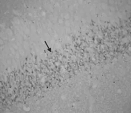
图1 正常对照组海马CA3区SYNⅠ阳性免疫反应产物呈现棕黄色颗粒状或点状,主要位于神经毡内,数量较多,排列密集,染色深↑。SYNⅠ免疫组化染色SABC×400Figure 1 Hippocampus CA3 area of normal control group.SYN I positive immune response product presents the yellowish brown color granulated or punctiform, Mainly is located in the neuropil quantity are more, the arrangement is crowded, dyeing depth↑.SYNⅠimmunohistochemical staining SABC×400

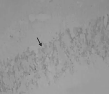
图2 低剂量组海马CA3区SYNⅠ阳性免疫反应产物呈现棕黄色颗粒状或点状,数量减少,染色稍淡,排列欠规则↑。SYNⅠ 免疫组化染色SABC×400Figure 2 Hippocampus CA3 area of low-dose group.SYN I The positive immune response product presents the yellowish brown color granulated or punctiform,quantity reduces few, the dyeing is slightly pale, the arrangement owes the rule↑.SYNⅠimmunohistochemical staining SABC×400

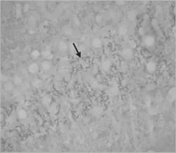
图3 高剂量组海马CA3区SYNⅠ阳性免疫反应产物呈现棕黄色颗粒状或点状,数量明显减少,染色淡,排列稀疏↑。SYNⅠ免疫组化染色SABC×400Figure 3 Hippocampus CA3 area of high-dose group.SYNI the positive immune response product Presents the yellowish brown color granulated or punctiform,Quantity reduces obviously,dyes palely, the arrangement is sparse↑.SYNⅠimmunohistochemical staining SABC×400

