
The diagnostic value of tenascin-C in acute aortic syndrome
2024-04-29 11:31:58MingMAWeiCHENHaiLongCAOJunPANQingZHOUXinLongTANGDongJinWANG
Journal of Geriatric Cardiology 2024年3期
Ming MA,Wei CHEN,Hai-Long CAO,Jun PAN,Qing ZHOU,Xin-Long TANG,✉,Dong-Jin WANG,✉
1.Department of Thoracic and Cardiovascular Surgery,Nanjing Drum Tower Hospital Clinical College of Nanjing Medical University,Nanjing,China;2.Department of Thoracic and Cardiovascular Surgery,Nanjing Drum Tower Hospital,Affiliated Hospital of Medical School,Nanjing University,Nanjing,China;3.Department of Thoracic surgery,Kunshan Hospital of Traditional Chinese Medicine,Jiangsu,China
ABSTRACT OBJECTIVES Misdiagnosis of acute aortic syndrome (AAS) significantly increases mortality.Tenascin-C (TN-C) is an extracellular matrix glycoprotein related to cardiovascular injury.The elevation of TN-C in AAS and whether it can discriminate suddenonset of acute chest pain in Chinese remains unclear.METHODS We measured the plasma concentration of TN-C by ELISA in a cohort of 376 patients with chest or back pain.Measures to discriminate AAS from acute coronary syndrome (ACS) were compared and calculated.RESULTS From October 2016 to September 2021,376 undiagnosed patients with chest or back pain were enrolled.166 of them were finally diagnosed as AAS,100 were ACS and 110 without cardiovascular diseases (NCV).TN-C was significantly elevated in AAS at 18.18 ng/mL (IQR: 13.10-27.68) compared with 7.51 ng/mL (IQR: 5.67-11.38) in ACS (P < 0.001) and 3.68 ng/mL (IQR:2.50-5.29) in NCV (P < 0.001).There was no significant difference in TN-C level among the subtypes of AAS.Of the 166 AAS patients,the peaked level of TN-C was at acute stage (P=0.012),then a slight of decrease was observed at subacute stage.The area under receiver operating characteristic curve for AAS patients versus NCV was 0.979 (95% CI: 0.964-0.994) for TN-C.At a cutoff level of 11.474 ng/mL,TN-C has a sensitivity of 76.0%,specificity of 85.5%,accuracy of 82.0%,positive predictive value (PPV) of 76.0%,negative predictive value (NPV) of 85.5%.Diagnostic performance of TN-C was superior to D-dimer and hs-cTnT.CONCLUSIONS The concentration of serum TN-C in AAS patients was significantly higher than that in ACS patients and NCV.TN-C could be a new biomarker to distinguish AAS patients in the early stage after symptoms onset from other pain diseases.
A cute aortic syndrome (AAS) is a fatal cardiovascular disease with high mortalit y,[1] and it includes aortic dissection(AD),intramural hematoma (IMH) and penetrating aortic ulcer (PAU).Half of the patients died 48 h after the symptom onset without immediate appropriate medical treatment.[2]The incidence of AAS is approximately 3 per 100,000 persons.[3]The accurate number might be much higher;25% of patients with AAS are not diagnosed because many doctors may not consider it in their initial diagnosis.[4]
Severe chest or back pain is the most common symptom of AAS.[5]Currently,the main challenge in the timely management of AAS patients is accurately distinguishing AAS from other disorders that present with sudden onset of severe chest or back pain,such as acute coronary syndrome (ACS).ACS patients show similar symptoms to ACS but require completely different treatments,including coronary angiography and thrombolytic drugs.A misdiagnosis of AAS as ACS greatly increases mortality and the rates of complications,especially when antiplatelet drugs are inappropriately used.[6]However,common examinations,such as ECGs and chest Xray,do not have enough sensitivity and specificity to distinguish AAS from ACS.[4]Computed tomography angiography (CTA) cannot be used in patients with a contrast medium allergy or renal insufficiency.Magnetic resonance imaging (MRI) takes more time to acquire,which makes it unsuitable for unstable patients.[7]
Pathologically,AAS is a disease involving the aortic medial layer.Lesions in the aortic medial wall are the key mechanism of the pathogenesis of AAS.[8]Several studies have investigated potential biomarkers related to the above disease process,such as smooth muscle myosin,[9]soluble elastin fragments,[10]soluble ST2,[11]troponins,[12]creatine kinases[13]and D-dimer.[14]D-dimer is the most commonly used biomarker in the real world,with a sensitivity of 94% and a varying specificity of 40%-100%in aortic dissection (AD).[15]The low specificity limits its clinical use.
Tenascin-C (TN-C) is an extracellular matrix glycoprotein that is barely expressed in normal tissues but is highly expressed in the process of embryonic development,trauma healing,and mechanical stress.[16-18]Several studies have shown that serum TN-C levels are associated with the prognosis and severity of myocardial injury and aortic disease.[19,20]
By sequencing the post-operative specimens of AAS patients (Raw data and processed data are available at the Gene Expression Omnibus (GEO accession:GSE153434))[21],we found that the expression of TNC was significantly increased in aortic tissue.This finding indicates that TN-C might be associated with aortic lesions.The aim of this study was to investigate the diagnostic value of serum TN-C levels in patients with AAS.
METHODS
Study Sample
This was a single-center retrospective cohort study including patients with acute chest or back pain.These patients were initially seen in community hospitals,and based on the standard procedures,including measurements of troponin,ECG or CT,the patients were triaged and transferred to different centers for further treatment.Some undiagnosed patients with aortic dilatation were transferred to our center,the Nanjing Drum Tower Hospital Clinical College of Nanjing Medical University (Nanjing,China),for further definitive diagnosis (n=606)between October 2016 and September 2021.The overall study design is shown in Figure 1.The exclusion criteria were as follows: pregnant women and patients who did not have complete information;patients with heart failure not due to AAS;acute and chronic liver failure not due to AAS;renal insufficiency not due to AAS;tumors in any part of the body;active infections or autoimmune diseases.
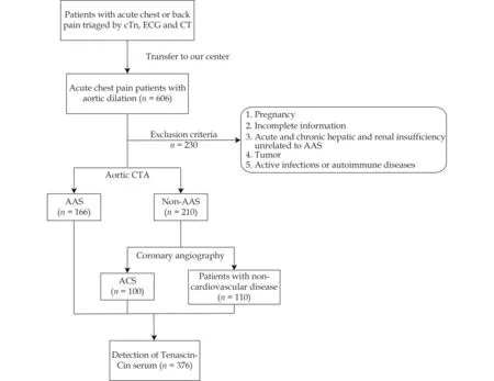
Figure 1 Study flow chart.AAS: acute aortic syndrome;ACS: acute coronary syndrome;CT: computed tomography;CTA: computed tomography angiography;cTn: troponin;ECG: electrocardiogram.
About 2-3 mL of abandoned whole blood after routine examines was placed in a sodium citrate anticoagulant tube immediately.The blood spin down at 2000 round/min for 20 min.Serum samples were stored at -80 ℃ before using.This study was approved by the Ethics Committee of Nanjing Drum Tower Hospital Clinical College of Nanjing Medical University,and all patients gave research authorization (No.2016-197).
Definition
All AAS subjects with medium and high pretest probabilities had imaging information from chest or abdominal computed tomography (CT).The AAS diagnoses were based on CTA (the 2014 European Society of Cardiology Guidelines for the Diagnosis and Treatment of Aortic Diseases).The ACS diagnoses were measured with coronary angiography by sophisticated cathlab cardiologists.Ultimately,376 patients were enrolled.One hundred and sixtysix patients were diagnosed with AAS,while 100 patients were diagnosed with ACS.Another 110 subjects were found to have no cardiovascular diseases (NCV) that needed further invasive treatment.In most of these patients,chest or back pain was caused by musculoskeletal,dyspeptic,or psychiatric factors such as anxiety or panic attack.
Measurements of TN-C
Serum levels of TN-C were tested using a Human TN-C Enzyme-linked immunosorbent assay(ELISA) kit (ab213831,USA).Briefly,prepare all reagents,samples,and standards as instructed.Add 100 ul standard or ample to appropriate wells.Discard plate content and add 100 ul biotinylated Antibody into all wells.Wash each well three times with 300 μL 0.01 M PBS.Add 100 μL ABC working solution.Wash each well five times with 300 μL 0.01M PBS and add 90 μL of prepared TMB.Add 100 μL TMB Stop Solution and read OD at 450 nm within 30 min.
Statistical Analysis
Continuous variables were summarized by mean± SD or median (interquartile range (IQR)) for skewed variables.Categorical variables were evaluated by the Fisher test.Gaussian numerical variables were compared with the Studentttest by the Mann-Whitney non-parametric test.AllPvalues are two tailed.The alpha level was set as 0.05.R software (version 4.2.1) was used for data analysis.R packages“gtsummary”,“pROC”and “ggplot2”were used for basic statistics and tables.
RESULTS
Clinical Features of the Patients
A total of 166 patients were finally diagnosed with AAS in the study.A total of 100 patients were ultimately diagnosed with ACS.Another 110 patients were diagnosed with NCV.The patient characteristics are shown in Table 1.These three groups were different in age (P=0.004),gender (P=0.044),smoker(P=0.017),hypertension (P< 0.001),diabetes (P<0.001),aspartate aminotransferase (P=0.020),triglyceride (P< 0.001) and low-density lipoprotein(P< 0.001).However,the median values of aspartate aminotransferase,triglyceride and low-density lipoprotein in each group were still within the normal ranges (the normal range of alanine aminotransferase in our center was 0-35 U/L,triglyceride was 0.56-1.7 mmol/L and low-density lipoprotein was 1.89-3.1 mmol/L),and the values had no clinical significance.
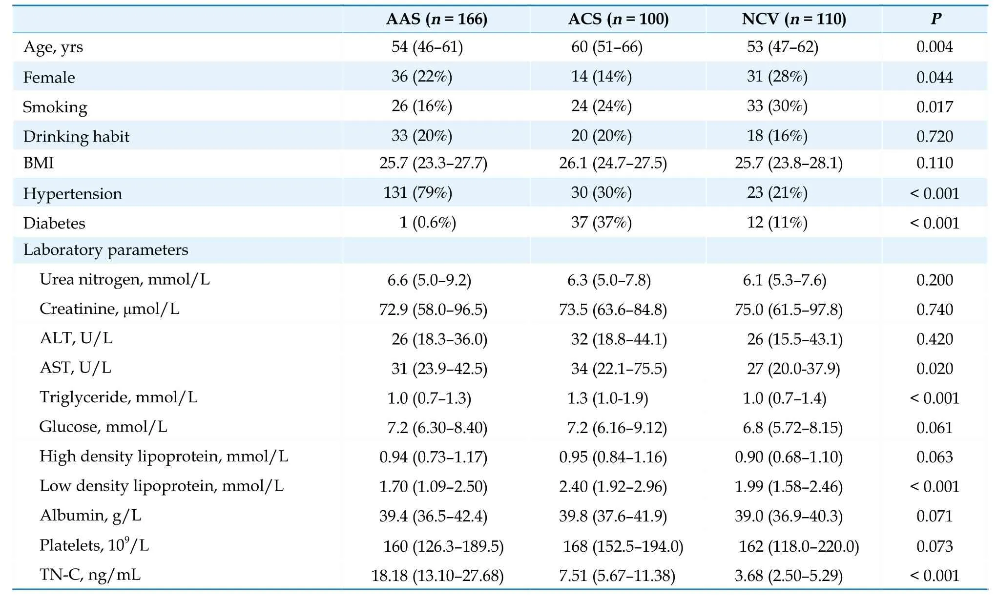
Table 1 Comparison of basal characteristics of the groups.
TN-C Distribution
The serum levels of TN-C were significantly higher in the AAS patients than in the ACS and NCV patients (P< 0.001).The TN-C levels were higher at 18.18 ng/mL (IQR: 13.10-27.68 ng/mL) in the AAS patients compared with 7.51 ng/mL (IQR:5.67-11.38 ng/mL) in the ACS patients.The TN-C concentration was 3.68 ng/mL (IQR: 2.50-5.29 ng/mL) in the NCV participants (Figure 2).
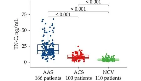
Figure 2 Comparison of admission TN-C between AAS,ACS and NCV patients.AAS: acute aortic syndrome;ACS: acute coronary syndrome;NCV: non-cardiovascular disease;TN-C:tenascin-C.
According to the AAS type,the patients were divided into three groups: 114 type A aortic dissection (TAAD),41 type B aortic dissection (TBAD)and 11 intramural hematoma (IMH).We did not enroll any penetrating aortic ulcer (PAU) patients during the entire study.The patient characteristics are shown in Table 2.In the different types of AAS,the TN-C concentration was 18.3 ng/mL (IQR:13.3-29.3) in the patients with TAAD,17.0 ng/mL(IQR: 13.1-24.3) in the TBAD patients,and 20.2 ng/mL (IQR: 12.2-24.9) in the IMH group.The IMH patients had a slightly higher TN-C concentration,but there was no significant difference between the three subtypes (P=0.810,Figure 3).

Table 2 Comparison of basal characteristics of different types of AAS.

Figure 3 Distribution of TN-C in different type of acute aortic syndrome.TAAD: type A aortic dissection;TBAD: type B aortic dissection;IMH: intramural hematoma.TN-C: tenascin-C.
Because the time of treatment is closely related to the prognosis of AAS,we analyzed the expression of serum TN-C in the AAS patients at different stages of symptom onset before admission.The patient characteristics are shown in Table 3.The hyperacute stage refers to the time of symptom onset within 24 h.The acute stage refers to the time of onset within 14 days.The subacute stage refers to an onset time of more than 14 days.The TN-C concentration was lower in the hyperacute stage at 17.3 ng/mL (IQR: 12.9-25.6 ng/mL) than in the acute stage at 24.8 ng/mL (IQR: 17.8-53.2 ng/mL,P=0.012).However,the serum TN-C levels between the acute and subacute stages were not significantly different (24.8vs.5.6 ng/mL withP=0.65,Figure 4).
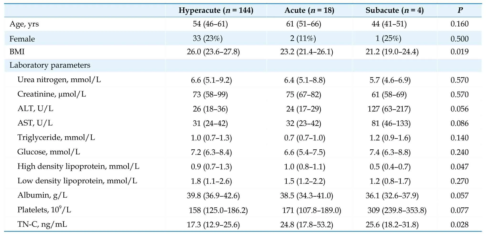
Table 3 Comparison of basal characteristics of the different stages of AAS.
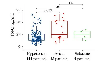
Figure 4 Distribution of TN-C according to time after onset in patients with AAS.AAS: acute aortic syndrome;TN-C: tenascin C.ns: P > 0.05
Diagnostic Performance for Discriminating AAS
The ROC analysis results indicated that TN-C for AAS patients versus NCV had an AUC of 0.979(95%CI: 0.964-0.994) and the cutoff level is 9.933 ng/mL (Figure 5A).The TN-C cutoff level of 11.474 ng/mL was the threshold leading to the maximum summation of the sensitivity and specificity in discriminating AAS from ACS (Figure 5B and Table 4).For D-dimer at a predefined cutoff level of 500 ng/ml,the corresponding sensitivities were 76.0%for TN-C and 67.1% for D-dimer,and the specificities were 85.5% for TN-C and 84.0% for D-dimer,resulting in 82.0% of the patients for TN-C and 77.6% of the patients for D-dimer being correctly classified.The positive and negative predictive values for TNC at 11.474 ng/mL were 76.0% and 85.5%,respectively.The positive and negative likelihood ratios forTN-C were 5.3 and 0.28,respectively.For another widely known test,hs-cTnT,at an hs-cTnT of 0.045 μg/L in our cohort,hs-cTnT had an accuracy of 76.2%with a sensitivity of 76.5%,a specificity of 76.1%,a positive predictive value of 65.8%,a negative predictive value of 84.4%,a positive likelihood ratio of 3.2,and a negative likelihood ratio of 0.31.The diagnostic performance of TN-C was superior to that of D-dimer and hs-cTnT.

Table 4 Diagnostic performance of patients with AAS versus ACS using TN-C compared with D-Dimer and hs-cTnT.
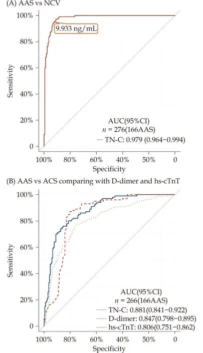
Figure 5 Receiver operating characteristic curves of TN-C.(A): AAS vs.NCV patients;(B): AAS vs.ACS patients comparing with D-Dimer.AAS: acute aortic syndrome;ACS: acute myocardial infarction;NCV: non-cardiovascular disease;TN-C:tenascin C.
DISCUSSION
This study analyzed the data of 376 participants with chest or back pain as the main symptoms before admission.There were 166 patients with AAS,100 patients with ACS,and 110 without cardiovascular diseases.Some promising results suggest that TN-C may be a new potential biomarker to distinguish AAS from ACS.This may contribute to the early diagnosis of AAS.
The most common symptoms of AAS are sudden and severe pain.[22]Chest pain,abdominal pain and back pain are the main clinical manifestations of most AAS patients.Even so,there are still some patients with none of the above classical symptoms,and AAS could be inadvertently found during other physical examinations or treatments.According to the guidelines,12-lead or 18-lead ECG,chest Xray,CT,MRI and transesophageal echocardiography (TEE) are the most common diagnostic methods for patients with suspected AAS.[23]CTA is the gold standard method to diagnose AAS with firstepisode chest pain.The sensitivity and specificity were both nearly 100%.However,the use of enhanced CT is limited because some patients have allergies to the contrast media,and there is the possibility of reinjury to renal function.[5]MRI requires more examination time and cannot be performed in patients with unstable circulation or intolerance to the closed environment.[7]Esophageal stimulation during TEE may exacerbate aortic lesions,which may greatly increase the risk of aortic rupture.However,the diagnostic efficiency of ordinary echocardiography is not ideal.[24]
In recent decades,circulating biomarkers have gained an exponential key player role in the diagnostic-prognostic pathways of cardiovascular diseases,namely ACS,heart failure (HF) and AAS.[25,26]Generally,these biomarkers can be roughly divided into five categories: (1) smooth muscle cell markers,like smooth muscle myosin heavy chain(smMHC),reflect the destruction of aortic media;(2) extracellular matrix proteins,such as matrix metalloproteinases (MMPs),reflects aortic injury;(3) coagulation markers,such as D-Dimer,reflect the dynamic coagulation state caused by AAS;[27](4)inflammatory biomarkers,such as ST2,are increased post-dissection,reflecting the extent of damage to the aorta and ongoing inflammation;and (5)some specific markers reflect cardiac stress and damage,such as CK-MB,[13]TNT.[2,12]In general,current studies have shown that extracellular matrix degradation induced by inflammatory factors is a potential mechanism for the pathogenesis of aortic syndrome.[2,28,29]
TN-C is an extracellular matrix protein.It is a large molecule of approximately 220-400 kDa as an intact monomer and is assembled as a hexamer.[17]From the viewpoint of aortic homeostasis and AAS pathogenesis,TN-C could be regarded as a stressactivated molecular damper.It is inactive under normal conditions,but once the tissue experiences high mechanical stress,TN-C can be activated and works to reinforce the tissue strength by inducing extracellular matrix (ECM) proteins and ameliorating the excessive proinflammatory response.[17,30]When destruction of the ECM occurs in the tunica media of the aorta,smooth muscle cells and fibroblasts release TN-C.[19]In pathological conditions,it is believed that TN-C plays an important role[31]as a transducer of signals for tissue repair and as a vascular wall protector through the modulation of inflammatory and fibrotic responses in cardiovascular diseases,including abdominal aortic aneurysm and AAD.[32]
In our study,we found that the magnitude of elevated TN-C can distinguish AAS patients from ACS patients.Some scholars believe that when destructive mechanical stress occurs in blood vessels,the blood vessels can protect tissues by inducing the synthesis of TN-C in vascular smooth muscle and helping to reduce the inflammatory response caused by the tissue injury.[19,33,34]In fact,we also found that TN-C levels were higher in ACS patients,but they were still lower than those in AAS,suggesting that the increase in TN-C in AAS may mainly be the result of smooth muscle cell stretching and inflammation rather than cardiomyocyte necrosis.Substantial aortic hemodynamic changes significantly increased the circulating TN-C levels in the aorta(largest artery) compared with the coronary artery(smaller artery).This result suggests that the release location may be another reason for the higher degree of TN-C in AAS.
We enrolled patients with different types of AAS in our study.We found no significant difference in the serum level of TN-C among patients with these different types.AD and IMH share similar clinical features and complications but have different pathophysiologic mechanisms.IMH could then progress to AD if the intimal layer ruptures,which is termed the entry tear.
Previous studies showed that the expression of TN-C becomes detectable within 24 h of the onset of infarction in experimental animal models,peaks on Days 3-5,and disappears by Day 28.[35]Previous clinical studies suggested that TN-C contributes to the progression of adverse ventricular remodeling.[36]AAS patients with high serum levels of TN-C in the acute stage were found to be at an increased risk of ventricular dilatation in the chronic stage and had a poor long-term prognosis.[35,37]An in-depth study also found that the expression of tenascins was different in the histological analysis of the occurrence and development of AD and was even involved in the reconstruction of the vascular wall in the chronic stage of AD.[17]Our study found that TN-C peaked 2-14 days after symptom onset,which is the subacute stage of AAS.However,the stage at which TN-C decreases and the role of TN-C in the process of the chronic stage of AAS need further research.
At present,D-dimer is a widely used serum biomarker in the diagnosis of AD.[38]However,it should be emphasized that normal levels of D-dimer may also exist in AAS patients.[39]Therefore,we included D-dimer for further comparison in our study.The results show that TN-C has better diagnostic performance than D-dimer.To our knowledge,this study indicates for the first time that TNC may become a new biomarker for AAS.One advantage of this study is that we found and verified the diagnostic performance of TN-C for AAS.
This study has some limitations.This study is a single-center study with a limited sample size.The area under the ROC curve and the positive and negative predictive values observed in our study may be exaggerated by our derivation.Therefore,as a single center study,the universality of our findings needs to be verified by a large prospective multicenter study to confirm the diagnostic efficacy and accuracy of this new biomarker.The absolute value of TN-C concentration may vary according to the determination method used,which may affect the recommended cutoff level.Finally,other diseases(such as pulmonary embolism) may also affect the diagnosis of AAS.However,due to the problem of sample size,further comparative analysis could not be carried out in this study.
In conclusion,TN-C showed a favorable overall diagnostic performance for AAS over D-dimer and hs-cTnT in patients with suspected AAS.It might be a useful biomarker for distinguishing AAS,but more research is needed for the application of AAS prognosis and outcomes.Therefore,TN-C could be a sensitive candidate that may provide a timely and cost-effective diagnostic examination to determine early AAS.
DISCLOSURE
Declarations of Interest
None
Fundings
This work was supported by the National Natural Science Foundation of China (No.81970401,No.82270346,No.82170496),Jiangsu Provincial Key Medical Discipline (ZD2021023),Nanjing Municipal Health Science and Education Key Project (ZKX 21021) and Nanjing Science and Technology Bureau Medical and Health International Joint Project(No.202002052).
Ethical Statement
The study was conducted at Nanjing Drum Tower Hospital Clinical College of Nanjing Medical University,and was approved by the Ethics Committee of Nanjing Drum Tower Hospital Clinical College of Nanjing Medical University,and all patients gave research authorization(No.2016-197).
Data Availability
The datasets used and/or analyzed during the current study are available from the corresponding author on reasonable request.
 Journal of Geriatric Cardiology2024年3期
- Journal of Geriatric Cardiology的其它文章
- Community-based prevention and treatment of cardiovascular diseases
- Significance of balloon aortic valvuloplasty as palliative procedure for symptom benefit in patients with severe aortic stenosis
- Sleep quality and characteristics of older adults with acute cardiovascular disease
- Association of acute glycemic parameters at admission with cardiovascular mortality in the oldest old with acute myocardial infarction
- Effects of loneliness and isolation on cardiovascular diseases:a two sample Mendelian Randomization Study
- Cardiovascular risk burden and disability: findings from the International Mobility in Aging Study (IMIAS)