Effects of Moringa oleifera aqueous extract on the growth performance,blood characteristics,and histological features of gills and livers in Nile tilapia
Mhmou A.Emm,Rmy M.Shourl,Wl N.El-Hwrry,Shm Y.Ao-Kor,Ftm Al-Monm G,Ashrf M.A El-ltif,Mhmou A.O.Dwoo
a Histology Department,Faculty of Veterinary Medicine,Benha University,Benha,13511,Egypt
b Department of Animal Husbandry and Animal Wealth Development,Faculty of Veterinary Medicine,Alexandria University,Alexandria,22758,Egypt
c Pharmacology Department,Faculty of Veterinary Medicine,Benha University,Benha,13511,Egypt
d Clinical Pathology Department,Faculty of Veterinary Medicine,Benha University,Benha,13511,Egypt
e Department of Aquatic Animal Diseases and Management,Faculty of Veterinary Medicine,Benha University,Benha,13511,Egypt
f Animal Production Department,Faculty of Agriculture,Kafrelsheikh University,Kafr El-Sheikh,33511,Egypt
g The Center for Applied Research on the Environment and Sustainability,The American University in Cairo,Cairo,11835,Egypt
Keywords: Growth performance Liver Gills Moringa oleifera Nile tilapia
ABSTRACT Moringa oleifera is well known as a highly nutritious plant and a water purifier in fish culture.Tilapia fish has many impressive qualities making them very suitable for aquaculture.Therefore,this study aimed to evaluate the effect of supplementation of Moringa oleifera aqueous extract (MOAE) at varying concentrations (0,100,200,and 400 mg/kg diet for 90 days) on growth performance,hematological,and some biochemical parameters of Nile tilapia.Additionally,histological changes and immunohistochemical expression of caspase 3 in gills and livers were evaluated.Significant enhancements (P <0.05) in the final weight,weight gain,specific growth rate,red blood cells,hemoglobin,packed cell volume,white blood cells,and total serum protein were observed in fish fed 200 mg MOAE/kg diet compared with other groups.However,a significant reduction (P <0.05) of liver and kidney function tests and non-significant changes in lipid profile were recorded.On the other hand,fish fed 400 mg MOAE/kg diet revealed disturbance in the corresponding parameters.Severe histological degenerative changes were detected in the gills and liver of fish fed 400 mg MOAE/kg diet.In comparison,fish fed 200 mg MOAE/kg diet showed the best histo-architectures of these organs compared with other groups.Concomitant immune expressions of caspase 3 were determined where gills and liver of fish fed 400 mg MOAE/kg diet revealed extensive number of positive caspase 3 cells indicating severe apoptotic features.Accordingly,a dietary MOAE at 200 mg/kg diet is recommended to improve the growth performance,hematological parameters,liver and kidney functions in Nile tilapia.Meanwhile,caution must be considered when using MOAE at a concentration over 200 mg/kg diet.
1.Introduction
Nile tilapia,Oreochromis niloticus,is omnivore fish that belong to the family Cichlidae.It is considered one of the most productive food fish in Egypt and all over the world (Modadugu &Belen,2004;Mugwanya et al.,2021).In fish aquaculture,using antibiotics and animal protein resources as growth promoters is becoming unaffordable (Shourbela et al.,2021).Hence,more attention has been paid to using alternative medicinal plants to replace the expensive growth promoters and avoid potential health and environmental hazards (Enayat Gholampour et al.,2020;Kumar et al.,2022).The nutritional and medicinal properties ofMoringa oleiferasuggest it as a good option for replacement (Puycha et al.,2017).
M.oleiferais a widely grown tree in tropical and sub-tropical areas(Fahey,2005).In Egypt,M.oleiferahas been grown in Aswan,North Sinai,and Al-Sharkyia.The different parts of such plants have nourishing importance (Leone et al.,2015).Leaves ofM.oleiferacontain significant quantities of vitamins,minerals,and protein (Afuang et al.,2003;Murro et al.,2003).However,seeds ofM.oleiferacontain 31.65%protein,7.54% fiber,and 6.53% ash contents (Anwar &Rashid,2007).Moreover,M.oleiferaseeds possess antimicrobial properties (Anhwange et al.,2004) due to their lipophilic compounds (Jabeen et al.,2008) and also are used as a water purifier in fish culture (Ayotunde et al.,2011a).In the last decade,M.oleiferahas been used as a growth promoter and increases feed conversion ratio in broiler chicken (Gakuya et al.,2014;Alabi et al.,2017).Phytochemically,M.oleiferais rich in rhamnose sugar,glucosinolates,isothiocyanates,and carotenoids (Bharah et al.,2003;Fahey,2005).It is noteworthy thatM.oleiferahas an antioxidant activity due to its phenolic (Sreelatha &Padma,2009) and flavonoid(DeiviArunachalam et al.,2021;Xu et al.,2019) contents.Although several benefits ofM.oleiferaseeds,few studies have been done on their toxicity to cultivated fish (Ayotunde et al.,2006;2011b).
Based on such background,the current study was designed to evaluate the dietary supplementation ofM.oleiferaseed aqueous extract(MOAE) at different concentrations on growth performance,hematological,and some biochemical parameters in Nile tilapia,O.niloticusunder static bioassay procedure.In addition,the histological alterations in the gill and liver were described,and the evaluation of apoptosis via immunohistochemical expression of caspase 3 in these organs was demonstrated.So,this study will serve as baseline information for applying theM.oleiferainO.niloticusfarming.
2.Material and methods
2.1.Chemicals
Biochemical kits for aspartate aminotransferase (AST),alanine aminotransferase (ALT),total cholesterol (TC),high-density lipoprotein cholesterol (HDL-C),low-density lipoprotein cholesterol (LDL-C),total protein (TP),albumin (Alb),triglyceride (TG),blood urea nitrogen(BUN) and creatinine (Cr) were purchased from Bio-Diagnostic Co.(Cairo,Egypt).
2.2.Preparation of MOAE and diet preparation
Mature seeds ofM.oleiferawere gathered from Abu-Hammad,Al-Sharkyia,Egypt,dried,and grounded using mortar and pestle.The extraction procedures were done according to Fernandez (1990).The groundedM.oleiferaseeds were soaked in distilled water using a 1:2 ratio (weight/volume) for 24 h,then filtered and evaporated in a rotary evaporator (60◦C).The concentrated extract was kept in a sterile container and refrigerated (4◦C) until needed.Test diets were prepared by mixing MOAE with the basal diet (commercial feed,30% crude protein,Skretting,El Sharqia Governorate,Egypt) at 100,200,and 400 mg MOAE/Kg diet.
2.3.Experimental design
Healthy three hundred overwintered monosexOreochromis niloticusfingerlings were obtained from a private fish hatchery at Kafr El Sheikh Governorate,Egypt.The fish were transferred into polyethylene bags filled with oxygen to the Lab of Fish Breeding and production,Faculty of Veterinary Medicine,Alexandria University,Egypt.Fingerlings were divided into 4 equal groups (50 fish/group divided into two replicates;25 fish per replicate) in rectangular white plastic tanks (500 L capacity)for acclimation 2 weeks before the experiment.Each tank was supplied with 300 L of underground freshwater and equipped with individual biofilters and a constant air supply.During the adaptation period,fish were fed twice daily (08:00 a.m.;03:00 p.m.) with a diet previously formulated to obtain a 30% crude protein.
After the acclimation,the group I was fed on a control diet containing no MOAE.Group II,III,and IV received a 100,200,and 400 mg MOAE/Kg diet,respectively.The end of the feeding experiment was (90 days).
2.4.Growth performance
At the end of the feeding experiment,all fish were weighted to calculate final weight (FW),weight gain (WG),specific growth rate(SGR),and feed conversion ratio (FCR).Also,the initial weight (IW) was calculated before the experiment.Calculations were conducted using the following formulae:
2.5.Blood sampling
At the end of the experiment,blood samples were collected from the caudal vein of fish and divided into two parts,one part of the blood was collected on EDTA containing tubes for hematological analysis,including red blood cells (RBCs),white blood cells (WBCs) count,packed cell volume (PCV),hemoglobin (Hb),mean corpuscular volume(MCV),mean corpuscular hemoglobin (MCH) and mean corpuscular hemoglobin concentration (MCHC).Another part of the blood was collected on plain tubes and allowed to clot at room temperature,then centrifuged at 3000 g for 15 min then sera samples were collected and stored at 20οC for all biochemical analysis including AST and ALT activities,TC,HDL-C,LDL-C,TP,Alb,TG,BUN,and Cr.
2.6.Hematological analysis
All hematological parameters,including RBCs and WBCs count,Hb concentration,PCV valve,and red blood cell indices (MCV,MCH,and MCHC),were estimated according to the hematological procedures for fish (Feldman et al.,2000).
2.7.Biochemical analysis
Serum AST and ALT (Reitman &Frankel,1957),TG (Fredrickson et al.,1967),TC (Allain et al.,1974),HDL-C (Finely et al.,1978),LDL-C(Friedewald et al.,1972),TP and Alb (Lowry et al.,1951;Doumas et al.,1971 respectively) were measured.Serum globulin was determined by subtracting the Alb value from the TP value of the same sample,and consequently,the albumin to globulin ratio (A/G) was calculated.BUN and Cr levels were measured according to Patton and Crouch (1977) and Henry (1974),respectively.
2.8.Histological examination
At the end of the experiment,ten random fish from each group were selected for the histological examination.The fish were dissected to obtain the gills and livers.Small tissue specimens from these organs were fixed in 10% neutral buffered formalin for 48 h,then dehydrated in alcohol,cleared in xylene,and embedded in paraffin wax.Sections of 5-μm thickness were cut and stained with hematoxylin and eosin for general histo-architecture.The fixation and staining were done as outlined by Bancroft and Gamble (2008).
2.9.Semi-quantitative scoring method
The histological changes of each fish tissue were photographed using a Leica DM3000 microscope with a digital camera.The changes were evaluated by a semi-quantitative score as outlined by Benli et al.(2008).Scores were– to +++depending on the degree and extent of the changes as follows: (-) no histological changes,(+) histological changesin <20% of fields,(++) histological changes in 20–60% of fields,(+++)histological changes in >60% of fields.Five slides were observed for each organ and treatment.
2.10.Immunohistochemical localization of caspase 3
Paraffin sections from the different organs were collected on positive charge slides.Avidin-biotin peroxidase method,as outlined by Emam et al.(2018),was used to localize caspase 3.Dewaxing,rehydration,and blocking of endogenous peroxidase were done.Antigen retrieval was done at 90◦C with citrate buffer pH 6 for 30 min.Next,the sections were incubated with monoclonal antibody anti-caspase-3 (Santa Cruz Biotechnology Inc.,Dallas,TX,USA,1:150 dilutions) for 1 h at room temperature (RT).The sections were incubated with anti-mouse IgG secondary antibodies (Abcam,Boston,USA) for 30 min at RT.Ultimately,the stain was evident with 3,3-diaminobenzidine tetrahydrochloride (DAB) (Dako,Japan),and counterstained was with hematoxylin.
2.11.Statistical analysis
One-way ANOVA was used to analyze the data statistically.Duncan’s multiple range tests were applied to detect any anticipated significant differences between treated fish groups at a significant level of 95%.All values are expressed as mean ±SE and deemed significant atP≤0.05.
3.Results
3.1.Growth performance findings
Concerning feeding MOAE supplemented diet,significant increases in growth performance were observed in group III (200 mg MOAE/Kg)compared with other groups.However,group IV (400 mg MOAE/Kg)revealed disturbance in those parameters.In comparison to the control group,FW,WG,and SGR were significantly increased (P<0.05) in groups supplemented with 100 and 200 mg MOAE/Kg diet (Table 1).

Table 1 Growth performance of O.niloticus after 90 days exposed to different concentrations of dietary M.oleifera seeds aqueous extract.
3.2.Hematological findings
Fish from group III revealed significant enhancement in RBCs counts,Hb concentration,and PCV values compared to the control group.Conversely,those parameters showed significant decreases in group IV compared to control one.Meanwhile,group II (100 mg MOAE/Kg)expressed non-significant alterations in those parameters (Table 2).

Table 2 Hematological parameters (Mean ± SE) of O.niloticus exposed to different concentrations of dietary M.oleifera seeds aqueous extract.
Concerning red blood cell indices,MCV showed a significant decrease in fish from group III with a significant increase in group IV when both groups compared to control.However,group II revealed a non-significant change in MCV compared to control.Meanwhile,MCH showed non-significant alteration in different treated groups compared to the control group.Also,MCHC expressed a non-significant change in fish groups II and III compared to control.On the other hand,fish from group IV showed a significant decrease compared to the control(Table 2).
Regarding WBCs counts,group III revealed significant increases in WBCs counts compared to the control group.Meanwhile,groups II and IV revealed non-significant alteration (Table 2).
3.3.Biochemical findings
Lipid profiles including TC,HDL-C,LDL-C,and TG revealed nonsignificant changes in fish groups II and III compared to the control group.On the other hand,there were significant increases in TC,LDL-C,and TG levels with non-significant alteration in HDL-C level in group IV(Table 3).
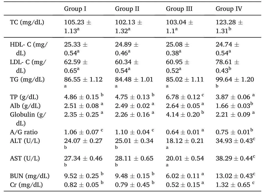
Table 3 Biochemical parameters (Mean ± SE) of O.niloticus exposed to different concentrations of dietary M.oleifera seeds aqueous extract.
There was a significant elevation in TP level in fish from group III compared to the control.Conversely,group IV exhibited a significant reduction in TP compared to control.Meanwhile,there was a nonsignificant alteration in their level in fish from group II compared to control (Table 3).
Non-significant changes were observed in albumin level in groups II and III compared to control,but group IV showed a significant decrease in its level compared to control.Meanwhile,globulin level expressed a significant elevation in group III compared with control and nonsignificant alteration in its level in both groups II and IV compared to control.A/G ratio revealed a significant reduction in fish from groups III and IV compared to control but,non-significant change was recorded in group II compared to control (Table 3).
Concerning the results of ATL,AST,BUN,and Cr,there were significant decreases in ALT and AST activities and BUN and Cr levels in group III.However,group IV revealed significant increases in ALT and AST activities besides BUN and Cr levels.On the other hand,fish from group II expressed non-significant changes in these parameters(Table 3).
3.4.Histological findings
The observed histological findings in gills and livers of Nile tilapia,O.niloticusfrom different groups were summarized in Table 4 and presented in Figs.1–2.Additionally,immunohistochemical expressions of caspase 3 in these organs were presented in Figs.3–4.
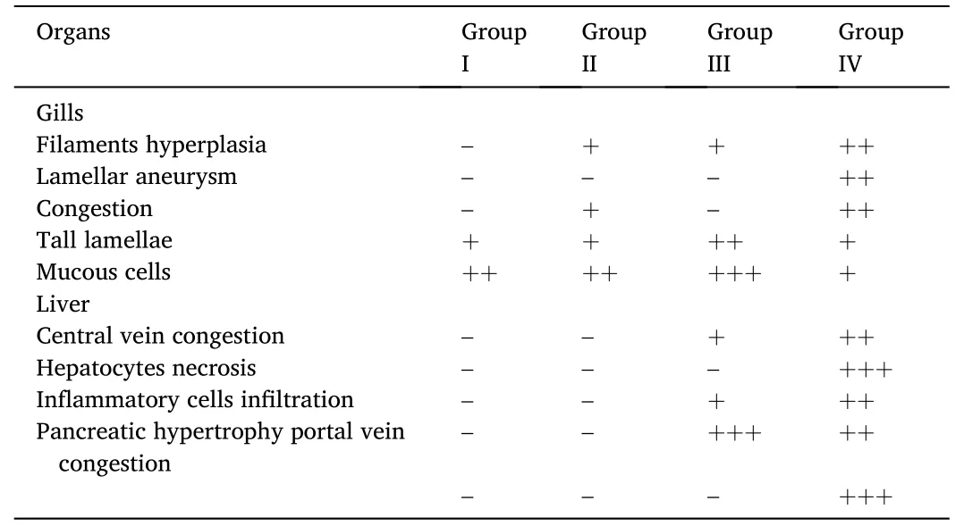
Table 4 Summary of histological changes observed in O.niloticus exposed to different concentrations of dietary M.oleifera seeds aqueous extract.
3.5.Gills
Histological sections of the gills inO.niloticusof the control group showed normal histo-architecture.Each gill had numerous gill filaments,which carried many regular gill lamellae shorter than other groups.Each filament was lined with epidermal epithelium,while each lamella consisted of pillar cells enclosing blood capillaries (Fig.1A).However,gills from group II showed mild hyperplasia in the epithelia of filaments and lamellae with mild congestion (Fig.1B).The longest gill lamellae with increased hyperplasia of filaments epithelium were observed in fish from group III (Fig.1C).Meanwhile,congestion and dilatation of blood vessels of filaments,lamellar aneurysm,degeneration,and sloughing of lamellae,as well as hyperplasia of filaments epithelium,were the characters of gills in group IV (Fig.1D).
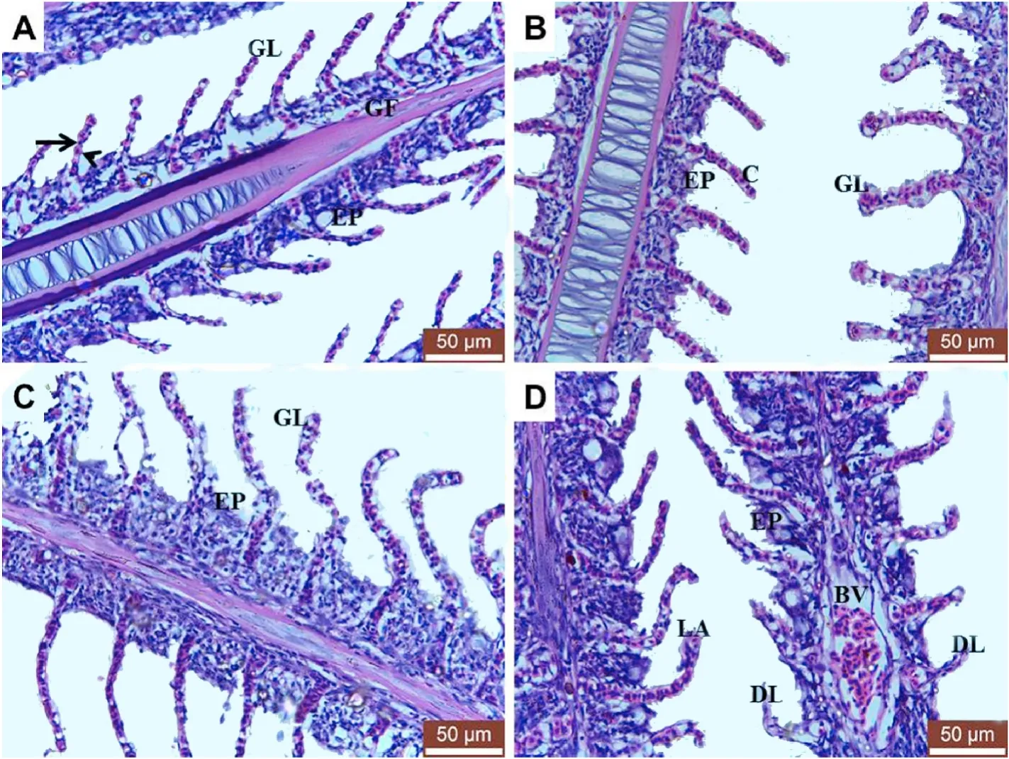
Fig.1.Photomicrograph of H&E stained gills sections from mature O.niloticus fed 0,100,200 and 400 mg MOAE/kg diet (A,B,C and D) respectively.A:showing normal gills histoarchitecture.GF,gill filaments;GL,gill lamellae;EP,epidermal epithelium;arrow,pillar cells;arrowhead,blood capillaries.B:showing mild hyperplasia in the epithelia of filaments(EP) and lamellae (GL) with mild congestion (C).C:showing long gill lamellae (GL) with increased hyperplasia of filaments epithelium (EP).D: showing congestion and dilatation of blood vessels (BV),lamellar aneurysm (LA),hyperplasia of filaments epithelium (EP),degenerated and sloughed lamellae(DL).
3.6.Liver
Histological sections of the liver inO.niloticusfrom groups I and II showed normal histological features of both hepatic and pancreatic tissues (Fig.2A,a,b).Normal hepatic parenchyma,including normal central vein,blood sinusoids,hepatic cords,and lipid droplets within the cytoplasm of hepatocytes (Fig.2A and B),were observed as well as normal hepatopancreas which consisted of group acinar cells around a portal vein (Fig.2a and b).However,fish from group III showed dilatation and congestion of the central vein,inflammatory cells infiltration,and hypertrophy of pancreatic acinar cells (Fig.2C,c).Rupture of the central vein wall with extravasation of erythrocytes among the hepatocytes (Fig.2D) were the characteristics for fish from group IV.Additionally,necrosis of hepatocytes began to appear.Necrotic cells had condensed cytoplasm and pyknotic nuclei,especially around the ruptured central vein.Moreover,vacuolar degeneration of the hepatocytes was noticed (Fig.2D).On the other hand,the hepatopancreas showed hypertrophy of pancreatic acinar cells as well as severe congestion of the portal vein of (Fig.2d).
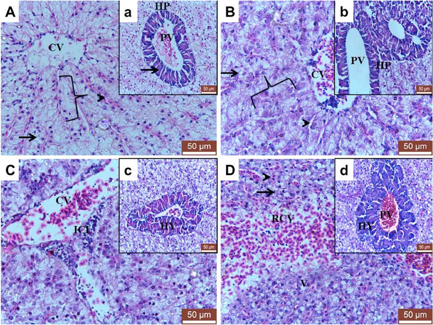
Fig.2.Photomicrograph of H&E stained liver sections from mature O.niloticus fed 0,100,200 and 400 mg MOAE/kg diet (A,B,C and D) respectively and their insets.A and B: showing normal histological feature of hepatic tissue.CV,Central vein;arrowhead,blood sinusoid;arrow,lipid droplets within the cytoplasm of hepatocytes;Brace,hepatic cords.Insets (a and b): showing normal histological feature of hepatopancreas (HP).PV,portal vein;arrow,acinar cells.C: showing dilatation and congested central vein (CV) and inflammatory cells infiltration (ICI).Inset c: showing hypertrophy of pancreatic acinar cells (HY).D: showing severe toxic effect of 400 mg MOAE/kg diet.RCV,ruptured central vein,arrowhead,extravasation of erythrocytes among the hepatocytes;arrow,necrotic cells with condensed cytoplasm and pyknotic nuclei;V,vacuolar degeneration of the hepatocytes.Inset d: showing severe congestion of the portal vein of hepatopancreas (PV)and hypertrophy of pancreatic acinar cells (HY) were characteristics for fish fed 400 mg MOAE/kg diet.
3.7.Immunohistochemical findings
Gills from groups I,II,and III showed weak caspase 3 (Fig.3A–C);however,group IV showed potent caspase 3 expression (Fig.3D).
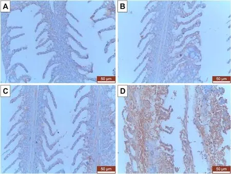
Fig.3.Immunohistochemical expression of caspase 3 in gills from mature O.niloticus fed 0,100,200 and 400 mg MOAE/kg diet (A,B,C and D) respectively.Strongest expression was noticed in fish fed 400 mg MOAE/kg diet (D).
Both hepatic and pancreatic tissues from groups I,II,and III showed weak caspase 3 (Fig.4A–C);however,the same tissues from group IV showed potent caspase 3 expression (Fig.4D).
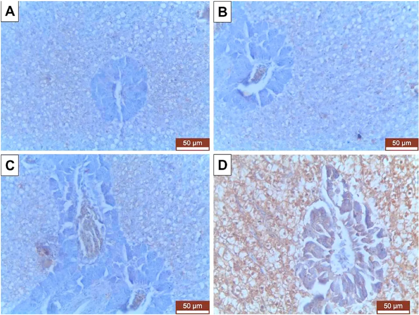
Fig.4.Immunohistochemical expression of caspase 3 in livers from mature O.niloticus fed 0,100,200 and 400 mg MOAE/kg diet (A,B,C and D) respectively.Strongest expression was noticed in fish fed 400 mg MOAE/kg diet (D).
4.Discussion
In the last decade,M.oleiferaseeds have been used in aquaculture as a growth promoter (Sherif et al.,2014),antimicrobial agent (Ruttarattanamongkol &Petrasch,2015) as well as an enhancer for reproductive performance (Gad et al.,2019).Therefore,the current study aimed to investigate the effect of MOAE dietary supplementation at different concentrations on growth performance,clinicopathological,histological,and immunohistochemical pictures of some vital organs in Nile tilapia,O.niloticus.
In the current study,feeding of MOAE supplemented diet revealed significant growth performance parameters (FW,WG,and SGR) in fish fed 200 mg MOAE/kg diet compared with other groups.However,fish fed 400 mg MOAE/kg diet revealed disturbance in those parameters.Recently,Shourbela et al.(2020) also clarified thatO.niloticusfingerlings fed on MOAE containing diet showed an enhanced growth performance.Additionally,Heidarieh et al.(2013) indicated thatM.oleiferahad improved the food digestibility and absorption capacity ofO.mykiss.On the contrary,Dongmeza et al.(2006) reported a considerable reduction in feed intake,feed utilization,and fish performance after receiving dietaryM.oleiferaleaf extracts.
The hematopoietic system is considered a mirror of the body that reflects the health status of fish (Burgos-Aceves et al.,2019;Rehulka,2000).The current study revealed that fish fed a 200 mg MOAE/kg diet showed significant enhancement in RBCs counts,Hb concentration,and PCV values compared to the control group.These results partially agreed with Sherif et al.(2014),who reported that hemogram including RBC,Hb,PCV,MCV,MCH,and MCHC ofO.niloticussupplemented with different concentrations ofM.oleiferais significantly improved.This owed toM.oleiferacauses enhancement in health status (Nascimento et al.,2016) as it contains saponin (Khalil &Eladawy,1994),alkaloids,and flavonoids which elevates RBCs,HB,and PCV (Anwar et al.,2007).On the other hand,fish fed 400 mg MOAE/kg diet expressed significant decreases in RBC,Hb,PCV,MCHC with an increase in MCV resulting in macrocytic hypochromic anemia.This result goes in harmony with Bbole et al.(2016),and this anemia may be attributed to high anti-metabolites,which are toxic for fish,especially tannin (Dienye &Olumuji,2014).Our study revealed an increase in WBCs count in dose 200 mg MOAE/kg diet that confirms the results of Eyiwunmi et al.(2018),who reported that African catfish that received moringa shows an elevation in WBCs.The elevated WBCs count may be owed to the immunomodulatory capacity ofM.oleiferain fish (Stadtlander et al.,2013).This WBC elevation may refer to high immunity,which will be helpful against infections (Hrubec et al.,2000).
Concerning biochemical analysis,fish fed 200 mg MOAE/kg diet revealed significant decreases in ALT and AST activities besides BUN and Cr levels.These results agreed with Shourbela et al.(2020),which may be attributed to hepatoprotective (Nascimento et al.,2016) and nephron-protective effects of moringa (Sharma &Paliwal,2012),which are owing to its antioxidant properties and ability to scavenge reactive oxygen species (Cao et al.,2016).Also,this group showed elevation in TP and globulin that goes in harmony with the results of Shourbela et al.(2020).
On the other hand,fish fed 400 mg MOAE/kg diet exhibited significant elevation in TC,TG,LDL-C,BUN,and Cr levels and ALT and AST activities along with a reduction in TP albumin levels.The elevations in serum BUN and Cr levels have been used in fish to indicate gill and kidney dysfunction (Adham et al.,2002).Additionally,BUN is primarily associated with gill and liver disturbance.So that increases in serum levels of BUN and Cr may be due to gill dysfunction,which leads to disturbance in urea diffusion between the blood and the water.Elevation in ALT and AST are considered essential markers for hepatic impairment as when the cell membrane of the hepatocyte is damaged or permeability of hepatocyte increased;these enzymes typically present in the cytosol so that they are released into the blood stream resulting in an elevation in their activities (Gabriel &George,2005).Elevated lipid parameters with a decrease in TP and Alb resulted from liver damage at this dose of MOAE,and histological findings confirmed this damage.
Histological examination is used to indicate the effects of various toxicants on fish (Dawood,2021;Vajargah et al.,2021).The present study revealed that the dietary MOAE (0 and 100 mg/kg diet) has no marked histological changes in the liver and gills ofO.niloticus.However,the dietary 400 mg MOAE/kg diet demonstrated gills and liver tissues.The observed gill lamellar hyperemia and hypertrophy,as well as disarrangement of the hepatic cell,necrosis,and vacuolation,were similar to Ayotunde et al.(2011b),Sirimongkolvorakul et al.(2012),and Akinjide et al.(2017).These histological changes of the gills can negatively affect its oxygen uptake and osmoregulation functions(Babatunde et al.,2001).It was worthy to note the decreased number of mucous cells in the gills of this group.
On the other hand,fish fed a 200 mg MOAE/kg diet showed good histological signs for these organs,especially at the level of their growth,development,and,consequently,functions.It was worthy to note for the increased number of mucous cells in the gill lamellae of this group which may resist the entrance of pathogens into the gill and protects it against toxic substances (Sirimongkolvorakul et al.,2011) and aid in ionic and osmotic regulation and excretion (Rogers &Wood,2004).This may be due to the high content of unsaturated fatty acids,vitamins,and protein inM.oleiferaseeds which are essential in growth and development(Soliva et al.,2005).
Immunohistochemically,caspase 3 expressions were strong in organs from fish fed 400 mg MOAE/kg diet indicating an increase of apoptosis.This finding may support Ayotunde et al.(2011b),who correlated the increased intensity of cell damage to the increasingM.oleiferaconcentration.
5.Conclusion
All in all,growth performance,hematological,biochemical,histological,and immunohistochemical findings of the current study revealed that dietary MOAE at dose 200 mg/kg in Nile tilapia aquaculture has a good effect;meanwhile;the dietary 400 mg MOAE/kg has an adverse effect.So,this study recommends using dietaryM.oleiferaseeds extract but with caution.
CRediT authorship contribution statement
Mahamoud A.Emam:Conceptualization,Formal analysis,Funding acquisition,Investigation,Project administration,Writing– original draft.Ramy M.Shourbela:Conceptualization,Formal analysis,Funding acquisition,Investigation,Project administration,Writing– original draft.Waleed N.El-Hawarry:Conceptualization,Formal analysis,Funding acquisition,Investigation,Project administration,Writing–original draft.Seham Y.Abo-Kora:Conceptualization,Formal analysis,Funding acquisition,Investigation,Project administration,Writing–original draft.Fatma Abdel-Monem Gad:Conceptualization,Formal analysis,Funding acquisition,Investigation,Project administration,Writing– original draft.Ashraf M.Abd El-latif:Conceptualization,Formal analysis,Funding acquisition,Investigation,Project administration,Writing– original draft.Mahmoud A.O.Dawood:Conceptualization,Formal analysis,Funding acquisition,Investigation,Project administration,Writing– original draft.
Declaration of compteting interest
All authors declare that they have no confilict of interest.
 Aquaculture and Fisheries2024年1期
- Aquaculture and Fisheries的其它文章
- Editorial special issue: Friendly nutritional strategies for sustainable aquaculture
- Hematobiochemical and histopathological alterations in Nile Tilapia(Oreochromis niloticus) exposed to ethidium bromide: The protective role of Spirulina platensis
- Evaluation of the nutritional value of Artemia nauplii for European seabass(Dicentrarchus labrax L.) larvae
- Antioxidant effects of the aqueous extract of turmeric against hydrogen peroxide-induced oxidative stress in spotted seabass (Lateolabrax maculatus)
- Marine yeast Rhodotorula paludigena VA 242 a pigment enhancing feed additive for the Ornamental Fish Koi Carp
- Microbial,immune and antioxidant responses of Nile tilapia with dietary nano-curcumin supplements under chronic low temperatures
