Major royal-jelly proteins intake modulates immune functions and gut microbiota in mice
Hng Wu,Shin Zhou,Wenjun Ning,Xio Wu,Xioxio Xu,Zejin Liu,Wenhu Liu,Kun Liu,Lirong Shen,Junpeng Wng,*
a Infection and Immunity Institute and Translational Medical Center of Huaihe Hospital,Henan University,Kaifeng 475000,China.
b College of Biology Science and Engineering,Hebei University of Economics and Business,Shijiazhuang 050061,China.
c College of Biosystems Engineering and Food Science,Zhejiang Key Laboratory for Agro-Food Processing,Zhejiang University,Hangzhou 310058,China
Keywords:Major royal-jelly proteins Immunity Estrogen Gut microbiota Cytokines
ABSTRACT In this study,we investigated the effects of major royal jelly proteins (MRJPs) on the estrogen,gut microbiota,and immunological responses in mice.Mice given 250 or 500 mg/kg,not 125 mg/kg of MRJPs,enhanced the proliferation of splenocytes in response to mitogens.The splenocytes and mesenteric lymphocytes activated by T-cell mitogens (ConA and anti-CD3/CD28 antibodies) released high levels of IL-2 but low levels of IFN-γ and IL-17A.The release of IL-4 was unaffected by MRJPs.Additionally,splenocytes and mesenteric lymphocytes activated by LPS were prevented by MRJPs at the same dose as that required for producing IL-1β and IL-6,two pro-inflammatory cytokines.The production of IL-1β,IL-6,and IFN-γ was negatively associated with estrogen levels,which were higher in the MRJP-treated animals than in the control group.Analysis of the gut microbiota revealed that feeding mice 250 mg/kg of MRJPs maintained the stability of the natural intestinal microflora of mice.Additionally,the LEfSe analysis identified biomarkers in the MRJP-treated mice,including Prevotella,Bacillales,Enterobacteriales,Gammaproteobacteria,Candidatus_Arthromitus,and Shigella.Our results showed that MRJPs are important components of royal jelly that modulate host immunity and hormone levels and help maintain gut microbiota stability.
1. Introduction
Both innate and adaptive immunities play key roles in controlling and eliminating pathogenic infection,oncogenesis,and maintaining immune homeostasis in humans[1].The immune cells protect the host against infectious agents in the environment (e.g.,bacteria,fungi,viruses,and parasites) and other harmful substances[2].Immune cells can be injured by malnourishment,stress,an unhealthy lifestyle,and exposure to extreme environmental conditions[3].Healthy food can optimize the host immunity and are superior to chemicals and drugs as immunity-boosting agents since they do not have any associated side effects.
Royal jelly (RJ) is a creamy substance secreted from the mandibular and hypopharyngeal glands of the nursing workers of the honeybee (Apis mellifera)[4].It has many biological activities,for example,it can increase the lifespan of fruit flies,and it also possesses antioxidant,anti-microbial,anti-cancer,anti-inflammatory,anti-aging,and antihypertensive properties[5,6].Proteins are the main components of RJ and account for nearly 50% of the dry matter[7].The major royal-jelly proteins (MRJPs),a family of nine members (MRJP1–9) with high homology,comprise about 82%–90% of RJ proteins[8].Besides promoting DNA synthesis,proliferation,and apoptosis in rat hepatocytes,MRJP1 facilitates the differentiation of bee castes[9].Some MRJPs (except for MRJP1)collectively affect the differentiation of the queen[9–11].Highly pure dietary freeze-dried and powdered MRJPs can promote follicle development and early puberty in immature female mice[12],increase the life span ofDrosophila[6],and enhance spatial memory in aging rats[13].Additionally,a recent study showed that RJ could exhibit an anti-depression-like effect;the oral administration of RJ or a single intracerebroventricular injection decreased the duration of inactivity in the tail suspension experiment[14].
The gastrointestinal (gut microbiota) community represents the highest density and total quantity of microorganisms in humans,with microbiota living on almost every host surface accessible to them[15].The dynamic reciprocity between animals and their colonized bacterial populations,which contribute to the function and development of the immune system,has been demonstrated by several studies over the past 10 years on the association between gut microbiology and immunology[16].The daily diet can modulate gut microecology.The rapid transition from eating pre-agricultural wild foods for hundreds of years to processed meals has interfered with the evolution and adaptation of symbiotic microbial species in the human intestinal tract,significantly affecting gut health[17].For example,the microbial-gut-brain axis can become inflamed due to a high-sugar diet[18].By influencing gut microorganisms and the metabolism of amino acids,sodium oligomannate (GV-971) can slow down the onset of Alzheimer’s disease[19].Bacteroidetes and Firmicutes can change the production of fatty acids and are closely linked to obesity[20].Food intervention is an effective strategy to control the equilibrium of intestinal microbes and maintain human health.Therefore,dietary intervention can be performed to effectively regulate the equilibrium of intestinal microbes and maintain human health.
RJ can modify gut microbiota,elevate blood IL-10 levels,increase the number of regulatory T cells,and promote antioxidative activities[21,22].Additionally,RJ can regulate steroid hormones in young rats[23].Whether MRJPs are involved in this process is undetermined.Using healthy mice,we investigated the impact of MRJPs on the peripheral and local immune response,the estrogen levels,and the diversity and abundance of gut microbiota.
2. Materials and methods
2.1 Preparation and analysis of MRJPs
Fresh RJ was used as the raw material for preparing the MRJP powder,which was then freeze-dried,separated using ultrafiltration technology,and kept at–80 °C until use.The lyophilized MRJP powder was provided by the College of Biosystems Engineering and Food Science,Zhejiang University.The protein content of the powder was analyzed by performing an SDS-PAGE assay.Following the methods used in published studies[12,24],total protein,sugar,and 10-hydroxy-trans-2-decenoic acid (10-HDA) were quantified.
2.2 Animals and treatment
Female C57BL/6 mice (6–8 weeks old) were provided by the Nanjing Institute of Biomedical Research,Nanjing University.Four groups of six mice each were created by randomly dividing the mice: The mice in the control group were gavaged with normal saline;those in the M125 group were given 125 mg/kg body weight of MRJPs via gavage;the M250 mice were given 250 mg/kg of MRJPs via gavage;finally,the M500 mice received 500 mg/kg of MRJPs.These doses were selected based on our previous study[6].Throughout the experiment,the mice were given MRJPs or regular saline daily and weighed weekly.All animals followed a 12:12 h dark:light cycle.The Ethical Committee of Huaihe Hospital of Henan University confirmed that all experimental procedures were performed following the guidelines of the NIH for the use of experimental animals.
2.3 Cell populations
After giving mice MRJPs orally for four weeks,we collected the spleen and mesenteric lymph node (mLN) and then isolated splenocytes and mesenteric lymphocytes as previously described[25]to determine the effect of MRJPs on the lymphocyte population.After the cells were blocked with anti-CD16/CD32 Ab (eBioscience,San Diego,CA),they were stained with fluorescence-conjugated anti-CD3,anti-F4/80,and anti-NK1.1 Abs (all from eBioscience).The data were acquired by performing flow cytometry (Canto Plus,BDbioscience,San Diego,CA),and the FlowJo 10.0 software was used to analyze the results.
2.4 Cell Proliferation
After giving mice MRJPs orally for four weeks,spleens and mesenteric lymph nodes were collected for preparing single-cell suspensions.We seeded the cells into 96-well round-bottom plates at a density of 3 × 10⁵cells per well and stimulated them for 72 h with ConA (1.5 µg/mL,Sigma-Aldrich) or plate-precoated anti-CD3 (5 µg/mL) and soluble anti-CD28 (1 µg/mL) Abs(anti-CD3/CD28,both from BD Bioscience) for 72 h,or for 24 h with LPS (1 µg/mL,Sigma-Aldrich).We measured cell proliferation using a microplate reader (SpectraMax i3x,Molecular Devices,US) in the final 3 h by administering the CCK-8 solution to each well.The formula for cell proliferation is as follows:
2.5 Measurement of cytokines
After giving mice MRJPs orally for four weeks,a single-cell suspension was prepared from the spleen and mLN,and the cells were stimulated with ConA or anti-CD3/CD28 for 48 h to determine the T-cell secreting IFN-γ,IL-2,IL-4 (all from BD Bioscience),and IL-17(eBioscience),or LPS for 24 h for measuring the pro-inflammatory cytokines IL-1β (RND system,Inc.) and IL-6 (eBioscience).
2.6 Serum hormone measurement
After giving mice MRJPs orally for four weeks,we collected the serum.Following the manufacturer’s instructions,the Mouse/Rat Estradiol kit (Calbiotech,USA) was used to measure the serum estrogen level.
2.7 Gut microbiota analysis
After collecting fresh fecal samples from mice treated with MRJPs for four weeks,they were euthanized with CO2.The cecal contents were also obtained from the euthanized mice.All samples were kept at–80 °C until further use.Sequencing services were provided by Shanghai Personal Biotechnology Co.Ltd.,Shanghai,China.Whole genomic DNA was extracted from the mouse feces and cecal contents following published methods[26],with some alterations.The EZNA ® (stool/soil) DNA isolation kit was used to extract bacterial DNA (OMEGA,USA).The 16S rRNA gene V3-V4 regions were amplified by performing PCR using the forward primer(5’-ACTCCTACGGGAGGCAGCA-3’) and the reverse primer(5’-GGACTACHVGGGTWTCTAAT-3’).The PCR products were extracted using 2% agarose gel electrophoresis (Axygen Biosciences,CA,USA) and purified using the AP-GX-500 DNA Gel Extraction Kit.After establishing the library,the acquired products were sequenced using a MiSeq sequencing device (Illumina,San Diego,USA).The adorned and installed sequences from every sample were uniformed to the Greengene 16S rRNA database set 10 based on the greatest hit classification choice to classify the taxonomy abundance in QIIME.
Following demultiplexing,we combined the quality filtered using the fastp (0.19.6) tool and the resulting sequences with FLASH(v1.2.11).Then,using the recommended appropriate parameters,the DADA2 plugin was used to de-noise the high-quality sequences in the Qiime2 pipeline,resulting in single-nucleotide resolution due to error profiles within the samples.The DADA2 de-noised sequence amplicon sequence variants are known as ASVs.The sequence number was rarefied for each sample to an appropriate amount,which minimized the effect of sequencing depth onα-andβ-diversity measures.
Theα-diversity was evaluated to determine the diversity of the microbial community,including microbial community richness(Chao1) and diversity (Shannon,Simpson).The diversity was evaluated based on the Bray-Curtis algorithm and expounded according to the principal coordinate analysis.The R stats package was used to statistically compare the abundances of the taxa at the phylum,class,order,family,genus,and species levels among samples or groups.The linear discriminant analysis effect size (LEfSe) is an analytical method that combines nonparametric Kruskal-Wallis and Wilcoxon rank-sum tests with linear discriminant analysis(LDA) effect quantity.It helps to search for strong differential species between groups or biomarkers and is not restricted to the investigation of community composition variations in different sample groups.Shanghai Personal’s Genescloud platform was used to perform all analyses.
2.8 Statistical analysis
We used the GraphPad Prism 8.0 software to determine significant differences among three or more groups by performing one-way ANOVA,followed by Tukey’s multiple comparisons tests.The results are presented as the mean ± standard deviation in the figures.The level of significance was set atP<0.05.
3. Results
3.1 Composition of the MRJP powder
The MRJP powder isolated from fresh RJ was quantified using the ultrafiltration method.The results showed that its total protein,total sugar,and 10-HDA contents were 75.85%,9.35%,and 0.86%,respectively.There were more than four bands,MRJP1,MRJP2,MRJP3,and MRJP5,with 25–87 kDa (Fig.S1),which was consistent with the results of our previous study[12].We hypothesized that the content of MRJPs,which included glycoproteins withN-linked glycosylation sites,might consist of >85% proteins and sugars.
3.2 Effect of MRJPs on the change in body weight and organ weight
Similar to the results of our previous study[12],we found no significant differences in body weight among the four groups(Fig.S2).Additionally,we calculated the splenic and hepatic indices(organ weight/body weight) and the spleen and liver weights.The weight of the liver and spleen,and the organ index did not differ significantly among the four groups (Fig.S2).
3.3 Effect of MRJPs on the proliferation of splenic and mesenteric lymphocytes
As shown in Fig.1A-C,the proliferation of splenocytes induced by ConA,anti-CD3/CD28,and LPS increased after the administration of 250 and 500 mg/kg of MRJPs,but not 125 mg/kg of MRJPs.However,the different doses of MRJPs did not alter the proliferation of mesenteric lymphocytes triggered by mitogens (Fig.1D-F).
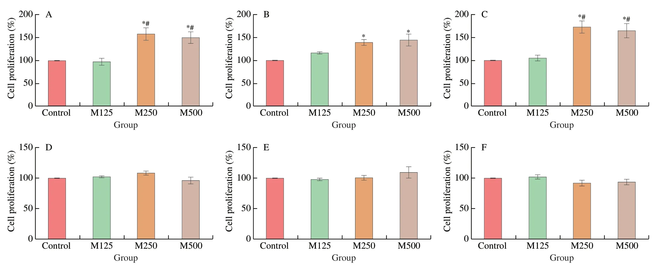
Fig.1 The effects of MRJPs on the proliferation of splenocytes and mesenteric lymphocytes.On day 28,the spleen (A to C) and mesenteric lymph node(mLN;D to F) from mice treated with different doses of MRJPs were collected to evaluate the proliferation of splenocytes and mesenteric lymphocytes,triggered by ConA (A and D),anti-CD3/CD28 (B and E),and LPS (C and F);*P <0.05 and **P <0.01 vs.the control group;#P <0.05 vs.the M125 group.Control: mice received normal saline;M125: mice received 125 mg/kg of MRJPs;M250: mice received 250 mg/kg of MRJPs;M500: mice received 500 mg/kg of MRJPs.MRJPs: major royal-jelly proteins.
3.4 Effect of MRJPs on the population of immune cells in the spleen and mesenteric lymph node
The proportion and quantity of total T lymphocytes (CD3+) in the spleen increased considerably in the M500 group compared to the control group (Fig.2A and 2D).Additionally,splenic NK cells(NK1.1+) were unaffected after supplementation with MRJPs (Fig.2B and 2E).Also,the number of macrophages (F4/80+) among the four groups did not differ.However,mice that were orally administered 125 mg/kg of MRJPs had more macrophages (F4/80+) than mice in the M250 or M500 groups (Fig.2C and 2F).
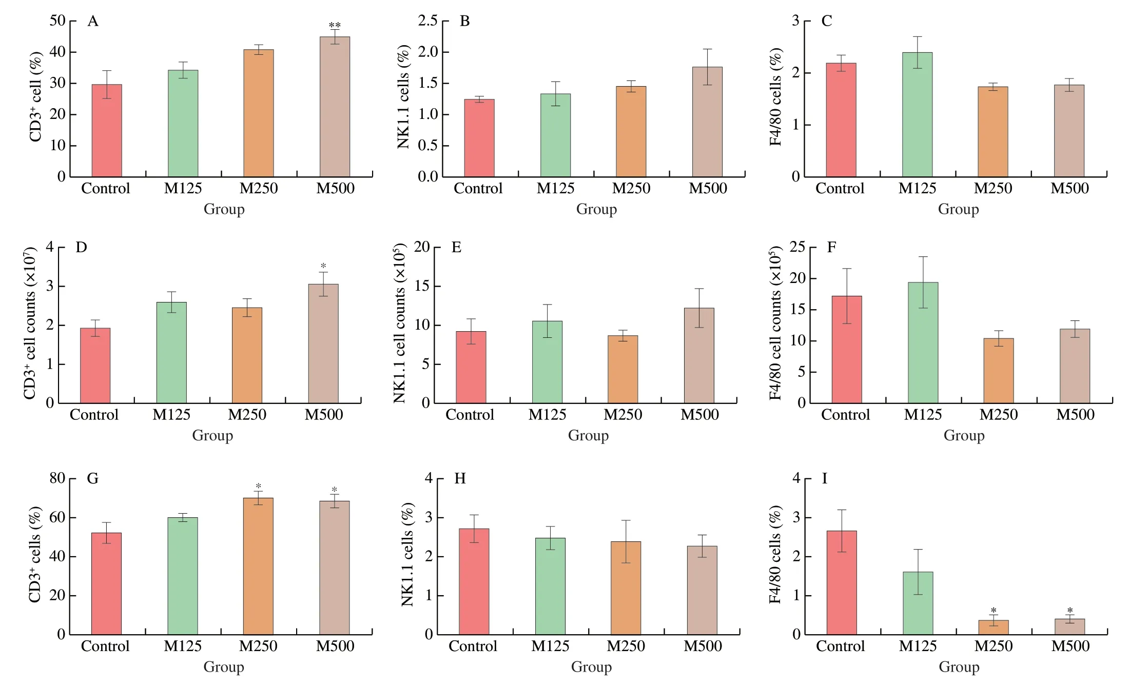
Fig.2 The effects of treatment with MRJPs on the murine spleen and mesenteric lymph node cell populations.On day 28,the spleen (A to F) and mesenteric lymph node (G to I) from mice treated with different doses of MRJPs were collected to determine the immune cell populations,including T cells (CD3+;A,D,and G),NK cells (NK1.1;B,E,and H),and macrophages (F4/80;C,F,and I);*P <0.05 and **P <0.01 vs.the control group;# P <0.05 vs.the M125 group.Control:mice received normal saline;M125: mice received 125 mg/kg of MRJPs;M250: mice received 250 mg/kg of MRJPs;M500: mice received 500 mg/kg of MRJPs.MRJPs: major royal-jelly proteins.
Mice treated with 250 or 500 mg/kg of MRJPs had a higher percentage of total T cells in the mLN (Fig.2G) and fewer macrophages (Fig.2I) than mice in the control group.No difference between the M250 and M500 groups was detected in T cells or macrophages.Finally,the percentage of NK cells between any of the four groups did not differ significantly (Fig.2H).The gates for the T cells,NK cells,and macrophages are illustrated in Fig.S3.
3.5 Effect of MRJPs on T-cell secreting cytokines in the spleen and mesenteric lymph node
The mice that were administered 250 or 500 mg/kg of MRJPs produced more IL-2 when splenic T cells were activated by ConA or anti-CD3/CD28 Abs (Table 1) compared to the mice in the control group.However,the M250 or M500-treated mice had considerably lower IFN-γ and IL-17A from T-cell mitogen-stimulated splenocytes than those in the control group.Additionally,splenocytes activated with anti-CD3/CD28 Abs did not produce different level of IL-4 in any of the four groups (Table 1).Between the control and M125 groups,we did not detect any change in cytokine production.

Table 1 Effect of MRJPs on cytokine production of splenocytes and mesenteric lymphocytes stimulated with mitogens.
Mice administered 500 mg/kg of MRJPs generated more IL-2 in the mLN than control mice when T cells were activated with ConA or anti-CD3/CD28 Abs (Table 1).Additionally,mice administered 250 or 500 mg/kg of MRJPs had considerably fewer IFN-γ than those administered normal saline when their splenocytes were triggered with ConA or anti-CD3/CD28 Abs,but this difference was not found between the control and M125 groups.However,there was no significant difference in IL-4 among the four groups (Table 1).
3.6 Effect of MRJPs on inflammatory cytokine production in the spleen and mesenteric lymph node
The mice orally administered 250 and 500 mg/kg of MRJPs for four weeks generated less IL-1β and IL-6 in the LPS-stimulated splenocytes than the mice in the control group (Table 1).Additionally,the mice administered 250 and 500 mg/kg of MRJPs showed lower level of IL-6 released by LPS-mesenteric cells compared to those in the control group (Table 1).
3.7 Effects of MRJPs on the estrogen levels in the serum of mice
Since host immunity is closely related to estrogen alteration[27]and administration of MRJPs can increase the serum estrogen levels in immature female ICR mice,as found in our previous study[12],we wanted to determine whether treatment with MRJPs could affect serum estrogen levels in female C57BL/6 mice.We evaluated the blood estrogen levels after mice received MRJP supplementation for four weeks and determined the effect of this supplementation on the hormone level.The serum estrogen levels of M125,M250,and M500 were (10.6 ± 2.80),(13.64 ± 3.09),and (6.54 ± 0.43) pg/mL,respectively,which were 103%,161%,and 25% higher than those of the mice in the control group ((5.22 ± 0.34) pg/mL) (Fig.3).Similar to the results of our previous study[12],treatment with 250 mg/kg of MRJPs significantly increased the serum estrogen levels (P<0.05).
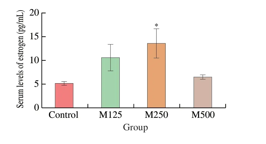
Fig.3 The effects of MRJPs on the serum estrogen levels in mice;*P <0.05 and **P <0.01 vs.the control group;# P < 0.05 vs.the M125 group.Control:mice received normal saline;M125: mice received 125 mg/kg of MRJPs;M250: mice received 250 mg/kg of MRJPs;M500: mice received 500 mg/kg of MRJPs.MRJPs: major royal-jelly proteins.
3.8 Correlation analysis between the estrogen in serum and cytokine levels in mice
Since the administration of 250 mg/kg of MRJPs considerably increased the serum estrogen levels,we conducted a correlation analysis between MRJPs at this dosage and cytokine levels.The results showed that the cytokines,including IFN-γ,IL-6,and IL-1β,were negatively correlated with the estrogen in serum when mice received 250 mg/kg of MRJPs (Fig.4).
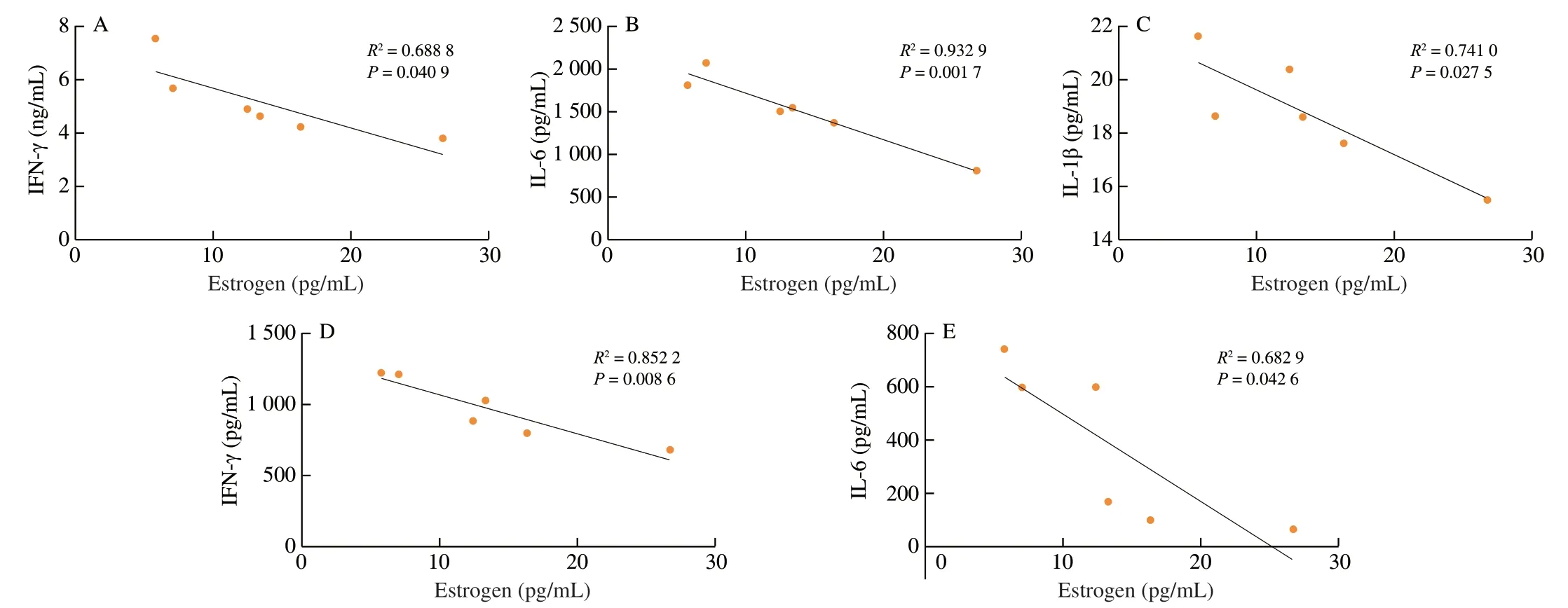
Fig.4 The correlation between the estrogen levels in the serum and the cytokines secreted by lymphocytes (n =6);*P <0.05,**P <0.01,and ***P <0.001 vs.the control group.
3.9 Variation in the gut microbial communities of mice
To determine whether MRJPs can influence the gut microbiota composition,we performed 16S rRNA gene sequencing from feces and cecal contents of mice fed a diet without MRJPs or with 250 mg/kg of MRJPs.We identified high-quality reads across all 24 samples.These reads were clustered into 56 671 ASVs,while 25 833 (45.6%)and 19 104 (33.7%).ASVs were unique to feces and cecal contents,respectively (Fig.5A).In the fecal samples,7 640 (20.3%) ASVs were shared between the MRJPs and control groups,while 15 625(41.6%) and 14 302 (38.1%) ASVs were unique to the MRJPs and control groups,respectively (Fig.5B).Additionally,4 866 (15.8%)ASVs in the cecal contents were shared across the MRJPs and control groups,while 11 674 (37.9%) and 14 298 (46.3%) ASVs were unique to the MRJPs and control groups,respectively (Fig.5C).At the phylum level,Firmicutes and Bacteroidetes dominated the feces and cecal contents;Bacteroidetes was more abundant in the feces,and Firmicutes was more abundant in the cecal contents (Fig.5D).At the genus level,AllbaculumandLactobacillusdominated the feces and cecal contents;Allbaculumwas more abundant in the feces,andLactobacilluswas more abundant in the cecal contents (Fig.5E).
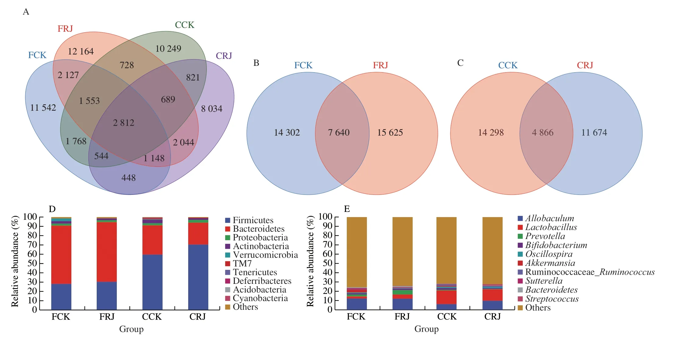
Fig.5 A comparison of the microbial composition between the MRJPs and Control groups.(A) A Venn diagram shows ASV richness and the overlap in microbial communities in the fecal and cecal contents between the MRJPs and Control groups.(B) A Venn diagram shows ASV richness and the overlap in microbial communities in the feces between the MRJPs (left) and Control groups (right).(C) A Venn diagram shows ASV richness and the overlap in microbial communities in the cecal contents between the MRJPs (left) and Control groups (right).(D) The phylum-level relative abundance of the microbial composition.(E) The genus-level relative abundance of the microbial composition.FCK,feces from control mice;FRJ,feces from MRJPs-treated mice;CCK,cecal contents from control mice;CRJ,cecal contents from MRJPs-treated mice.
A rarefaction curve reflects the impact of sequencing depth on the diversity of observed samples.As shown in Fig.6A,new microbiome types might be discovered as the number of sequencing increases.Within-sample (α) phylogenetic diversity analysis for four indices(Chao1,Faith_pd,Goods_coverage,and Observed_spectes) showed that there were notable differences in feces between the two groups(FRJ and CRJ) (Fig.6B).At the ASV level,the PCA showed that the gut microbial composition of the MRJP group in the fecal and cecal samples did not differ substantially from the control group in the feces and cecal contents (Fig.6C).
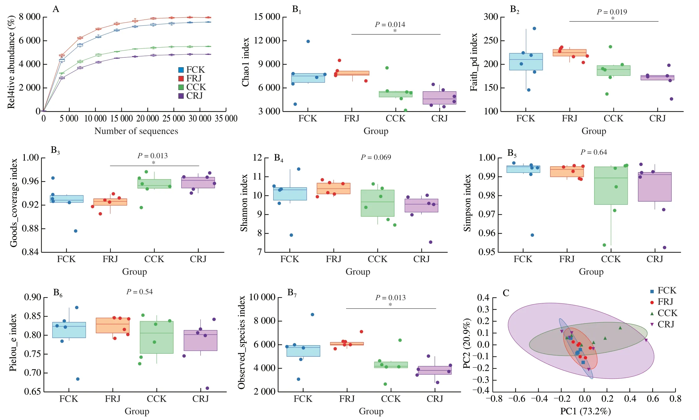
Fig.6 The effect of treatment with MRJPs on the α-and β-diversity of the microbiota.(A) The observed species dilution curve.(B) A comparison of the α-diversity indices.(C) A principal component analysis.The percentage variation in the interpretation of each principal coordinate is indicated on the axes.FCK,feces from control mice;FRJ,feces from MRJPs-treated mice;CCK,cecal contents from control mice;CRJ,cecal contents from MRJPs-treated mice.
Additionally,we compared the composition of the top 10 microorganisms in the MRJPs and control group at the phylum and genus levels.LEfSe was used to identify the species or communities that showed notable differences in sample classification and to determine the differences in the biomarkers between groups.Our results showed thatPrevotella,Paraprevotellaceae,and Bacillales were the different biomarkers of the feces from the MRJPs-treated group,and Enterobacteriales,Gammaproteobacteria,Candidatus_Arthromitus,andShigellawere the different biomarkers of the cecal contents for MRJPs-treated group (Fig.7).Other microorganisms showed negligible differences,except for a considerably low abundance of Tenericutes in cecal contents from the MRJPs-treated group,suggesting that MRJPs had little impact on the gut microbiota.

Fig.7 LEfSe was used to find the biomarker of intergroup difference.(A) The cladogram shows the evolutionary branches of different species.The radiation circle from the inside to the outside represents the classification level from the door to the genus (or species).The size of the small circle represents the average relative abundance of the taxon.The hollow circle represents the taxon with no significant difference between groups,while the small circle in a different color shows a significant difference between groups,and the abundance is higher in the grouped samples represented by this color.(B) The histogram shows the distribution of the LDA values of different species.The length of the histogram represents the contribution of different species.FCK,feces from control mice;FRJ,feces from MRJPs-treated mice;CCK,cecal contents from control mice;CRJ,cecal contents from MRJPs-treated mice.
4. Discussion
Apart from honey,RJ is the primary bee product,with complex chemical composition and a rich biological activity,and is beneficial to human health.We showed that MRJPs are essential components of RJ with anti-aging and reproduction-promoting properties[6,12,13].MRJPs make up more than 80% of the proteins in RJ,primarily MRJP1–9.Their molecular weight varies from 49 to 87 kDa,and amino acid sequence homology ranges from 60% to 70%[6,28].In mammalian cells,the MRJP1 oligomer is essential for cell proliferation[29].Our results showed that supplementation with MRJPs altered the mucosal and peripheral immune response,boosting mitogenstimulated T cell proliferation and IL-2 production while reducing the production of pro-inflammatory cytokines (IFN-γ and IL-17)in splenocytes and lymphocytes.MRJP2 and its subtype X1 can substantially alleviate hepatitis by lowering the level of TNF-α[30].MRJP3 can function as an anti-inflammatory mediator by preventing activated macrophages from producing pro-inflammatory cytokines[31].Thus,they represent a new nutritious protein source that needs further investigation.
Immune cell-mediated immunity is essential for controlling allergy,cancer,and inflammation[32].The first study on systemic lupus erythematosus in children showed a significant improvement in symptoms after three months of RJ treatment through increased regulatory T cells and decreased T cell apoptosis[21].Furthermore,10-HDA can boost immunity in immunosuppressed mice[33].Similarly,RJ was found to lower histamine levels in the plasma and anti-β-lactoglobulin IgE and IgG levels in the serum of mice allergic to hypersensitive substances[21].In this study,supplementation with MRJPs promoted splenic T cell proliferation but did not affect mesenteric T cell expansion,triggered by T-cell mitogens.IL-2 is a vital mediator in promoting T cell survival,growth,and expansion[34].Notably,dietary MRJPs also increased the content of IL-2 secreted by T-cell mitogen-activated splenocytes and mesenteric lymphocytes,indicating that IL-2 might be involved in the expansion of splenic T-cells,but not mesenteric T-cells.Surprisingly,dietary MRJPs were the first to cause LPS-induced B-cell proliferation in splenocytes rather than mesenteric lymphocytes.
Cytokines can mediate the immunological response in cells by binding to their corresponding receptors[35].Oka found that RJ could improve Th1/Th2 cell response,restore macrophage function,and suppress the production of antigen-specific IgE and histamine release in mast cells[36].IL-6 can control the development and differentiation of various cells and is crucial for the immunological response to infections[37].The aberrant expression of IL-6,specifically an increase in the IL-6 levels,contributes to various diseases.Th17-related cytokine IL-17,a potent pro-inflammatory cytokine,has similar characteristics as IL-1β and TNF-α in humans and mice[38].They can damage and cause inflammation in joints[39].We found that treatment with MRJPs decreased proinflammatory cytokines such as the Th1-related cytokine IFN-γ and Th17-related cytokines IL-17,IL-6,and IL-1β.In vitro,IFN-γ production by T cells can be inhibited by MRJP3[28],indicating that MRJPs have sophisticated biological functions and delicate immunomodulatory capacity.However,further studies are required to determine whether a single compound isolated from RJ has multifunctional properties.
Estrogen is a G18 steroid hormone produced by the conversion of androgens,which influence the reproductive system under the catalysis of aromatase.Estrogen works primarily by binding to estrogen receptors.Estrogen receptors,which might be divided into ER1 and ER2,are the products of different genes with 56%homology[40].Estrogen levels play a crucial role in influencing the differences in immunity between sexes,indicating that estrogen might be a sensitive factor in the immune response.Estrogen also has a strong effect on the immune system[27].Females have more powerful cellular and humoral immune responses than males[41].Women are also more likely than men to develop autoimmune diseases,with the severity of these conditions depending on a woman’s estrogen levels,particularly during the menstrual cycle and pregnancy[42,43].
In another study,we found that oral MRJPs could increase serum hormone levels,including estrogen and progesterone,and the expression of estrogen receptors in immature female ICR mice[12].Consistent with the findings of that study,we found that in female C57BL/6 mice,250 mg/kg of MRJPs caused a significant increase in serum estrogen levels.However,500 mg/kg of MRJPs did not further increase the estrogen levels,suggesting that prolonged exposure to a high dose of MRJPs might blunt an elevation in host estrogen.However,this hypothesis needs to be confirmed.Furthermore,estrogen fluctuations are directly related to the host’s immunity[27].The effects of multiple autoimmune diseases,such as multiple sclerosis,rheumatoid arthritis,inflammatory bowel diseases,systemic lupus erythematosus,and autoimmune hepatitis,are reduced due to the ability of estrogen to increase regulatory T cells and Th2 cell polarization and suppress Th1/Th17 cells and inflammatory cytokine production[44–46].These findings suggested that orally administered MRJPs might increase estrogen through the modulation of immune responses.The results of the correlation analysis showed that the increase in estrogen levels mediated by MRJPs was negatively correlated with IFN-γ,IL-6,and IL-1β,indicating that MRJPs might have an estrogen-like effect on the immune response.However,additional studies need to be performed to determine the role of MRJPs in preventing or treating inflammatory disorders such as autoimmune diseases.
The gut microbiome is a critical link between nutrition and immunity.Intestinal symbiotic bacteria and their metabolites can promote anti-inflammatory responses by regulating T cell differentiation and the immunological response in the intestinal tract[47].The gut flora is important for maintaining immunological homeostasis and preserving a robust immune system[48].Moreover,the gut flora directly affects immune cell function in the lamina propria[49],which regulates local and systemic immunological responses[49,50].A misalignment of the microbiota might result in immunological malfunction,leading to the onset and development of disorders linked to inflammation.It can also influence the maturation and stability of the immune system[51].Since microbial communities in the gastrointestinal tract are significantly different,possibly due to the differences in the growth and propagation of microbial organisms(including those of mammals),depending on their surrounding environments[52,53],we analyzed feces and cecal contents to investigate changes in microbiota for minimizing inconsistencies in the fecal microbiome as much as possible.The supplementation with MRJPs did not significantly affect the gut microbiome,but the immunological function of mice improved.Additionally,we did not find any noticeable change in theβ-diversity between the two groups of mice.Therefore,the ecological balance of the microbial community in mice remained undisturbed after supplementation with MRJPs.Next,LEfSe was used to find differences between the groups.Prevotellais one FRJ group biomarker that might increase antioxidant capacity[54].
Furthermore,gram-positive Bacillales bacteria are vital producers of anti-microbial compounds that might be used in food,medicine,or agriculture[55].We found significant differences in the microbial composition between the fecal and cecal contents,raising concerns about the validity of the approach of only using fecal samples to represent intestinal flora.The effect of RJ on intestinal function in mice needs to be further investigated as a food supplement.
A limitation of this study was its inability to assess the effects of microflora changes in other intestinal regions and the immunological response of lymphatic tissues in the lamina propria of mice following treatment with MRJPs.In this study,we only investigated the effects of MRJPs on the functions of splenic and mesenteric lymphocytes and the changes in the intestinal flora.We found that MRJPs influenced estrogen levels and the flora in the intestinal tract;however,we did not determine whether they affected certain metabolites such as amino acids,proteins,and fatty acids.Another limitation of this study was that we only administered MRJPs to mice to test their effects on gut flora and immunity.Future studies should compare the effects of MRJPs and RJ or use a positive control protein like casein.
5. Conclusion
In conclusion,we found that orally administered MRJPs have immunomodulatory effectsin vivo,although these effects might change depending on whether immunological organs are local(mucosal) or peripheral (expanding in response to mitogens).Furthermore,dietary MRJPs can maintain the stability of the gut microflora in mice.Hence,they might be used as a source of functional food proteins for enhancing host immunity and modulating hormone levels.These findings might be relevant for preventing and/or treating chronic inflammatory diseases.
Conflict of interests
Lirong Shen is an editorial board member forFood Science and Human Wellnessand was not involved in the editorial review or the decision to publish this article.All authors declare no conflict of interest.
Acknowledgments
This work was financially supported by the National Natural Science Foundation of China (U2004104),the Natural Science Foundation of Henan Province (202300410080),the Key Project of Henan Education Committee (21A310005),the Internal Fund of Hebei University of Economics and Business (2020ZD10),and the Postgraduate “Talent Program” of Henan University (SYL20060187 and SYL20060189).
Appendix A.Supplementary data
Supplementary data associated with this article can be found,in the online version,at http://doi.org/10.26599/FSHW.2022.9250038.
- 食品科学与人类健康(英文)的其它文章
- Modifications in aroma characteristics of ‘Merlot’ dry red wines aged in American,French and Slovakian oak barrels with different toasting degrees
- Effect of different drying methods on the amino acids,α-dicarbonyls and volatile compounds of rape bee pollen
- Dynamic changes in physicochemical property,biogenic amines content and microbial diversity during the fermentation of Sanchuan ham
- A comparison study on structure-function relationship of polysaccharides obtained from sea buckthorn berries using different methods:antioxidant and bile acid-binding capacity
- Yolk free egg substitute improves the serum phospholipid profile of mice with metabolic syndrome based on lipidomic analysis
- Underlying anti-hypertensive mechanism of the Mizuhopecten yessoensis derived peptide NCW in spontaneously hypertensive rats via widely targeted kidney metabolomics

