
The interventional effect of Polygonatum cyrtonema Hua polysaccharide on atherosclerosis in mice of different sexes
2024-02-16 07:14:44AnjunGuoXueyingLiLihuPnQingmingLiJinpingLuoXueqingZh
食品科学与人类健康(英文) 2024年1期
Anjun Guo,Xueying Li,Lihu Pn,Qingming Li,Jinping Luo,b,Xueqing Zh,b,c,*
a Engineering Research Centre of Bioprocess of Ministry of Education,Hefei University of Technology,Hefei 230009,China
b School of Food and Biological Engineering,Hefei University of Technology,Hefei 230009,China
c Key Laboratory of Metabolism and Regulation for Major Disease of Anhui Higher Education Institutes,Hefei University of Technology,Hefei 230009,China
Keywords:Polygonatum cyrtonema polysaccharide Anti-atherosclerosis Sex
ABSTRACT In the present study,we investigated the intervention effects of a purified Polygonatum cyrtonema polysaccharide (PCP) on high-fat diet (HFD)-induced atherosclerosis in male and female LDLr−/− mice.Results showed that HFD caused severe dyslipidemia,atherosclerotic lesions,oxidative damages and inflammation in male and female mice,and these effects seemed to be more pronounced in males than in females.However,the above variations could be dose-dependently reversed by PCP treatment,and the intervention effects on males were greater than those on females.Nuclear factor kappa-B (NFκB),mitogen-activated protein kinase (MAPKs) and protein kinase B (Akt) are 3 pivotal signaling pathways mediating the development of atherosclerosis.Consistently,PCP was also found to significantly decrease the phosphorylation of p65,p38,extracellular-regulated kinase 1/2 (ERK1/2) and Akt,and increase the protein expression of inhibitor of NF-κB (IκB) in the aortas of male and female mice induced by HFD.Taken together,these findings indicated that PCP could be effective for the prevention of atherosclerosis,and the intervention effect of PCP on male mice was more obvious than that of female mice.
1. Introduction
Cardiovascular diseases (CVDs) are still a serious health problem throughout the world,of which the morbidity and mortality are rising rapidly year by year.According to the statistics of World Health Organization in 2019,CVDs take the lives of 17.9 million people annually,which accounts for nearly 32% of total global deaths[1].Although significant progress has been made in the treatment or prevention of CVDs nowadays,it has been predicted that the annual death toll from CVDs will be estimated to reach 23.6 million by 2030[2].Atherosclerosis is a progressive inflammatory disorder of vasculature and the primary underlying cause of CVDs.The pathogenesis of atherosclerosis is considerably complex,including dyslipidemia,oxidative stress and inflammation[3,4].Generally,it is believed that dietary,lifestyle behavioral and environmental factors are the control keys in the progression of atherosclerotic CVDs.Latest evidence highlighted that sex difference should be considered as a key factor in the prevention of CVDs[5,6].Epidemiological and experimental studies demonstrated that there were differences in many classical risk factors and clinical characteristics of CVDs between men and women,which may be related to the different gene combinations carried on X chromosome and Y chromosome,the modification of DNA and histone,and the changes of sex hormones[7].Human studies have indicated the risks of atherosclerosis in premenopausal women are significantly lower than that in age-matched men,but the protective effect is lost after menopause,and the risk is similar to or higher than that in men[8].Likewise,in LDLr-deficient or apoEdeficient mice,the differences in CVDs between males and females have also been documented and widely reviewed.Therefore,sex makes a great impact on the occurrence and development of atherosclerosis,early identification and active intervention are very important for the treatment of atherosclerosis.However,sex dimorphism is rarely considered in current anti-atherosclerotic therapy.
Statins are widely applied in clinic to improve the symptoms of atherosclerotic CVDs,but long-term use of these drugs have been proved to trigger a variety of detrimental effects,such as muscle weakness and liver damage[9].Thereby,to try to develop practical and cost-effective agents against atherosclerosis from ediblemedicinal foods or herbs is undoubtedly of great significance.Polygonatum cyrtonemaHua,a medicinal and edible perennial plant,is extensively used in traditional Chinese medicine for the effects of protecting the cardiovascular systems,resisting fatigue,tonifying spleen and nourishing lung[10,11].Polysaccharide is one of the main active constituents inP.cyrtonema,which may become a potential therapeutic option for patients with atherosclerosis due to its hypolipidemia,anti-inflammatory and antioxidant functions[12-14].Recently,we obtained a purified polysaccharideP.cyrtonemapolysaccharide (PCP) with anti-atherosclerotic effect fromP.cyrtonema.TheP.cyrtonemapolysaccharide has a relative molecular weight of 8 500 Da.Monosaccharide composition analysis showed that PCP was mainly composed of fructose and glucose in a molar ratio of 28:1.The structure of PCP was characterized as a repeating unit consisting of →6)-β-D-Fruf-(2→,→1,6)-β-D-Fruf-(2→,→1)-β-D-Fruf-(2→,β-D-Fruf-(2→,and →6)-α-D-Galp-(1→[15].To date,despite some polysaccharides have been demonstrated to have anti-atherosclerotic activity,few studies have tried to evaluate the intervention effect of polysaccharides on atherosclerosis from the perspective of sex.LDL-receptor knockout (LDLr−/−) mice,a wellrecognized atherosclerosis animal model,have been widely used as a model to mimic the progression of human atherosclerosis[16,17].Compared to wild-type mice,LDLr−/−mice are highly susceptible to atherosclerosis when fed a high-fat diet[18,19].In the present work,we investigated the interventional effect of PCP on atherosclerosis induced by high-fat diet in male and female LDLr−/−mice.
2. Materials and methods
2.1 Materials and chemicals
The rhizomes ofP.cyrtonemaHua were obtained from Jiuhua Mountain (Anhui Province,China).The PCP was obtained from the rhizomes ofP.cyrtonemaHua according to our previous report[15].DEAE-cellulose 52 and Sephadex G-100 were purchased from Sigma-Aldrich (St.Louis,MO,USA).The kits for the detection of total cholesterol (TC),triglyceride (TG),high-density lipoprotein cholesterol (HDL-C),low-density lipoprotein cholesterol(LDL-C),superoxide dismutase (SOD),glutathione peroxidase(GSH-Px) and malondialdehyde (MDA) were purchased from Jiancheng Technology Co.,Ltd.(Nanjing,China).Bicinchoninic acid (BCA) protein assay kit and bovine serum albumin (BSA)were purchased from Sangon Biotech Co.,Ltd.(Shanghai,China).ELISA kits were purchased from Elabscience Biotechnology Co.,Ltd (Wuhan,Hubei,China).The primary antibodies against NF-κB p65 (1:1 000),phospho-NF-κB p65 (p-p65;1:1 000),p38 MAPK(1:1 000),phospho-p38 MAPK (1:1 000),inhibitor of NF-κB(IκB;1:1 000),extracellular-regulated kinase 1/2 (ERK1/2;1:1 000),phospho-ERK1/2 (1:1 000),protein kinase B (Akt;1:1 000),phosphor-Akt (1:1 000) and glyceraldehyde 3-phosphate dehydrogenase (GAPDH;1:1 000) were purchased from Abcam(Cambridge,MA,UK).The secondary antibody was purchased from Boster Biological Technology Co.,Ltd.(Wuhan,China).All other analytical-grade reagents were purchased locally.
2.2 Animals
The 8-week-old male and female low density lipoprotein receptor deficiency (LDLr−/−) mice ((20 ± 2) g) were purchased from the Model Animal Research Center of Nanjing University (Nanjing,China).All mice were kept under specific pathogen-free conditions(22−24 °C,12-h light/dark cycles,50%−55% relative humidity) with free access to food and water.The protocols for animal experiments were strictly followed by the national guidelines for the care and use of laboratory animals and approved by the Animal Ethics Committee of Hefei University of Technology (2018-0808-001).
2.3 Experimental procedure
After one week of adaptive feeding,150 mice were randomly divided into two batches with 75 mice in each batch.Each batch was randomly divided into 5 groups (Fig.1A),including normal diet group (ND,n=15),high-fat diet group (HFD,n=15),PCPlow-dose group (L-PCP,65mg/(kg·day),n=15),PCP-high-dose group (H-PCP,260 mg/(kg·day),n=15) and Atorvastatin Calcium Tablets group (ACT,2 mg/(kg·day),n=15).In the early stage,the doses of PCP to mice have been optimized and published in a previous report[20].The ND group was fed a normal diet,while the other groups were fed a high-fat diet (Table S1).PCP and ACT were dissolved in physiological saline and orally administered at the same time every day.The ND and HFD groups were orally administered with the same volume of physiological saline.After 16 weeks of feeding,the mice were euthanized with CO2.Serum,hearts and aortas were collected for the analysis of blood biochemistry,pathology,real-time quantitative polymerase chain reaction (qRTPCR) and Western blot.
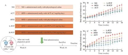
Fig.1 PCP intervention strategy (A) and variations in body weight of female and male mice during the experiment (B-C).
2.4 Biochemical parameter analysis
The serum levels of TC,TG,LDL-C and HDL-C were detected by automatic biochemical analyzer (Accu-check Performa,Roche,Germany).The activity of SOD,GSH-Px and the content of MDA in serum and aorta were measured using commercial kits according to the manufacturer’s instructions.The levels of cytokines TNF-α,IL-1β,IL-10 and IL-6 in serum were analyzed by ELISA kits according to the manufacturer’s protocols.
2.5 Pathological examination of the aorta
According to the previous methods[21],the arterial tree was fixed,stained with Oil Red O,rinsed in 70% ethanol and photographed using a digital camera (Nikon DXM1200,Tokyo,Japan).The hearts were fixed in 4% paraformaldehyde,dehydrated in increasing concentrations of ethanol,embedded in paraffin and cut into 5 μm thick sections.The sections of the aortic sinus were stained with hematoxylin and eosin (H&E) and Masson Trichome (MT) using standard procedures to evaluate inflammation and fibrosis.All images were observed and captured under the optical microscope.
2.6 qRT-PCR analysis
Total RNA was isolated using TRIzol reagent from aortic tissue according to the manufacturer’s instruction.qRT-PCR was performed as described previously[22].All primer sequences tumor necrosis factor-α (TNF-α),interleukin-1β (IL-1β),IL-6,IL-10,andGAPDHare listed in Table S2.
2.7 Western blot
Total proteins were extracted from aortic tissue with RIPA lysate.The protein concentration was detected by Bradford protein assay reagent.The protein in the sample was denatured for 10 min at 100 °C and stored at −80 °C until analysis.The levels of p65,IκB,p-p65,p38,p-p38,ERK1/2,p-ERK1/2,Akt,p-Akt and GAPDH were measured by Western blot analysis as previously described[23].
2.8 Statistical analysis
Each experiment was performed independently in triplicates.The data were presented as mean ± standard deviation (SD) values and statistically analyzed with one-way analysis of variance,followed by Tukey’s multiple-comparisons tests.P<0.05 was considered statistically significant.
3. Results
3.1 PCP attenuated the weight gains of HFD-fed male and female mice
The body weight of mice in each experimental group was monitored once a week.As shown in Figs.1B-C and S1,after 16 weeks of HFD feeding,the total weight gains of male and female mice in model groups were (15.01 ± 1.23) g and (10.40 ± 0.91) g,respectively.However,PCP treatment significantly decreased the body-weight gains in both male and female mice,compared with the HFD groups.When PCP dosage reached 260 mg/(kg·day),the HFD-induced weight gains of male and female mice were reduced by 43.96% and 37.27%,indicating that PCP could attenuate the weight gain in male and female mice caused by HFD.
3.2 PCP down-regulated serum lipid levels of HFD-fed male and female mice
Hyperlipidemia,a disorder of lipid metabolism,is a wellestablished predictor for atherosclerosis.It has been recognized that HFD can lead to hyperlipidemia,characterized by elevated levels of TC,TG,LDL-C and decreased HDL-C levels.LDL-C is known as a primary pro-atherogenic factor,which promotes the formation and progression of atherosclerotic plaques via transporting and accumulating cholesterols to the arterial wall.On the contrary,HDL can exert anti-atherosclerotic activity through reverse cholesterol transport[23,24].As shown in Figs.2A-D,compared with the ND groups,the levels of TC,TG,LDL-C were significantly elevated and the level of HDL-C was significantly decreased in the serum of male and female mice fed HFD.However,these changes were remarkably inhibited by the treatment of PCP and ACT.Especially when PCP dosage reached 260 mg/(kg·day),the serum TC,TG,LDL-C were decreased by 42.05%,55.20%,70.22% and 29.96%,26.37%,50.69%,and the HDL-C levels were increased by 85.04% and 50.03% in male and female mice,when compared to those of model groups.To make a proper evaluation of lipidrelated atherosclerosis risk factors,the atherosclerotic index (AI)was further calculated[25].As shown in Fig.2E,compared with the ND groups,the AI values of male and female mice in the HFD groups were significantly higher than the atherosclerosis diagnostic limit of 4.0.However,the alterations of AI values in HFD-fed male and female mice could be dose-dependently reduced by PCP.Compared to those of model groups,the AI values of male and female groups treated with H-PCP were decreased by 79.94% and 64.39%,respectively.

Fig.2 Effects of PCP on serum TC (A),TG (B),HDL-C (C),LDL-C (D) and AI (E) levels of HFD-fed female and male mice.**P <0.01 (vs.ND group);# P <0.05,## P <0.01 (vs.HFD group);$ P <0.05 (Female group vs.Male group).
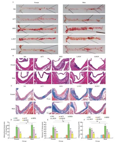
Fig.3 Effects of PCP on pathological changes in the aortas of HFD-fed female and male mice.(A,D) Oil Red O staining and semi-quantitation of the aortic tree;(B,E) H&E staining and semi-quantitation of the aortic sinus;(C,F) Masson staining and semi-quantitation of the aortic sinus.** P <0.01 (vs.ND group);# P <0.05,## P <0.01 (vs.HFD group);$ P <0.05,$$ P <0.01 (Female group vs.Male group).

Fig.4 Effects of PCP on serum MDA (A),GSH-Px (B) and SOD (C) levels and aorta MDA (D),GSH-Px (E) and SOD (F) levels of HFD-fed female and male mice.* P <0.05,** P <0.01 (vs.ND group);# P <0.05,## P <0.01 (vs.HFD group);$ P <0.05 (Female group vs.Male group).
3.3 PCP prevented atherosclerosis progression of HFD-fed male and female mice
To evaluate the protective effects of PCP against atherosclerosis in male and female mice,we utilized LDL receptordeficient (LDLr−/−) mice fed a HFD for 16 weeks as a model for atherosclerosis.The atherosclerotic lesions were determined byen faceanalysis of the arterial tree and cross-sectional analysis of the aortic sinus[26,27].As shown in Figs.3A and D,the entire aortic plaque areas were significantly enhanced in HFD-fed male and female mice,and lesions in the aortas of male mice were more severe than in their female counterpart.However,it could be found that the aortic pathological areas stained with Oil Red O were notably reduced by PCP and ACT administration.When the dose of PCP reached 260 mg/(kg·day),the Oil Red O stained lesion area was reduced by 82.21% and 58.75% in male and female mice in comparison to those of model groups (Figs.3A and D).In addition,we assessed the intimal areas of aortic sinus by H&E staining,and found after administration of high-dose PCP,the aortic wall thickness induced by HFD in male and female mice decreased by 66.82% and 57.60%,respectively (Figs.3B and E).In the meantime,Masson’s trichrome staining revealed that the atherosclerotic plaque lesions in HFD-treated male and female mice contained higher percentage of collagen fibers when compared with those of ND-fed groups.Nevertheless,PCP and ACT administration inhibited the deposition of collagen fibers in the vascular wall in a dose-dependent manner.Compared to those of model group,deposition of collagen fibers in vascular wall was decreased by 68.05% and 51.89% in the H-PCP groups of male and female mice.The effect of H-PCP on male mice was not significantly different from that of the ACT treatment group(Figs.3C and F).These results supported that PCP could alleviate HFD-induced atherosclerotic lesions in male and female mice.
3.4 PCP enhanced the antioxidant capacities of HFD-fed male and female mice
It is well-accepted that oxidative stress,caused by the excessive production of reactive oxygen species (ROS),is strongly involved in the pathogenesis of atherosclerosis[28,29].As we know,the antioxidant enzymes GSH-Px and SOD play critical roles in scavenging the free radicals and restraining the lipid peroxidation in the endogenous defense system[30].MDA is one of the key products of lipid peroxidation,the level of which can reflect the severity of oxidative injury stimulated by free radicals indirectly[31].In the current study,as shown in Figs.4D-F,compared with the ND groups,HFD could significantly suppress the activities of GSH-Px and SOD,and remarkably increase the levels of MDA in the aortas of male and female mice.However,the alteration of GSH-Px,SOD and MDA levels could be significantly reversed by PCP and ACT.In male and female mice,the GSH-Px and SOD activities of H-PCP groups were increased by 89.72%,42.30% and 93.80%,42.79%,and MDA was decreased by 70.82% and 44.37%,compared to those of model groups (Figs.4D-F).Similar phenomenon was also observed in the serum antioxidative systems of male and female mice(Figs.4A-C).These data indicated that the increment of antioxidant capacity might be one of the essential mechanisms for PCP to prevent atherosclerosis.
3.5 PCP inhibited inflammation of HFD-fed male and female mice
In addition to oxidative stress,inflammation was considered as another pivotal etiological contributor for the pathogenesis of atherosclerosis[32-34].Hence,the effects of PCP on the inflammation of HFD-induced atherosclerosis in male and female mice were investigated.As shown in Figs.5A-D,compared with the ND groups,HFD could significantly up-regulate the levels of pro-inflammatory cytokines TNF-α,IL-1β and IL-6,and remarkably down-regulate the level of anti-inflammatory cytokine IL-10 in serum of male and female mice.However,the changes of these cytokines could be reversed by PCP and ACT.In male and female mice,the levels of TNF-α,IL-1β and IL-6 in serum of H-PCP groups were reduced by 64.36%,50.15%,53.11 and 35.77%,32.56%,29.46%,and the level of IL-10 was raised by 98.30% and 38.46%,compared to those of model groups (Figs.5A-D).Similarly,as shown in Figs.5E-H,compared with model groups,the mRNA levels ofTNF-α,IL-1βandIL-6of H-PCP groups were reduced by 65.40%,63.54%,61.72%and 44.00%,42.52%,37.78%,andIL-10mRNA level was raised by 81.61% and 40.97% in male and female mice (Figs.5E-H).These data were consistent with the phenomenon that PCP could evidently halt HFD-induced inflammatory cells infiltration,as observed in H&E staining.Accordingly,there was no doubt that PCP could limit the inflammatory response in HFD-fed male and female mice.
3.6 PCP played an anti-atherosclerotic role by regulating inflammatory pathways
It has been established that the activation of NF-κB,MAPKs and Akt signaling pathways has a tight relation to inflammatory responses by triggering the secretion of cytokines,chemokines,and other stimulatory molecules in response to oxidative stress,thus accelerating the process of atherosclerosis[35-37].As shown in Figs.6A-F,HFD could significantly increase the phosphorylation of p65,ERK1/2,p38 and Akt,and significantly reduce the protein expressions of IκB in male and female mice.However,the changes of these proteins could be reversed by PCP and ACT.In male and female mice,the protein levels of p-p65,p-ERK1/2,p-p38 and p-Akt of H-PCP groups were reduced by 60.62%,55.54%,45.44%,49.84% and 42.78%,40.65%,32.80%,31.95%,and IκB protein levels w e r e increased by 1.14-fold and 0.71-fold,compared to those indicators of model groups (Figs.6A-F).These findings suggested that PCP could attenuate the inflammatory reactions in atherosclerotic mice by regulating NF-κB,MAPK and Akt signals,and this anti-inflammatory effect of PCP on male mice was better than that of female mice.
4. Discussion
In this study,we found that a homogeneous polysaccharide PCP could delay the progression of atherosclerosis in HFD-induced male and female LDLr−/−mice by suppressing NF-κB/MAPKs/Akt-mediated inflammatory responses.It is widely recognized that hyperlipidemia is a major causative factor closely related to the occurrence and development of atherosclerotic cardiovascular disease.Thus,the lipid-lowering agents have been widely utilized to treat these diseases for decades[38].In general,hyperlipidemia is characterized by abnormally elevated serum levels of total cholesterol and triglycerides.These lipids are then carried by atherogenic lipoproteins,mainly including LDL and very low-density lipoprotein,and transported to the artery wall,resulting in the accumulation of lipids and ultimately causing atherogenesis[39].In this study,we found that PCP could significantly reduce the serum levels of TC(Fig.2A) and TG (Fig.2B) of male and female mice compared to the model groups.This finding suggested that PCP possessed good hypolipemic potential.In addition,PCP could also down-regulate the serum levels of LDL-C,which constitutes a critical risk factor in atherosclerotic progression.Numerous studies have confirmed that LDL can be oxidized by ROS to the form oxidized LDL (ox-LDL),which can induce endothelial dysfunction.Moreover,ox-LDL can be taken up by macrophages through scavenger receptors,leading to the production of macrophage-derived foam cells and eventually triggering atherosclerosis[40,41].To further elucidate these events,we assessed the lipid peroxidation status by detecting the markers of oxidative stress.Here,we found that PCP could significantly enhance the activities of GSH-Px (Figs.4B and E) and SOD (Figs.4C and F),and remarkably decrease the levels of MDA (Figs.4A and D) in the aortas and serum of male and female mice,compared with that of HFD-fed mice.The results revealed that PCP could also resist lipid peroxidation.Reportedly,HDL-C is regarded as an important protective factor against atherosclerosis that can transport cholesterol and promote cholesterol metabolism[42].Results showed that oral administration of PCP could up-regulate the serum levels of HDL-C in male and female mice,compared with HFD-fed mice(Fig.2C).
Current evidence supports that inflammation plays a central role in all phases of atherosclerosis,from foam cells to plaque formation to rupture and finally to thrombosis[43].It is reported that ox-LDL is an important initiator of inflammation,because it can cause endothelial dysfunction by enhancing the expression of adhesion molecules and the recruitment of inflammatory cells to early atherosclerotic lesions[38].In the atherosclerotic lesions,endothelial cells and smooth muscle cells can secrete proinflammatory cytokines,thereby activating the differentiation of monocytes into macrophages.These macrophages were then transformed into lipid-laden foam cells by taking up ox-LDL,thus amplifying the inflammatory reactions[44-46].It is well established that IL-1β,TNF-α and IL-6 are the typical proinflammatory cytokines highly associated with the pathogenesis of atherosclerosis.IL-1β is a classic inflammatory cytokine that contributes to atherosclerosis by promoting the recruitments of inflammatory cells to atherosclerotic lesion sites,whereas inhibition of IL-1β expression significantly reduced aortic plaque area in ApoE−/−mice[47].TNF-α is a strong inflammatory cytokine secreted by monocytes/macrophages and plays an atherogenic role via promoting the secretion of adhesion molecules and chemokine in the arterial wall,which has been confirmed in TNF-α−/−ApoE−/−mice[48].IL-6 is also considered to be a pro-atherogenic cytokine,which can trigger vascular inflammation and promote the expansion of lipid-rich necrotic areas[49,50].Consistent with these findings,after 16 weeks of HFD feeding in male and female LDLr−/−mice,we observed that the levels of TNF-α (Figs.5A and E),IL-1β(Figs.5B and F) and IL-6 (Figs.5C and G) were elevated in serum and their gene expressions were enhanced in the aortas,whereas these elevations could be reversed by PCP treatment.IL-10 is known as a potent anti-inflammatory cytokine by inhibiting the synthesis of IL-1β,IL-6 and TNF-α.Moreover,IL-10 was reported to play a pivotal role in the inhibition of atherosclerosis[51].In the meantime,we also observed that PCP could significantly increase the levels of IL-10 in serum and aortas (Figs.3D and H)of male and female mice,compared to the model groups.These results suggested that PCP could attenuate local and systemic inflammatory reactions in the progression of atherosclerosis.To elucidate the underlying mechanism of PCP in preventing atherosclerosis,we further investigated the intracellular signals involved in the regulation of inflammatory responses.NF-κB is a pivotal transcription factor,which can activate transcription of various pro-inflammatory genes involved in different processes of inflammatory and immune responses.It has been reported that NF-κB is a heterodimeric complex,most commonly composed of two subunits: p50 and p65.In resting cells,NF-κB dimers are sequestered in the cytoplasm by the IκB proteins.Upon stimulation,IκB is phosphorylated by IκB kinase,resulting in the degradation of IκB.Subsequently,NF-κB is released and translocates into the nucleus to activate the transcription of target genes[52,53].Importantly,NF-κB has been well recognized as a crucial regulator of inflammation that controls the transcription of series of genes,promoting the production of proinflammatory cytokines in atherosclerotic areas.Likewise,dysregulated activation of NF-κB pathway was also frequently detected in a variety of human atherosclerotic plaques[54-56].In contrast,blocking the activation of NF-κB could strongly ameliorate atherosclerosis in ApoE-knockout mice fed with a cholesterol-rich diet[57].In our study,it was observed that the phosphorylation of NF-κB p65 was increased and the expression of IκB was decreased by HFD,but these changes were inhibited by the treatment of PCP (Figs.6A,B and C).Consistently,similar changes were also found in serum levels and aortic gene expressions ofIL-1β,TNF-αandIL-6(Fig.5).These data proved that PCP could play a crucial role in suppressing NF-κB activation.Additionally,the MAPK was known to contribute to atherosclerosis through promoting the recruitment of monocytes into arterial intima and inflammatory response.Conversely,inhibition of MAPK was confirmed to have beneficial effects in atherosclerosis by suppressing adhesion molecules and inflammation,so as to improve the stability of carotid plaques[58,59].The Akt pathway has also been demonstrated to be involved in the pathogenesis of atherosclerosis,and Akt activation plays a major role in the aggravation of the atherosclerotic lesion in atherosclerosis mice.In contrast,when Akt pathway was blocked,atherosclerosis regressed[60,61].Notably,the expressions of adhesion molecules and NF-κB were demonstrated to be decreased by suppressing the phosphorylation in MAPK or Akt pathways directly,and the suppression of MAPKs or Akt resulted in reduced levels of adhesion molecules and lipids,thus inhibiting plaque formation in animal models of atherosclerosis[62,63].In the current study,PCP could also significantly decrease HFD-induced phosphorylation of ERK1/2,p38 and Akt in male and female LDLr−/−mice (Fig.6).Together,these results indicated the potential mechanisms of anti-atherosclerosis of PCP might be associated with the inhibition of NF-κB,MAPKs and Akt signals.
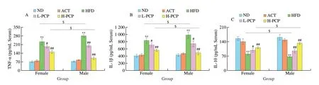
Fig.5 Effects of PCP on serum TNF-α (A), IL-1β (B), IL-10 (C) and IL-6 (D) levels and mRNA levels of TNF-α (E),IL-1β (F),IL-10 (G) and IL-6 (H) in the aortas of HFD-fed female and male mice.** P <0.01 (vs.ND group);# P <0.05,## P <0.01 (vs.HFD group);$ P <0.05 (Female group vs.Male group).
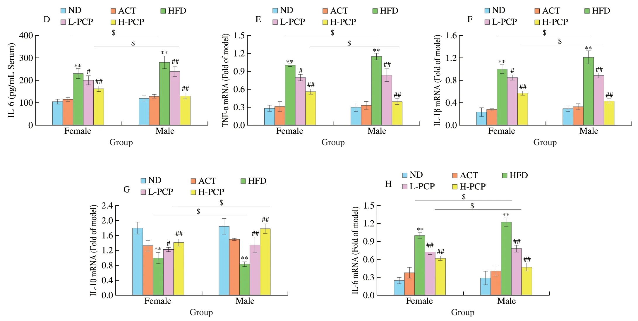
Fig.5 (Continued)
Increasing data have shown that gender has an important impact on the incidence of atherosclerosis.Studies found that the prevalence of atherosclerosis in men was almost twice as high as age-matched premenopausal women.The authors suggested that estrogens are closely related to gender differences in the prevalence of atherosclerosis[64].The potential mechanisms of estrogens protecting against cardiovascular diseases were mainly manifested in the following aspects: (1) Regulate the secretion of angiotensin and NO and causes rapid vasodilation,thus reducing the inflammatory response triggered by endothelial cell injury[65];(2) Reduce the oxidation level and inhibit cytochrome C by regulating the PI3K/Akt signaling pathway;(3) Reduce cholesterol storage by promoting hepatic surface apolipoprotein expression and lowering LDL levels;(4) Inhibit platelet aggregation and reduce thrombosis[66].Further studies showed that the combination of estrogen and estrogen receptor could induce macrophages to express and release HSP27,which in turn activated NF-κB to enter the nucleus and regulate cellular inflammatory response,thereby interfering with atherosclerosis[67,68].Tsuda et al.found that after 6 weeks of high-fat diet feeding,significant atherosclerotic lesions were observed in the proximal aorta of both male and female ApoE−/−mice,accompanied by an increase in oxidative stress,and these changes were more pronounced in male mice[69].In line with these findings,our study also found that atherosclerotic lesions were more severe in males than in females of HFD-fed LDLr−/−mice(Fig.3).Meanwhile,PCP treatment could significantly prevent atherosclerosis,and the intervention effect of PCP on male mice is more obvious than that of female mice,but the possible mechanism for difference of anti-atherosclerotic effect of PCP in different sex mice still needs to be further investigated.
5. Conclusions
In summary,as shown in Fig.7,our results reflected that PCP could exhibit good anti-atherosclerosis activity in both male and female LDLr−/−mice induced by HFD,and the anti-atherosclerosis capacity of PCP was more significant in males than females.The protective mechanism of PCP in preventing atherosclerosis might be related to the inhibition of NF-κB/MAPKs/Akt-mediated inflammatory responses.Based on the current investigation,PCP may have the therapeutic potential to delay the occurrence of atherosclerosis.
Declaration of Interest Statement
The authors declare no conflict of interest.
Acknowledgments
This study was financially supported by the National Natural Science Foundation of China (32 072176;31271814);the Outstanding Youth Funds of Anhui Province (2208085J31);and the Fundamental Research Funds for the Central Universities (JZ2022HGQA0232;JZ2022HGTA0316).
Appendix A.Supplementary data
Supplementary data associated with this article can be found,in the online version,at http://doi.org/10.26599/FSHW.2022.9250031.

- 食品科学与人类健康(英文)的其它文章
- Modifications in aroma characteristics of ‘Merlot’ dry red wines aged in American,French and Slovakian oak barrels with different toasting degrees
- Effect of different drying methods on the amino acids,α-dicarbonyls and volatile compounds of rape bee pollen
- Dynamic changes in physicochemical property,biogenic amines content and microbial diversity during the fermentation of Sanchuan ham
- A comparison study on structure-function relationship of polysaccharides obtained from sea buckthorn berries using different methods:antioxidant and bile acid-binding capacity
- Yolk free egg substitute improves the serum phospholipid profile of mice with metabolic syndrome based on lipidomic analysis
- Underlying anti-hypertensive mechanism of the Mizuhopecten yessoensis derived peptide NCW in spontaneously hypertensive rats via widely targeted kidney metabolomics