Histological Assessment and Transcriptome Analysis Provide Insights into the Toxic Effects of Perfluorooctanoic Acid to Juvenile Half Smooth Tongue Sole Cynoglossus semilaevis
ZHAN Min, SHIKunpeng, ZHANG Xue, FAN Qingxin, XU Qian, LIU Xinbao,LI Zhujun, LIU Hongning, XIA Yanting, and SHA Zhenxia, 2), *
Histological Assessment and Transcriptome Analysis Provide Insights into the Toxic Effects of Perfluorooctanoic Acid to Juvenile Half Smooth Tongue Sole
ZHAN Min1), #, SHIKunpeng1), #, ZHANG Xue1), FAN Qingxin1), XU Qian1), LIU Xinbao1),LI Zhujun1), LIU Hongning1), XIA Yanting1), and SHA Zhenxia1), 2), *
1),,266071,2),,266237,
Perfluorooctanoic acid (PFOA) is a widespread synthetic persistent organic pollutant that may enrich along the food chain and affect the growth, development, reproduction, and lipid metabolism of aquatic organisms, particularly the benthic organisms. How- ever, the toxic effects of PFOA on the half-smooth tongue sole, a commercial benthic fish in China, have rarely been reported. Because juvenile fish are sensitive to environmental pollutants, in the present study, histological assessment and tran- scriptome sequencing were performed to determine the short-term impact of PFOA on juvenile half-smooth tongue soles. Histologi- cal analysis showed that PFOA exposure caused hepatocyte rupture, intestinal villi breakage, increased goblet cell count, and brain ab-normal. Transcriptome results showed that some interesting signaling pathways, such as glycolysis/gluconeogenesis, PPAR signaling pathway and GABAergic synapse signaling pathway, were enriched after PFOA exposure. In addition, some metabolic, immune and neural genes were differentially expressed, which including,and, and they were up-regulated after 14 days of exposure. Transcriptome results also indicated that half-smooth tongue sole might improve energy metabolism in response to PFOA toxicity after 7 days of exposure. These findings provide a basis for studying the ecological effects of PFOA on marine benthic fishes.
; histological assessment; perfluorooctanoic acid; transcriptome analysis; toxic effect
1 Introduction
Perfluorooctanoic acid (PFOA), a perfluoroalkyl sub-stance (PFAS), has a unique chemical structure, strong ther- mochemical stability, hydrophobic and oleophobic proper-ties, and is widely used in industry (Liu, 2017b; Dong, 2019; Kang, 2019). The research has shown that PFOA can easily be released into the surrounding environ-ment and become a persistent organic chemical pollutant (Yang, 2017).
In recent decades, PFOA has attracted significant atten- tion, and various regulatory measures have been taken world-wide to control and limit the production and application of PFOA. However, PFOA can still be detected in dust, sedi- ments, soil, surface water, and various organisms. In parti- cular, PFOA has been detected in ocean waters with theconcentrations usually ranging from pgL−1to ngL−1(Yama- shita, 2005; Wan, 2017; Yu, 2022). For example, the concentration is 52pgL−1in the North Atlan- tic and 4534.41ngL−1in the Bohai Sea (Yamashita, 2008; Wang, 2014). Moreover, owing to its persistentand non-degradable characteristics, PFOA will gradually enter aquatic organisms and enrich the food chain. Previ- ous studies have shown that PFOA accumulated in the li- ver, plasma, and kidneys of animals and humans, leading to oxidative stress and even cancer (Abe, 2017; Li, 2018; Miranda, 2020).
Currently, most studies concerning PFOA have focused on humans and other animals, and have concentrated on theeffects of PFOA on reproduction, lipid metabolism, growth and development, liver carcinogenesis (Mayilswami, 2016; Seyoum, 2020). Previous studies have focus- ed on other freshwater fishes such as zebrafish, common carp, crucian carp, rare minnow, medaka, tilapia, rainbow trout, and marine Atlantic salmon. These studies have main- ly concentrated on PFOA enrichment and changes in fish tis- sues, as well as its effects on enzyme activities, cell viabi- lity and some key genes’ expressions (Wei, 2008; Fang, 2010; Mortensen, 2011; Giari, 2016; Tang, 2018). However, few studies have investigated the toxicity of PFOA on benthic marine fishes.
Half-smooth tongue sole () is a benthic marine fish that is distributed in the Yellow Sea and Bohai Sea of China, Korea Peninsula, Japan offshore waters, and is also an important aquaculture species in China (Shao, 2019). Since fishes are sensitive to the environment in the early life stage (Rial, 2010; Do and Tran, 2022), in this study, the histopathological changes and transcription analysis of juvenile half-smooth tongue soles were investigated after exposure to50mgL−1PFOA for 14 days. The liver, intestine, and brain were abnormal,several signaling pathways and genes were significantlyaffected, which are mainly related to lipids, metabolism,neuralsynapse and immunity. Our results provide a basis for studying the ecological effects of PFOA on benthic ma- rine fishes.
2 Materials and Methods
2.1 Experiment Animals and PFOA Exposure Treatment
Healthy juvenile half-smooth tongue soles (3.4cm±0.2cm, 400mg±30mg) were provided by Haiyang Yellow SeaAquatic Products Co., Ltd. (Shandong, China). Prior to the experiment, in order to adapt to the environment, juvenile half-smooth tongue soles were maintained in laboratorytanks (56cm×34cm×30cm) with aerated and filtered sea- water (pH 8.14±0.09, water temperature 21℃±1℃, sali- nity 24, dissolved oxygen 6–8mgL−1) for two weeks with a light/dark photoperiod cycle of 12h:12h. During this pe-riod, half-smooth tongue soles were fed brine worms daily.
After two weeks, a total of 300 half-smooth tongue soles were randomly divided into the PFOA-exposed group and the control group, three replicates were set in each group, with 50 fish in each replicate. The PFOA-exposed groups were exposed to seawater with the PFOA (CAS: 335-67-1, Shanghai Macklin Biochemical Co., Ltd.) concentration of50mgL−1for 14 days, and the control groups were cul-tured in seawater without PFOA for 14 days. The PFOA dose and the experimental period in this study were based on our preliminary experiments and previous researches (Yang, 2010; Han, 2012; Zhang, 2019; Shane, 2020; Zhong, 2020).
Samples were collected on 0, 3, 7 and 14 days, and threefish were randomly selected from each group. Before sam- pling, scissors, tweezers, and scalpels were soaked in 0.1% DEPC water for 12h to remove the RNA enzyme. During the sampling process, the whole fish was anesthetized with MS-222 and rinsed with PBS buffer, rapidly dissected and placed in a pre-cooled and numbered enzyme-free thread- ed cryovial, and preserved in liquid nitrogen. For RNA ex- traction, all samples were stored at −80℃.
2.2 Histological Assessment
To observe the liver, internal and brain tissue changesbetween the control and PFOA-exposed groups after 7 and 14 days of exposure, a set of parallel experiments were set up, and three biological replicates were set. The fish size, temporary rearing conditions, and experimental process were described in Section 2.1. The liver, intestine, and brain of the juvenile half-smooth tongue sole in each group were collected, and the thickness of the tissue was less than 5μm. The tissues were fixed with 4% paraformaldehyde for 24h, and routinely processed according to a previous report (Qi, 2021).
2.3 Library Construction and Sequencing
The Trizol kit (Invitrogen, USA) was used to extract theRNA from the whole tissues of juvenile half-smooth tongue sole separately, and the RNA quantity and purity of each sample were measured using NanoDrop ND-1000 spectro- photometer (Thermo Scientific, Rockland, USA) and Agi- lent 2100 bioanalyzer (Agilent Technologies, CA, USA). When RNA of individual tissue was too little for sequen- cing, equal amounts of RNA were mixed and sequenced. Subsequently, a total of twenty-one cDNA libraries were constructed and subjected to quality control analysis. Af- ter the libraries were qualified, they were sequenced on Illumina NovaseqTM6000 of LC-BIO (Hangzhou, China). After sequencing,the reads which contain ambiguous ‘N’, low quality reads (Q20 base ratio less than 50), and radap- tor sequences were removed from raw data (original off- line data), then the clean reads were obtained and used for downstream analysis.
2.4 Gene Annotation and KEGG Analysis
After clean reads were obtained, Hisat software was usedto compare the valid reads (clean reads) with the reference genome of half-smooth tongue sole (, txid244 447) from the NCBI database, and the valid reads of gene location information specified were counted using the ge- nome annotation file gtf. Subsequently, the value of FPKM (Fragments Per Kilobase of exon model per million mapp- ed fragments) was assembled and counted using the String- Tie software based on the results of the comparison (Pertea, 2015).
Differentially expressed genes (DEGs) were identified us- ing the edgeR package (https://www.bioconductor.org) ac-cording to |log2(Fold change)|>1 and q-value (FDR)0.05. Moreover, to reveal the biological functions of the DEGs, Gene Ontology (GO) enrichment analysis was performed using the Blast2 GO and WEGO software. Subsequently, KEGG database was used to analyze the signaling pathways involved in the DEGs. Statistical significance was set at<0.05. Heatmap analysis of the expression levels of key DEGs was performed using the OmicStudio heatmap tool (https://www.omicstudio.cn/tool/). In order to study PFOAtoxic effect to juvenile of half-smooth tongue sole, the keygenes were analyzed by OmicStudio tool (https://www. Omicstudio.cn/tool).
2.5 qRT-PCR
To validate the transcriptome data, eight genes were ran- domly selected and analyzed by quantitative real-time PCR (qRT-PCR). The specific primers used in this study were shown in Table 1. The details of RT-PCR reaction system and cDNA amplification steps were described in the fol- lowing reference (Fan, 2022). HiScript® III RT Super-Mix for qPCR (+gDNA waper) was used to obtain cDNA from RNA samples, and qRT-PCR was conducted using SYBR Green Real-time PCR kit according to the manufac-turer’s instructions (Vazyme, Nanjing, China). The PCR re-action was carried out as follows: 10μL 2×ChamQ SYBR Color qPCR Master Mix (Lox ROX Premixed), 0.4μL forward primer (10μmolL−1), 0.4μL reverse primer (10μmolL−1), 0.4μL 50×ROX Reference Dye 1, 1μL cDNA template and 7.8μL ddH2O. The PCR conditions were as follows: denaturation at 95℃ for 30s; followed by 40 cy- cles of amplification (95℃ for 10s, 60℃ for 30s) and amelting curve was analyzed by dissociation stage (95℃ for 15s, 60℃ for 1min, 95℃ for 15s). All samples were test- ed in triplicate. The CTvalue obtained by fluorescent quan- titative PCR were processed using ABI 7500 software v2.0.6.GraphPad Prism 9 was used for the graphical analysis. Andrelative expression was calculated using the 2−ΔΔCTmethod,expressed as the mean±standard deviation (Mean±SD). The results denoted the log2FC (fold change) relative to the control group.
3 Results
3.1 Histological Assessment
The results of the histological assessment of juvenile half- smooth tongue soles exposed to PFOA for 7 and 14 days were shown in Fig.1. In the control group, the hepatocytes (HC) were arranged in an orderly fashion with clear cellstructures and visible nuclei (Fig.1A), and the intestinal structure was tightly packed with intact intestinal villi (VI) and fewer goblet cells (GC) (Fig.1D). After 7 days of PFOAexposure, the liver showed vacuolation, hepatic macro- phages (Kupffer cells, KC) gradually increased in number, and hepatic sinuses (HS) became dilated (Fig.1B). The in- testinal structure of the PFOA-exposed group began to lax, with localized breaks in the VI (Fig.1E). After 14 days of PFOA exposure, the HC ruptured (Fig.1C), a large number of VI were broken, and the number of GC increased (Fig.1F).Moreover, during the experiment, the brain tissue of the ju- venile fish showed localized vacuolation and abnormalities after 14 days of exposure (Figs.1G–I).
3.2 Statistics Analysis of DEGs
A total of 1064977378 raw reads and 1012304674 clean reads were generated from the twenty-one libraries (Table 2). Subsequently, DEGs were identified in the PFOA-ex- posed group compared to the control group using String- Tie software and the edgeR package. The number of dif- ferentially expressed genes (DEGs) was shown in Fig.2A. DEGs after 3 days were more than that in other time points,and DEGs after 14 days were significantly higher than that after 7 days with exposure. Compared with the control group, there were 342, 32 and 86 DEGs after 3, 7 and 14 days of PFOA exposure, respectively (FDR0.05). Of these, there were 1 DEG common presented at the above three time points (as shown in Fig.2B), it was().
3.3 DEGs Enrichment Analysis
To explore the dynamic molecular responses to PFOA exposure, GO and KEGG pathway enrichment analyses of the DEGs were performed. GO analysis results were shownin Fig.3. At the molecular function level, GO analysis show- ed that the majority of DEGs were related to metal ion bind- ing, ATP binding and calcium ion binding in the PFOA-ex- posed group after 3 days of exposure; calcium ion binding, peptidase activity and hydrolase activity after 7 days of ex- posure; and metal ion binding after 14 days of exposure. Meanwhile, the percentage of genes in 7 days was signifi- cantly higher than that in 14 days of exposure (top 30 of GO terms). A total of 10 KEGG enrichment pathways were also detected (<0.05), and KEGG enrichment results showed that glycolysis/gluconeogenesis, carbon metabolism and synaptic vesicle cycle were significantly enriched after 3 days of exposure. The PPAR signaling pathway, p53 sig- naling pathway, glycolysis/gluconeogenesis, pentose phos-phate pathway, fructose and mannose metabolism, fat di- gestion and absorption and oxidative phosphorylation were significantly enriched after 7 days of exposure. For exam- ple, for PPAR signaling pathway,(,) and()were down-regulated after 7 days (Fig.4). In ad- dition, the nitrogen metabolism, phototransduction, ECM- receptor interaction, amino sugar and nucleotide sugar me- tabolism, GABAergic synapse were significantly enriched after 14 days of exposure (Figs.5A–C).
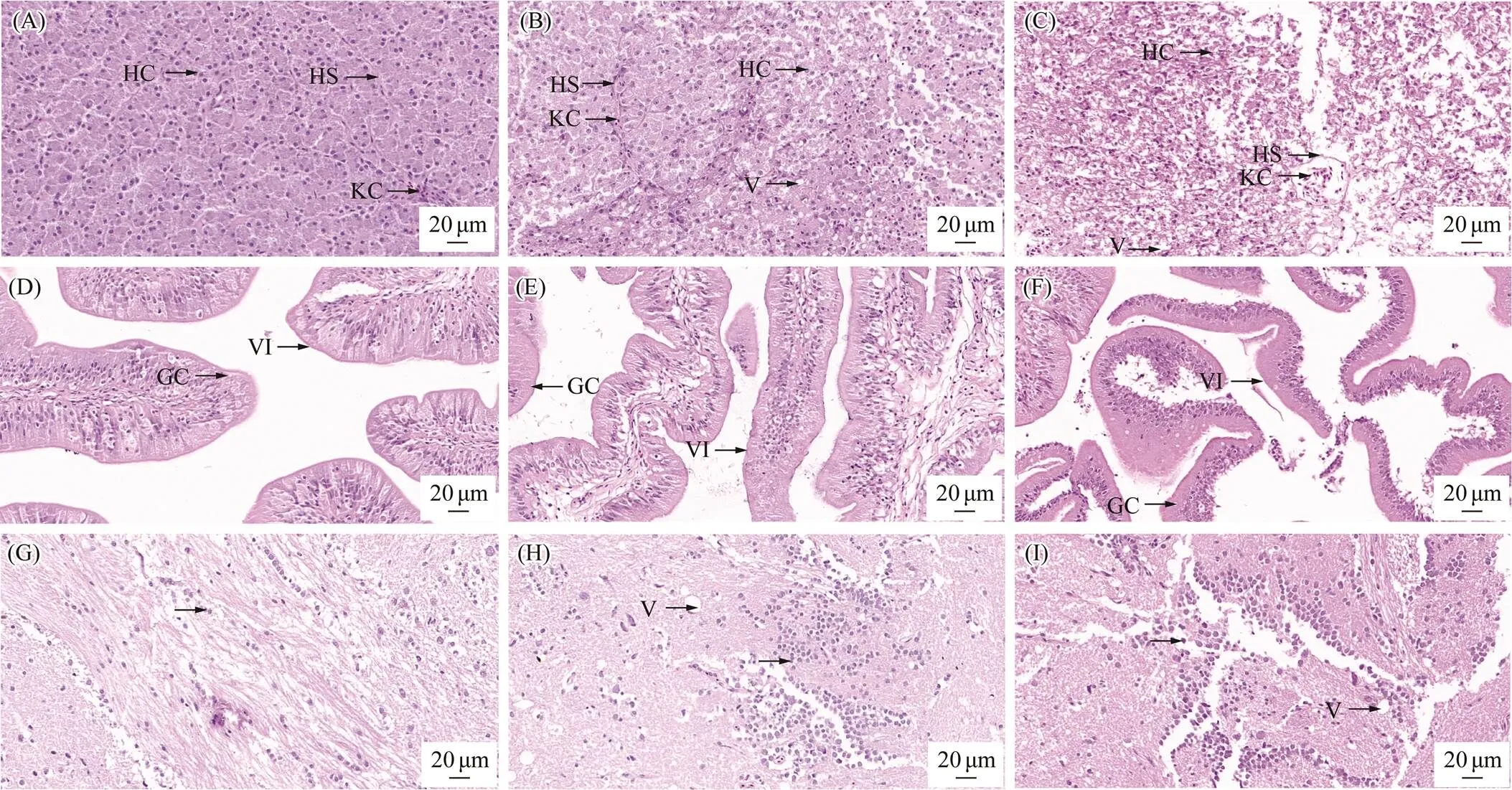
Fig.1 Liver, intestine and brain histological changes in half-smooth tongue sole after PFOA exposure. (A) The liver of control group (40×). (B) The liver of the PFOA-exposed group after 7 days of exposure (40×). (C) The liver of the PFOA- exposed group after 14 days of exposure (40×). (D) The intestine of the control group (40×). (E) The intestine of the PFOA-exposed group after 7 days of exposure (40×). (F) The intestine of the PFOA-exposed group after 14 days of exposure (40×). (G) The brain of control group (40×). (H) The brain of the PFOA-exposed group after 7 days of exposure (40×); (I) The brain of the PFOA-exposed group after 14 days of exposure (40×). HC, hepatocyte; HS, hepatic sinusoid; KC, Kupffer cells; V, vacuoles; GC, goblet cells; VI, intestinal villus. The arrows with no word indicated abnormal results or cell aggregation, scale bar=20µm.
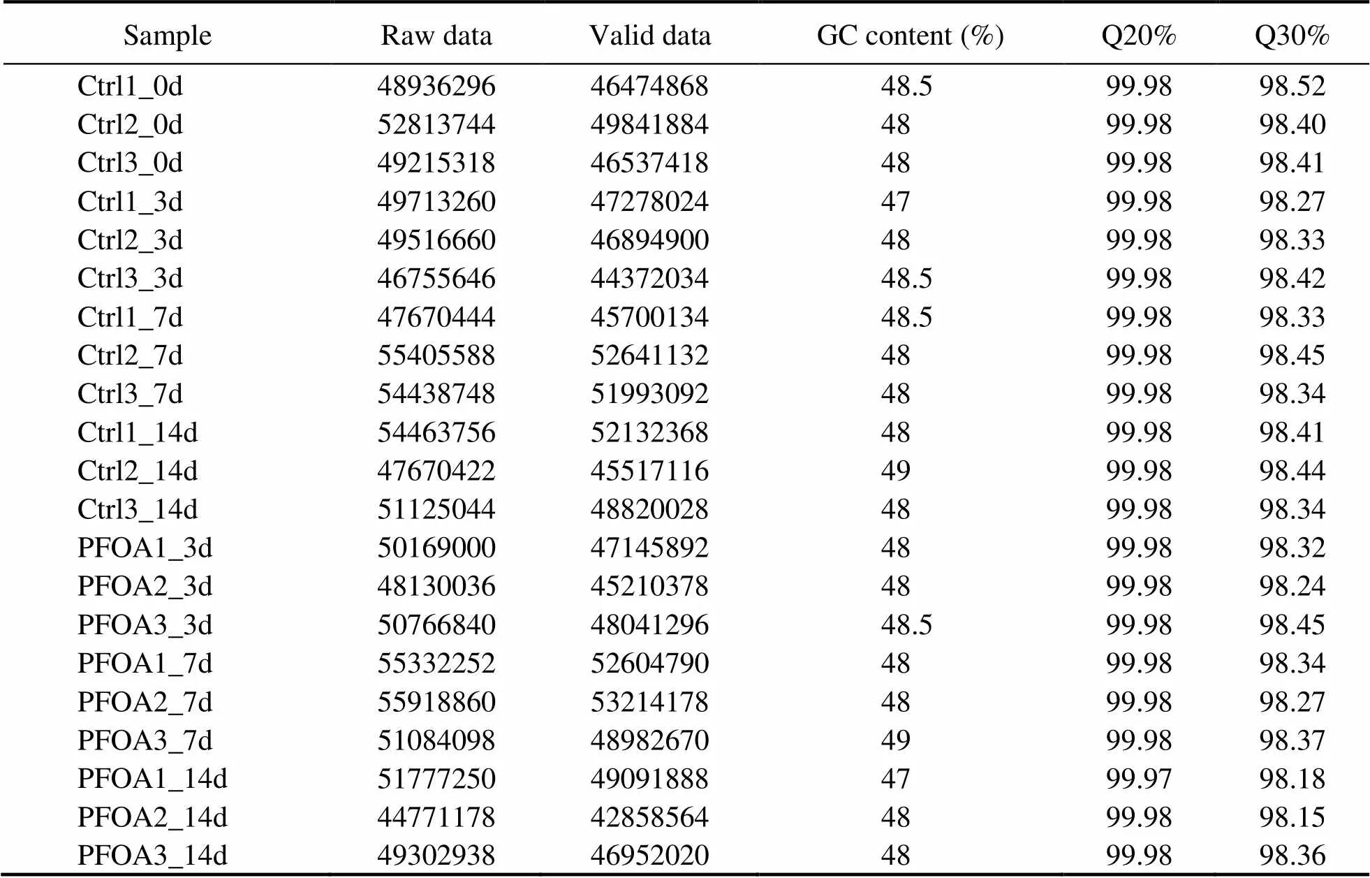
Table 2 Transcriptome information statistics of Cynoglossus semilaevis in different treatment groups
Note: Q20% and Q30% donate quality score≥20 base ratio (%) and quality score≥30 base ratio (%), respectively.
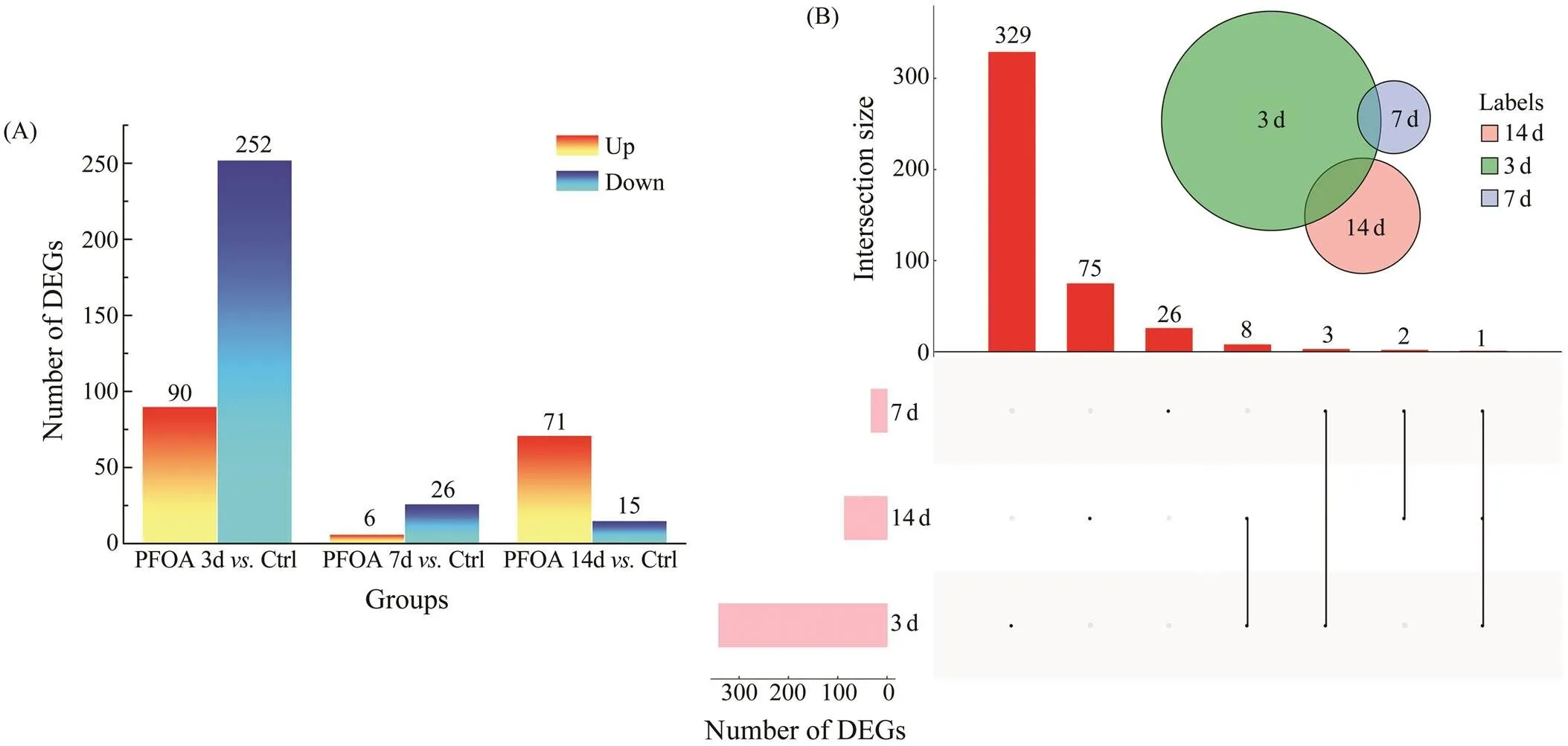
Fig.2 Statistics analysis of DEGs. (A) The number of DEGs between the PFOA-exposed group and the control group (Ctrl). (B) Venn diagram of DEGs. The pink columns below represented the number of DEGs in each group compared with the control group. The upper red column represents the number of intersection genes between groups. And the black dots and lines represented the intersections between groups.
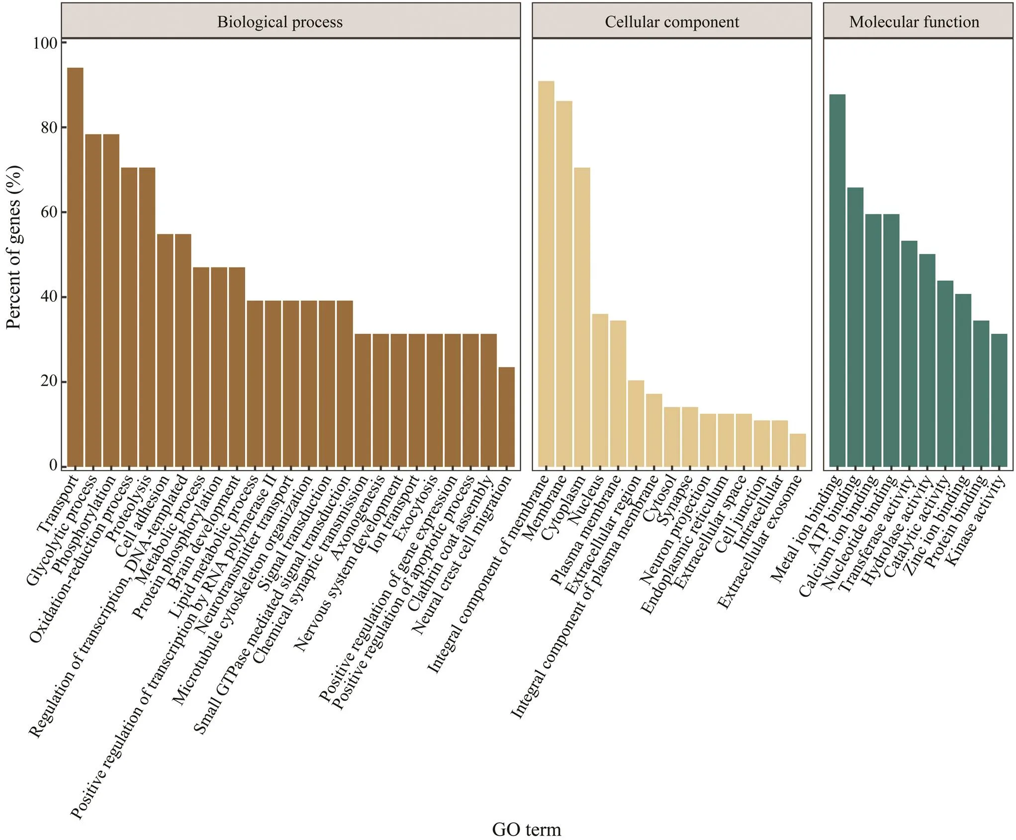
Fig.3 (A) GO enrichment analysis of DEGs after 3 days of exposure. The horizontal axis represents the GO terms. The vertical axis is the number of DEGs(to be continued).
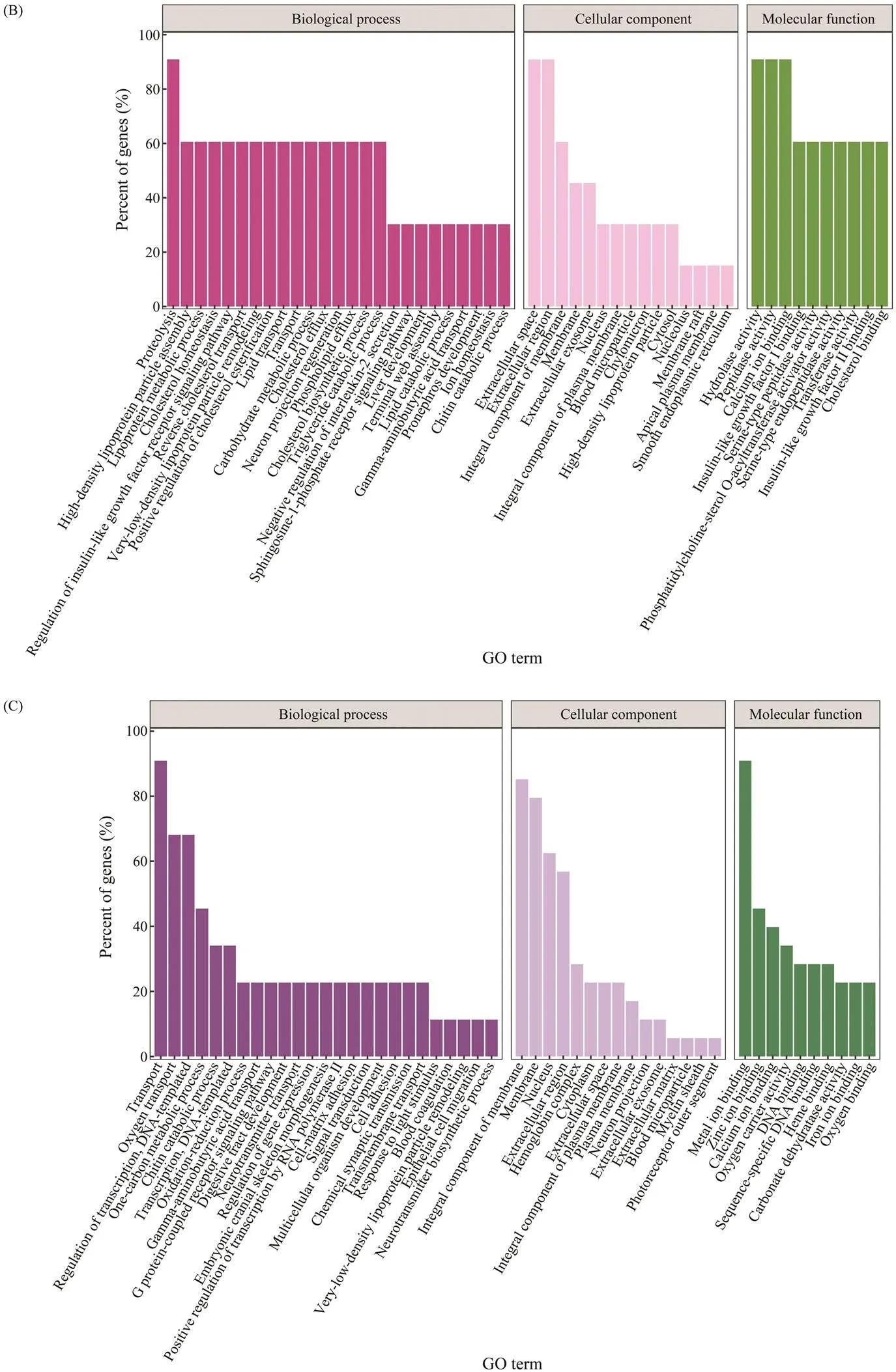
Fig.3 (continued) (B) GO enrichment analysis of DEGs after 7 days of exposure and (C) after 14 days of exposure. The horizontal axis represents the GO terms. The vertical axis is the number of DEGs.
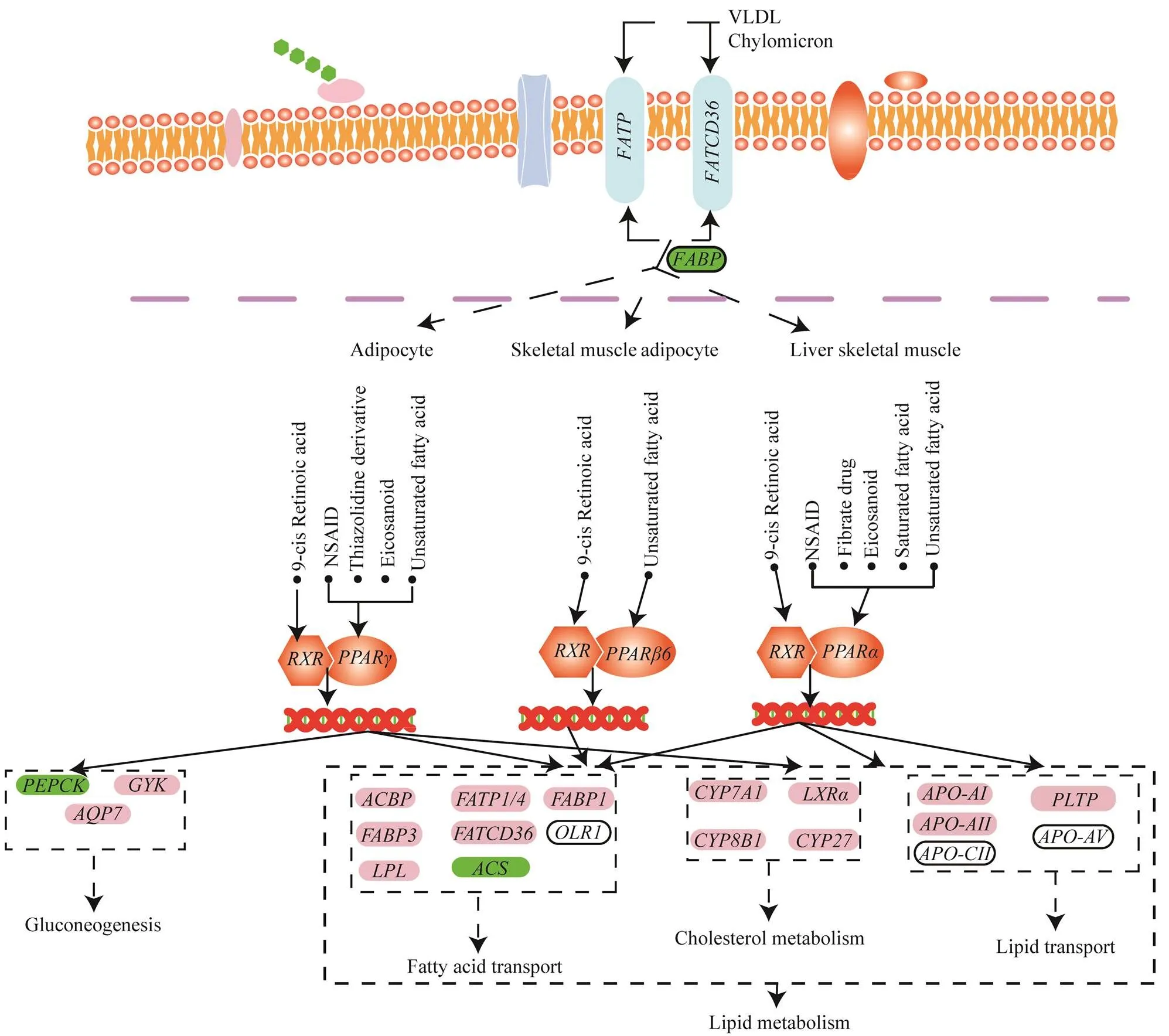
Fig.4 Significant changes in key genes of PPAR signaling pathway (part) after 7 days of exposure. Down-regulated DEGs are marked with green.
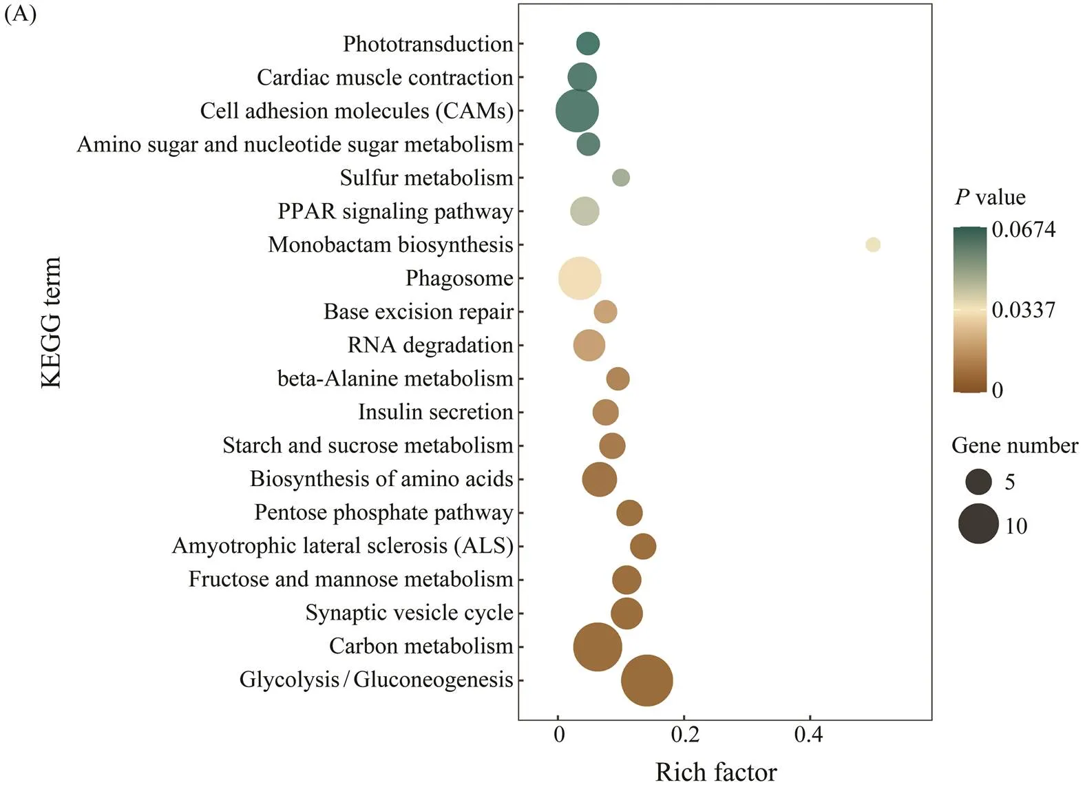
Fig.5 (A) KEGG enrichment analysis of DEGs after 3 days of exposure. The top 10 signaling pathways were analyzed. The horizontal axis was represented Rich factor. It was the ratio of the number of DEGs enriched in the signaling pathway to the total number of genes in the signaling pathway (to be continued).
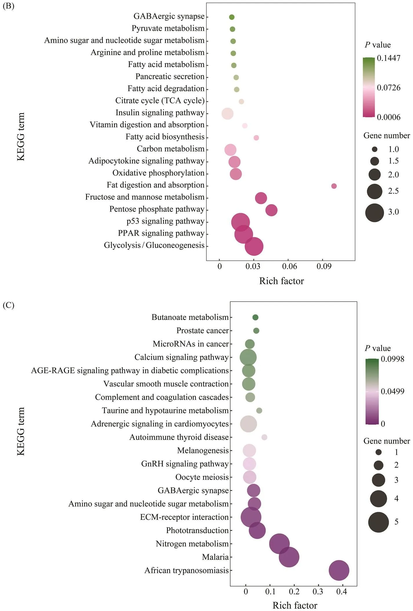
Fig.5 (continued) Same as Fig.5(A) but for KEGG enrichment analysis of DEGs after 7 days of exposure (B) and after 14 days of exposure (C).
3.4 Key Genes Analysis
To further study the DEGs of juvenile half-smooth tonguesoles after PFOA exposure, a heatmap analysis was perform- ed (Table 3). Some key genes were detected, as shown in Fig.6. Of these genes, energy transport-related genes were up-regulated after 7 days of exposure, such as. Some immune-related genes, such as(, mitochondrial) was up- regulated after 3 days of exposure and down-regulated after 7 and 14 days of exposure, immune-related genes like() and() were up-regulated after 14 days of exposure. Neural regulation genes were down- regulated after 3 days of exposure, then up-regulated after 7 days and 14 days of exposure, like(),() and(). The lipid metabolism-related gene() was down-regulated after 3 days and 7 days of exposure. Additionally, some PPAR signaling pathway-related genes were up-regulated after 14 days of exposure, such as() and(involved in lipid tran- sport).
To understand whether there was a relationship between the changes of key genes, key genes were further analyzedby OmicStudio tool. Some key genes were found to be cor- related. Such as there was a strong positive correlation be- tweenand(),and(),(Fig.7). Fur- thermore, there was a negative correlation betweenand()
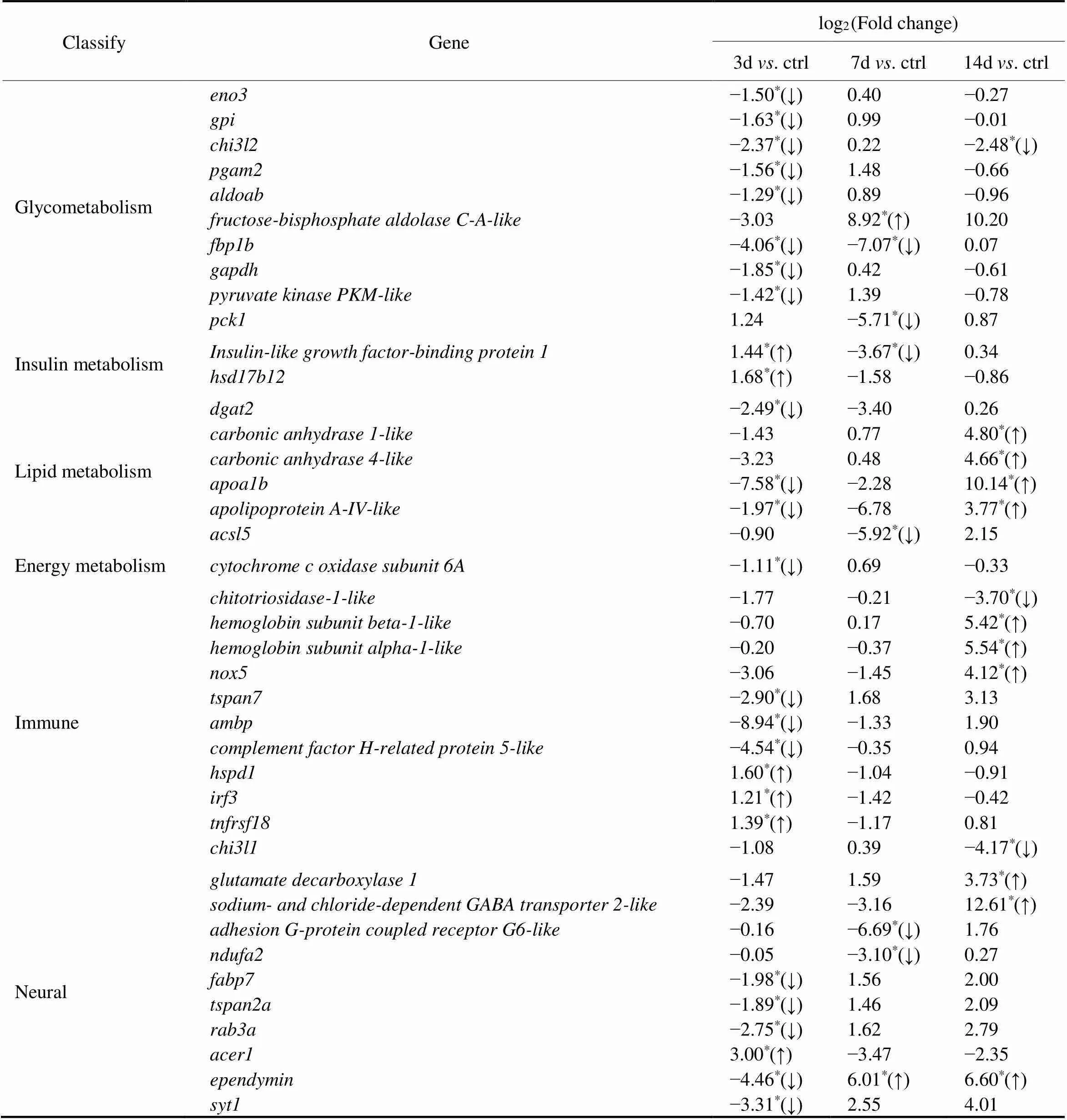
Table 3 log2FC of some key DEGs
Notes:*-value<0.05, ↑ up-regulated, ↓ down-regulated.
3.5 Validation of Sequencing Results
Transcriptome sequencing results were validated by qRT- PCR on eight randomly selected genes (Fig.8). The results showed that the expressions of(),(),(),,,(),() genes decreased significantly compared to those in the control group after 3 days of exposure (Fig.8A). Theexpressions of,,,andgenes increased significantly compared to those in the control group after 7 days of exposure, and the expressions ofandgenes decreased significantly compared to those in the control group (Fig.8B). The expressions ofandgenes decreased compared to those in the control group after 14 days of exposure, and the ex- pressions of,,(),,genes increased compared to that in the control group (Fig.8C). Results showed that the qRT-PCR data were consistent with the RNA-Seq, except for the expressions ofgene after 7 days of exposure andgene after 14 days of exposure. And Pearson’s corre- lation analysis showed that the gene expression changes of qRT-PCR were significantly correlated to the RNA-Seq re- sults with the correlation coefficient of 0.881 (<0.01). These results confirm the reliability of the RNA-Seq ana- lysis.
4 Discussion
PFOA is a widespread synthetic persistent organic pol- lutant (POP). Many studies have focused on its effects on the reproductive system and liver, as well as its immuno- toxicity in rats, mice and zebrafish (Cui, 2009; Na- kagawa, 2012; Liang, 2022). However, little is known about the toxic effects of PFOA on half-smoothtongue soles. In this study, the toxic effects of PFOA onhalf-smooth tongue soles were explored using transcriptome analysis and histopathological assessment.
Histopathological assessment revealed biochemical and physiological changes caused by toxicant exposure, which could be used as biomarkers of the effects (Guo, 2021).Previous studies have shown that PFOA caused liver da- mage, hepatocyte enlargement, local necrosis, and vacuo- lization in mice (Li, 2019a). However, histological changes in other organs after exposed to PFOA have rare- ly been reported. In this study, the histopathological resultsconfirmed that the liver, intestine, and brain of the juvenile half-smooth tongue sole were damaged after PFOA expo- sure. After 14 days of exposure, the hepatocytes were rup- tured, while large-scale rupture of intestinal villi and brain abnormal were also observed. Additionally, previous stud- ies have showed that PFOA-induced liver damage may be associated with the lipid metabolism-related genes (.,and) (Zhang, 2019). At the present study, the expressions of some lipid metabolism- related genes were affected significantly. Therefore, histo- logical changes of the juvenile half-smooth tongue may be associated with lipid metabolism-related and other genes expression.
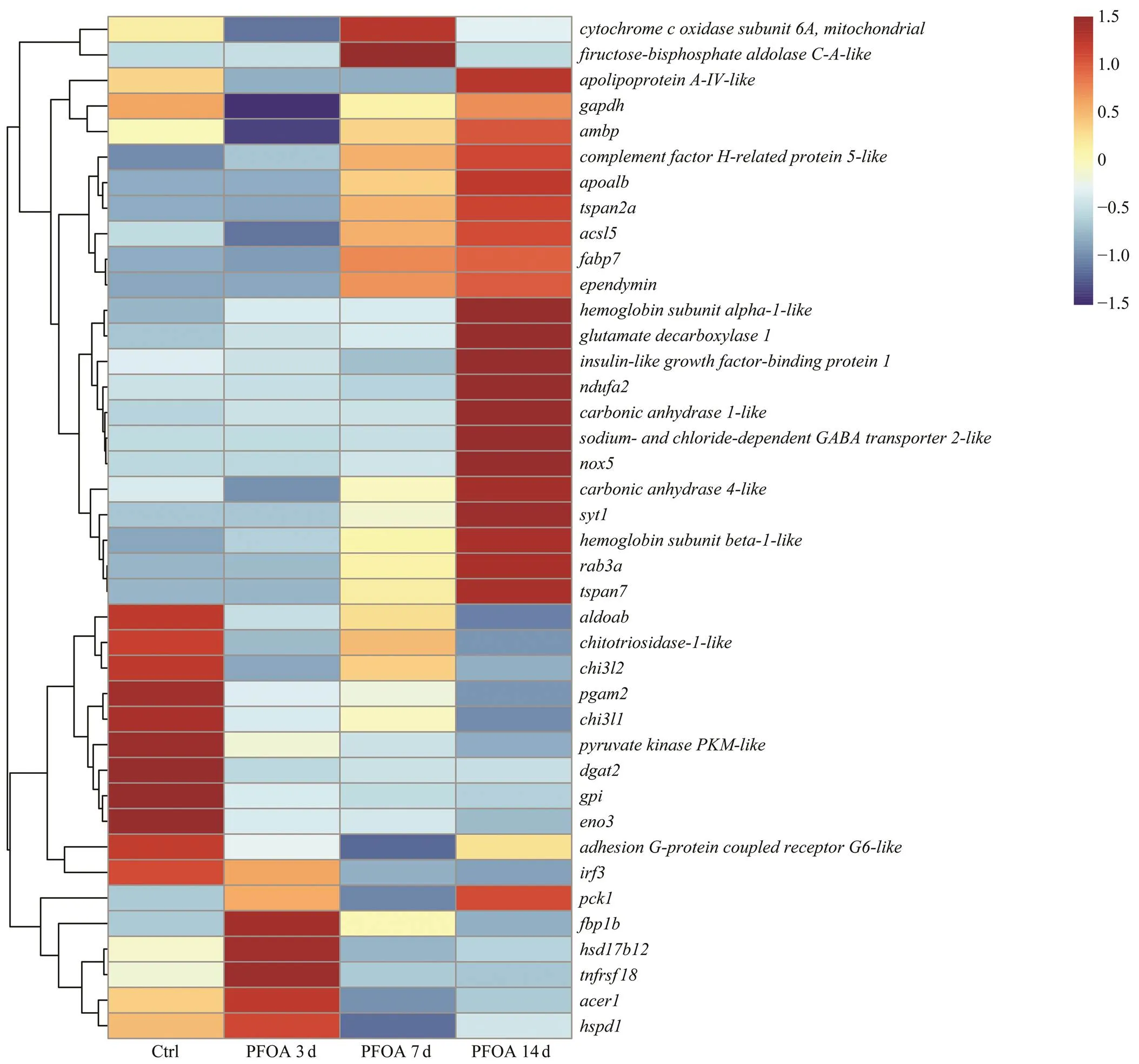
Fig.6 Heat map of some key DEGs. The change from blue to red stripes indicate an increase in gene expression levels. Ctrl, the control group; PFOA 3d, after 3 days of PFOA exposure; PFOA 7d, after 7 days of PFOA exposure; PFOA 14d, after 14 days of PFOA exposure.
Recently, transcriptome analysis has become a very use- ful research tool for quantifying DEGs in organisms and an excellent method for studying biological responses to en- vironmental changes (Hou, 2020; Wang, 2020). In this study, transcriptome analysis was also implemented to determine the short-term impact of PFOA exposure on juvenile half-smooth tongue soles. Results showed that DEGs after 14 days were significantly higher than those af- ter 7 days of exposure, but the percentage of genes after 7 days was significantly higher than that after 14 days of ex-posure (top 30 of GO terms). The GO enrichment results showed other genes (except top 30) might enriched in other GO terms (ranking lower than the top 30) in 14 days of ex- posure. Additionally, some metabolism-related DEGs werefound to be abnormal by transcriptome analysis. For instance,,andwere down-regulated after 7 days of exposure, and,were up-regulated after 14 days of exposure. Of these genes,is an important en- zyme in organisms. Research has shown that it is down- regulated in human hepatocellular carcinoma (HCC), and its overexpression inhibits the proliferation of HCC cells (Li, 2019b). In addition, PFOA exposure caused to- xicity by activating PPAR-α (Liu, 2017a). In this study,some genes related to the PPAR pathway were down-regu- lated after 7 days of exposure, such asand, some genes related to the PPAR pathway were up-regulated af- ter 14 days of exposure, likeand. Additionally, previous studies have also shown that there are significant differences in the responsiveness of regulated genes. After PFOA exposure, PPARs may al- so be down-regulated, but their regulated genes were strong- ly up-regulated (Abbott, 2012). These results indi-cated that the activation of PPAR by PFOA might causedifferent changes of downstream genes, and lead to abnor- mal metabolism in half-smooth tongue sole.
Mitochondrial oxidative phosphorylation (OXPHOS) pro- vides the majority of energy required for cell survival (Ji, 2022). In this study, OXPHOS signaling pathway was significantly enriched after 7 days of exposure. Many stud ies have shown that short-term stress leads to a reduction in the expression of oxidative phosphorylation-related genes, mainly based on toxic effects, and organisms show a gra- dual process of adaptation in response to stress (Zhao, 2014; Zeng, 2021). In this study, after 3 days of ex- posure, the expression of genes related to energy metabo- lism was down-regulated. After 7 days of exposure, the half- smooth tongue sole tries to gain more energy by upregu- lating the expression of oxidative phosphorylation-related genes for its own metabolism, immunity,. Additionally, studies have shown that improving energy metabolism playsa crucial role in protecting organisms against environmen- tal stress (Zeng, 2016). Therefore, when faced with the toxic stress of PFOA, half-smooth tongue soles might resist stress by improving energy metabolism.
Immune responses play an important role in fish exposedto external stresses. Interestingly, in the present study, someimmune-related genes, such aswasup-regulated af- ter 3 days of exposure and down-regulated after 7 and 14 days of exposure,andwere up-regu- lated after 14 days of exposure. As a kind of molecular cha- perone, heat shock proteins were closely related to immuneresponse and stress, and most of them were up-regulatedafter stress (Cheng, 2019; Waheed, 2020). In the present study, heat shock proteins were also up-regulated after 3 days of exposure, similar toAnother studycarried out an acute toxicity experiment of PFOS (Perfluo- rooctane Sulfonates, an analog chemical of PFOA), and the results showed that the expressions of immune-related genessuch asandregulated at 96 h, and immune dysfunction occurred after exposure to PFOS (Zhang, 2020). In this study, im- munosuppression might have occurred in the early stage of PFOA exposure, but the expression of some immune genes was up-regulated in the later stage of PFOA exposure.
In this study, some signaling pathways concerning neu- ral synapse activity were significantly enriched, such asGABAergic synapses. The expression of some genes relatedto these signaling pathways also showed an increasing trend. Of these genes,andwere down-regulated after 3 days of exposure and up-regulated after 7 and 14 days of exposure. Geneis a key mediator of neurotransmitter release in the central nervous system (Melland, 2022).Previous research also has indicated that variants ofmay lead to neurodevelopmental disorders (Bradberry, 2020). Abnormal neural synapse activity also affected neu- ronal health (Bell and Hardingham, 2011). In addition,is the main glycoprotein in cerebrospinal fluid of te- leost fishes, and plays a role in learning, memory and neu-ral plasticity (closely related to nerve excitability) (Ortil and Meyer, 1996). The present study found thatwas expressed significantly differently in each exposure point, which may be one of the reasons for brain abnormalities after 14 days of exposure. Thus, changes in the genes, such as,,and, may lead to abnormal neu- ral activity, resulting in abnormal neuronal excitation.
5 Conclusions
In conclusion, the toxic effects of PFOA on juvenile half- smooth tongue soles were explored by histological assess- ment and transcriptome analysis. The results of histology- cal analysis showed that PFOA exposure caused hepatocyterupture, intestinal villi breakage, an increase of goblet cells and brain abnormalities. Transcriptome results also show- ed that excessive PFOA may cause toxic effects by affect- ing PPAR signaling pathway and GABAergic synapses. To resist toxicity, metabolism was increased. The present study not only enriches the toxic theoretical basis of PFOA, but also provides a new perspective and insights for the toxi- city assessment of PFOA at the transcriptome level.
Acknowledgements
This work was supported by the National Key R&D Pro- gram (No. 2018YFD0900301-03), and the MNR Key La- boratory of Marine Eco-Environmental Science and Tech- nology, China (No. MEEST-2021-04).
Abbott, B. D., Wood, C. R., Watkins, A. M., Tatum-Gibbs, K., Das,K. P., and Lau, C., 2012. Effects of perfluorooctanoic acid (PFOA) on expression of peroxisome proliferator-activated re- ceptors (PPAR) and nuclear receptor-regulated genes in fetal and postnatal CD-1 mouse tissues., 33: 491-505, DOI: 10.1016/j.reprotox.2011.11.005.
Abe, T., Takahashi, M., Kano, M., Amaike, Y., Ishii, C., Maeda, K.,., 2017. Activation of nuclear receptor CAR by an envi- ronmental pollutant perfluorooctanoic acid., 91: 2365-2374, DOI: 10.1007/s00204-016-1888-3.
Bell, K. F. S., and Hardingham, G. E., 2011. The influence of synaptic activity on neuronal health., 21 (2): 299-305, DOI: 10.1016/j.conb.2011.01.002.
Bradberry, M. M., Courtney, N. A., Dominguez, M. J., Lofquist, S. M., Knox, A. T., Sutton, R. B.,., 2020. Molecular basis for synaptotagmin-1-associated neurodevelopmental disorder., 107:52-64, DOI: 10.1016/j.neuron.2020.04.003.
Cheng, D. W., Liu, H. X., Zhang, H. K., Soon, T. K., Ye, T., Li, S.K.,., 2019. Differential expressions ofgene between golden and brown noble scallopsunder heat stress and bacterial challenge., 94: 924-933, DOI: 10.1016/j.fsi.2019.10.018.
Cui, L., Zhou, Q. F., Liao, C. Y., Fu, J. J., and Jiang, G. B., 2009. Studies on the toxicological effects of PFOA and PFOS on ratsusing histological observation and chemical analysis., 56: 338-349, DOI:10.1007/s00244-008-9194-6.
Do, A. N. T., and Tran, H. D., 2022. Potential application of ar- tificial neural networks for analyzing the occurrences of fish larvae and juveniles in an estuary in northern Vietnam., 57: 813-831, DOI: 10.1007/s10452-022-09959-5.
Dong, H. K., Lu, G. H., Yan, Z. H., Liu, J. C., and Ji, Y., 2019. Mo-lecular and phenotypic responses of male crucian carp () exposed to perfluorooctanoic acid., 653:1395-1406, DOI: 10.1016/j.scitotenv. 2018.11.017.
Fan, Q. X., Shi, K. P., Zhan, M., Xu, Q., Liu, X. B., Li, Z. J.,.,2022. Acute damage from the degradation ofon the environmental microbiota, intestinal microbiota and tran-scriptome of Japanese flounder., 302: 119022, DOI: 10.1016/j.envpol.2022.11 9022.
Fang, X. M., Wei, Y. H., Liu, Y., Wang, J. S., and Dai, J. Y., 2010.The identification of apolipoprotein genes in rare minnow () and their expression following perfluorooc- tanoic acid exposure., 151: 152-159, DOI: 10.1016/j.cbpc.2009.09.008.
Giari, L., Vincenzi, F., Badini, S., Guerranti, C., Dezfuli, B. S., Fa-no, E. A.,., 2016. Common carpresponsesto sub-chronic exposure to perfluorooctanoic acid., 23: 15321-15330, DOI: 10.1007/s11356-016-6706-1.
Guo, H., Liang, Z., Zheng, P. H., Li, L., Xian, J. A., and Zhu, X. W., 2021. Effects of nonylphenol exposure on histologicalchanges, apoptosis and time-course transcriptome in gills of white shrimp., 781: 146731, DOI: 10.1016/j.scitotenv.2021.146731.
Han, Z. X., Liu, Y. R., Wu, D. D., Zhu, Z., and Lü, C. X., 2012. Immunotoxicity and hepatotoxicity of PFOS and PFOA in ti- lapia ()., 31: 424-430, DOI: 10.1007/s11631-012-0593-z.
Hou, L. B., Ma, Y. B., Cao, X. H., Gu, W., Cheng, Y. X., Wu, X. G.,., 2020. Transcriptome profiling of thethoracic ganglion under thechallenge., 524: 735257,DOI: 10.1016/j.aquaculture.2020. 735257.
Ji, W. H., Tang, X., Du, W., Lu, Y., Wang, N. X., Wu, Q.,.,2022. Optical/electrochemical methods for detecting mitochon- drial energy metabolism., 51: 71- 127, DOI: 10.1039/d0cs01610a.
Kang, J. S., Ahn, T. G., and Park, J. W., 2019. Perfluorooctanoic acid (PFOA) and perfluooctane sulfonate (PFOS) induce diffe-rent modes of action in reproduction to Japanese medaka ()., 368: 97-103, DOI: 10.1016/j.jhazmat.2019.01.034.
Li, D. Y., Zhang, L. C., Zhang, Y., Guan, S., Gong, X. C., and Wang,X. D., 2019a. Maternal exposure to perfluorooctanoic acid (PFOA) causes liver toxicity through PPAR-alpha pathway and lowered histone acetylation in female offspring mice., 26: 18866-18875, DOI: 10.1007/s11356-019-05258-z.
Li, X. Z., Bao, C. Y., Ma, Z. N., Xu, B. Q., Ying, X. Y., Liu, X. Q.,., 2018. Perfluorooctanoic acid stimulates ovarian cancer cell migration, invasion via ERK/NF-kappaB/MMP-2/-9 path- way., 294: 44-50, DOI: 10.1016/j.toxlet.2018. 05.009.
Li, Y. F., Li, T. T., Jin, Y. L., and Shen, J. W., 2019b.re- duces hepatocellular carcinoma malignancy via downregulationof cell cycle-related gene expression., 115: 108950, DOI: 10.1016/j.biopha.2019.108950.
Liang, L. Y., Pan, Y. L., Bin, L. H., Liu, Y., Huang, W. J., Li, R.,., 2022. Immunotoxicity mechanisms of perfluorinated com- pounds PFOA and PFOS., 291: 132892, DOI: 10. 1016/j.chemosphere.2021.132892.
Liu, H., Wang, J. S., Sheng, N., Cui, R. N., Pan, Y. T., and Dai, J. Y., 2017a. Acot1 is a sensitive indicator for PPARα activation after perfluorooctanoic acid exposure in primary hepatocytes of Sprague-Dawley rats., 42: 299-307, DOI: 10.1016/j.tiv.2017.05.012.
Liu, Z. Y., Lu, Y. L., Wang, P., Wang, T. Y., Liu, S. J., Johnson, A. C.,., 2017b. Pollution pathways and release estimation of perfluorooctane sulfonate (PFOS) and perfluorooctanoic acid (PFOA) in central and eastern China., 580: 1247-1256, DOI: 10.1016/j.scitotenv.2016.12. 085.
Mayilswami, S., Krishnan, K., Megharaj, M., and Naidu, R., 2016. Gene expression profile changes inchronically exposed to PFOA., 25:759-769, DOI: 10.1007/ s10646-016-1634-x.
Melland, H., Bumbak, F., Kolesnik-Taylor, A., Ng-Cordell, E., John, A., Constantinou, P.,., 2022. Expanding the geno- type and phenotype spectrum of-associated neurodeve- lopmental disorder., 24 (4): 880-893, DOI:10.1101/2021.07.21.21260857.
Miranda, A. F., Trestrail, C., Lekamge, S., and Nugegoda, D., 2020. Effects of perfluorooctanoic acid (PFOA) on the thyroid status,vitellogenin, and oxidant-antioxidant balance in the Murray Ri-ver rainbowfish., 29:163-174, DOI: 10.1007/ s10646-020-02161-z.
Mortensen, A. S., Letcher, R. J., Cangialosi, M. V., Chu, S. G., and Arukwe, A., 2011. Tissue bioaccumulation patterns, xenobio- tic biotransformation and steroid hormone levels in Atlantic sal- mon () fed a diet containing perfluoroactane sul- fonic or perfluorooctane carboxylic acids., 83: 1035-1044, DOI: 10.1016/j.chemosphere.2011.01.067.
Nakagawa, T., Ramdhan, D. H., Tanaka, N., Naito, H., Tamada, H., Ito, Y.,., 2012. Modulation of ammonium perfluoroocta- noate-induced hepatic damage by genetically different PPARα in mice., 86: 63-74, DOI: 10.1007/s00 204-011-0704-3.
Ortil, G., and Meyer, A., 1996. Molecular evolution of ependyminand the phylogenetic resolution of early divergences amongeuteleost fishes., 13 (4): 556- 573, DOI: 10.1093/oxfordjournals.molbev.a025616.
Pertea, M., Pertea, G. M., Antonescu, C. M., Chang, T. C., Men- dell, J. T., and Salzberg, S. L., 2015. StringTie enables improvedreconstruction of a transcriptome from RNA-seq reads., 33: 290-295, DOI: 10.1038/nbt.3122.
Qi, L. J., Chen, Y. D., Shi, K. P., Ma, H., Wei, S., and Sha, Z. X., 2021. Combining of transcriptomic and proteomic data to mine immune-related genes and proteins in the liver ofchallenged with.–, 39: 100864, DOI: 10.1016/j.cbd.2021.100864.
Rial, D., Beiras, R., Vazquez, J. A., and Murado, M. A., 2010. Acute toxicity of a shoreline cleaner, CytoSol, mixed with oil and ecological risk assessment of its use on the Galician Coast., 59: 407-416, DOI: 10.1007/s00244-010-9492-7.
Seyoum, A., Pradhan, A., Jass, J., and Olsson, P. E., 2020. Perfluo- rinated alkyl substances impede growth, reproduction, lipid me- tabolism and lifespan in., 737: 139682, DOI: 10.1016/j.scitotenv.2020.13 9682.
Shane, H. L., Baur, R., Lukomska, E., Weatherly, L., and Ander- son, S. E., 2020. Immunotoxicity and allergenic potential induced by topical application of perfluorooctanoic acid (PFOA) in a murine model., 136: 111114, DOI: 10.1016/j.fct.2020.111114.
Shao, P., Yong, P. Z., Zhou, W. L., Sun, J. H., Wang, Y. Z., Tang, Q. P.,., 2019. First isolation ofsubsp.from half-smooth tongue sole suffering from skin-ulceration disease., 511:734208, DOI: 10. 1016/j.aquaculture.2019.734208.
Tang, J., Lu, X. J., Chen, F. F., Ye, X. P., Zhou, D. R., Yuan, J. L.,., 2018. Effects of perfluorooctanoic acid on the associ- ated genes expression of autophagy signaling pathway oflymphocytes., 9: 1748, DOI: 10.3389/fphys.2018.01748.
Waheed, R., El Asely, A. M., Bakery, H., El-Shawarby, R., Abuo- Salem, M., Abdel-Aleem, N.,., 2020. Thermal stress ac- celerates mercury chloride toxicity invia up-regulation of mercury bioaccumulation andmRNA expression., 718: 137326,DOI: 10.1016/j.scitotenv.2020.137326.
Wan, Y., Wang, S. L., Cao, X. Z., Cao, Y. X., Zhang, L., Wang, H.,., 2017. Perfluoroalkyl acids (PFAAs) in water and sedi- ment from the coastal regions of Shandong Peninsula, China., 189: 100, DOI: 10. 1007/s10661-017-5807-8.
Wang, P., Lu, Y. L., Wang, T. Y., Fu, Y. N., Zhu, Z. Y., Liu, S. J.,., 2014. Occurrence and transport of 17 perfluoroalkyl acids in 12 coastal rivers in south Bohai coastal region of China with concentrated fluoropolymer facilities., 190: 115-122, DOI: 10.1016/j.envpol.2014.03.030.
Wang, Q., Hao, X. C., Liu, K. Q., Feng, B., Li, S., Zhang, Z. H.,., 2020. Early response to heat stress in Chinese tongue sole): Performance of different sexes, candidate genes and networks., 21: 745, DOI: 10.1186/s12864-020-07157-x.
Wei, Y. H., Liu, Y., Wang, J. S., Tao, Y., and Dai, J. Y., 2008. To- xicogenomic analysis of the hepatic effects of perfluoroocta- noic acid on rare minnows ()., 226: 285-297, DOI: 10.1016/j.taap. 2007.09.023.
Yamashita, N., Kannan, K., Taniyasu, S., Horii, Y., Petrick, G., and Gamo, T., 2005. A global survey of perfluorinated acids in oceans., 51: 658-668, DOI: 10.1016/j.mar- polbul.2005.04.026.
Yamashita, N., Taniyasu, S., Petrick, G., Wei, S., Gamo, T., Lam, P.K. S.,., 2008. Perfluorinated acids as novel chemical tracers of global circulation of ocean waters., 70: 1247- 1255, DOI: 10.1016/j.chemosphere.2007.07.079.
Yang, J. H., 2010. Perfluorooctanoic acid induces peroxisomalfatty acid oxidation and cytokine expression in the liver of male Japanese medaka ()., 81: 548-552,DOI: 10.1016/j.chemosphere.2010.06.028.
Yang, M., Ye, J. S., Qin, H. M., Long, Y., and Li, Y., 2017. Influ- ence of perfluorooctanoic acid on proteomic expression and cell membrane fatty acid of., 220: 532-539, DOI: 10.1016/j.envpol.2016.09.097.
Yu, J., Cheng, W. Q., Jia, M., Chen, L., Gu, C., Ren, H. Q.,., 2022. Toxicity of perfluorooctanoic acid on zebrafish early em- bryonic development determined by single-cell RNA sequen- cing., 427: 127888, DOI: 10. 1016/j.jhazmat.2021.127888.
Zeng, L., Li, W. C., Zhang, H., Cao, P., Ai, C. X., Hu, B.,., 2021. Hypoxic acclimation improves mitochondrial bioener- getic function in large yellow croakerun- der Cu stress., 224: 112688, DOI: 10.1016/j.ecoenv.2021.112688.
Zeng, L., Wang, Y. H., Ai, C. X., Zheng, J. L., Wu, C. W., and Cai, R., 2016. Effects of beta-glucan on ROS production and en- ergy metabolism in yellow croaker () un- der acute hypoxic stress., 42: 1395-1405, DOI: 10.1007/s10695-016-0227-1.
Zhang, H. J., He, J. B., Li, N., Gao, N., Du, Q. X., Chen, B.,., 2019. Lipid accumulation responses in the liver ofinduced by perfluorooctanoic acid (PFOA)., 167: 29-35, DOI: 10.1016/ j.ecoenv.2018.09.120.
Zhang, L. B., Sun, W., Chen, H. G., Zhang, Z., and Cai, W. G., 2020. Transcriptomic changes in liver of Juvenilefollowing perfluorooctane sulfonate exposure., 39: 556-564, DOI: 10. 1002/etc.4633.
Zhao, Y. Y., Xie, P., Fan, H. H., and Zhao, S. J., 2014. Impairment of the mitochondrial oxidative phosphorylation system and oxi-dative stress in liver of crucian carp (L.) ex- posed to microcystins., 29 (1): 30- 39, DOI: 10.1002/tox.20770.
Zhong, Y. C., Shen, L. L., Ye, X. P., Zhou, D. G., He, Y. Y., and Zhang, H. J., 2020. Mechanism of immunosuppression in ze- brafish () spleen induced by environmentally rele- vant concentrations of perfluorooctanoic acid., 249: 126200, DOI: 10. 1016/j.chemosphere.2020.126200.
(November 18, 2022;
January 9, 2023;
April 18, 2023)
© Ocean University of China, Science Press and Springer-Verlag GmbH Germany 2023
#The two authors contributed equally to this work.
. E-mail: shazhenxia@163.com
(Edited by Qiu Yantao)
 Journal of Ocean University of China2023年6期
- Journal of Ocean University of China的其它文章
- Effects of 5-Azacytidine (AZA) on the Growth, Antioxidant Activities and Germination of Pellicle Cystsof Scrippsiella acuminata (Diophyceae)
- Using Rn-222 to Study Human-Regulated River Water-Sediment Input Event in the Estuary
- Parameterization Method of Wind Drift Factor Based on Deep Learning in the Oil Spill Model
- YOLOv5-Based Seabed Sediment Recognition Method for Side-Scan Sonar Imagery
- A Metadata Reconstruction Algorithm Based on Heterogeneous Sensor Data for Marine Observations
- Large Active Faults and the Wharton Basin Intraplate Earthquakes in the Eastern Indian Ocean
