Study on pharmacodynamics and mechanism of nano-Kuiyangye in treating radiation esophagitis in rats
Li Kai-xuan, Wang Qin, Zheng Jia-bin, Teng Feng, Jia Li-qun
1.Graduate School of Beijing University of Chinese Medicine, Beijing 100010, China
2.Department of Integrated Traditional Chinese and Western Medicine Oncology, China-Japan Friendship Hospital, Beijing 100010
3.Department of Radiation Oncology, China-Japan Friendship Hospital, Beijing 100010, China
Keywords:
ABSTRACT Objective: To explore the curative effect of nano-Kuiyangye on radiation esophagitis in Wistar rats, and to explore its possible mechanism.Methods: Wistar rats were irradiated locally with 30Gy rays to establish an animal model of radiation esophagitis in rats.After irradiation, nano-Kuiyangye, traditional Chinese medicine ulcer solution, Kangfuxin solution, nano-hydrotalcite matrix, and distilled water were used to intervene continuously for 7 d, during which the body weight and food intake of the rats were recorded.On day 7, blood was collected from the abdominal aorta under anesthesia, and the serum was obtained by centrifugation.The expression levels of pain-related factors prostaglandin-2 (PGE-2), substance P (SP), and calcitonin gene-related peptide (CGRP) were detected by ELISA.Rats were sacrificed after blood collection, and full-length esophageal tissues were taken.Hematoxylin-eosin (HE)staining was performed to analyze the pathological changes of the rat esophagus, and Western Blotting was used to detect the expression of nuclear factor-κB (NF-κB) inflammatory protein.Results: Compared with the control group, the total food intake and body weight of the rats within 7 d after modeling were significantly decreased (P<0.05), and the expressions of pain-related factors PGE-2, SP, and CGRP were higher than those of the control group (P<0.05)., the esophageal pathological damage score increased (P<0.05), and the expression of NFκB inflammatory protein increased (P<0.05); after treatment, the total food intake of the rats in the nano-Kuiyangye intervention group within 7 days after modeling was higher than that in other groups (P<0.05), the expressions of pain-related factors PGE-2, SP, and CGRP were lower than those in the model group (P<0.05), the esophageal pathological damage score was lower (P<0.05), and the expression of NF-κB inflammatory proteins was lower (P<0.05).Conclusion: Nano-Kuiyangye increases the food intake of rats with radiation esophagitis,reduces esophageal tissue damage, and reduces the concentration of serum pain factors; the anti-inflammatory effect of nano-Kuiyangye may be related to the reduction of NF-κB inflammatory factor levels.
1.Introduction
Radiation esophagitis (RE) is a common adverse reaction of radiotherapy for cervical and thoracic tumors.It is a serious complication that limits treatment and affects quality of life.The treatment of radiation esophagitis lacks evidence-based treatment methods.The measures of modern medicine to prevent and treat radiation esophagitis mainly include antibiotics, glucocorticoids,local anesthetics, mucosal protective agents, proton pump inhibitors,vitamins and other drugs combined with symptomatic treatment.Such drug combinations have problems such as weak preventive effect, many side effects, and recovery after drug withdrawal[1].The advantages and potentials of Ulcer Liquid, a traditional Chinese medicine preparation in the China-Japan Friendship Hospital, are gradually emerging in the treatment of radiation esophagitis[2], but limited by problems such as insufficient adhesion, the traditional Chinese medicine preparation has yet to be optimized.In recent years, the combination of nano-carriers and traditional Chinese medicines in the treatment of radiation-induced esophagitis has revealed the dawn.The nano-hydrotalcite (nano-LDH) developed by the School of Chemistry, Beijing University of Chemical Technology has a simple synthesis method and relatively low cost.It has high drug loading efficiency and high drug cell membrane permeability.[3].At present, nano-LDH has been widely used as a new drug carrier in the fields of antibacterial, antitumor and biological imaging.nano-LDH has good biocompatibility, is easily taken up by cells, and is easily degraded under acidic conditions.Therefore, we chose nano-LDH as the carrier to prepare nano-ulcer solution and designed this study to explore the curative effect of nano-ulcer solution on radiation-induced esophagitis in Wistar rats and its effects on various pain factors and inflammation-related factors.
2.Materials and methods
2.1 Experimental animals and experimental environment
36 healthy and clean male wistar rats, weighing (215.71±4.71)g, were purchased from Beijing Weitong Lihua Experimental Animal Technology Co., Ltd., the experimental animal qualification certificate number: SYXK (Beijing) 2016-0043, and were bred in the China-Japan Friendship Hospital.Hospital animal SPF laboratory, license number: SYXK (Beijing)-2016-0043.The laboratory environment maintains a temperature of (22±2) ℃, a relative humidity of (55±10)%, and a 12 h light cycle with a day and night cycle.All animal experiments were performed under the permission of the Biomedical Ethics Committee of the Experimental Animal Center of the Clinical Research Institute of the China-Japan Friendship Hospital (animal experiment ethics number zryhyy21-20-09-5).
2.2 Experimental drugs
Ulcer solution: Traditional Chinese medicine decoction Ulcer solution is derived from the “Ulcer Oil” prescription from the Sino-Japanese Friendship Hospital with integrated traditional Chinese and Western medicine oncology.Japanese Friendship Hospital Outpatient Chinese Pharmacy.Add ulcer oil compound (raw rhubarb 75 g, comfrey 50 g, safflower 50 g, angelica 50 g, angelica 50 g) into distilled water, decoct 3 times, 1 hour for the first time, 0.5 h for the 2nd and 3rd time each time, combine the filtrates, and filter again Concentrate the medicinal solution to 1000 mL after passing through the dregs, and the active ingredient content of the crude drug is 0.275 g/mL; after sterilization, it is sealed and stored at 4 ℃.
Kangfuxin liquid: purchased from Sichuan Good Doctor Panxi Pharmaceutical Co., Ltd., specification: 100 mL bottle; production batch number: Z51021834.Store in a cool place after sealing.
Nano-hydrotalcite matrix: Preparation method: Weigh 0.1mol of magnesium salt (magnesium nitrate) and 0.05 mol of aluminum salt (aluminum nitrate), ensure that the ratio of Mg:Al is 2:1, and dissolve it with 500 mL of deionized water to obtain a salt solution.At the same time, prepare 0.3 mol of NaOH, dissolve it in 500 mL of deionized water, mix the two solutions by colloid mill method,and prepare nano-scale MgAl hydrotalcite in an oven at 80 ℃for 24 h, and wash it three times with deionized water.Nano-hydrotalcite concentration: Quantitatively obtain the concentration of nanohydrotalcite: 0.0202 g/mL.
Nano-ulcer solution: Mix 100 mL of nano-hydrotalcite matrix solution with a concentration of 0.101g/mL and 100mL of ulcer liquid decoction.Hydrotalcite is fully loaded with active ingredients of traditional Chinese medicine to obtain nano-ulcer liquid, the concentration of nano-hydrotalcite is 0.0505 g/mL, and the content of active ingredients of traditional Chinese medicine is 0.1375 g/mL.
2.3 Main reagents
Isoflurane, sodium pentobarbital, hematoxylin staining solution,eosin staining solution, xylene, 10% neutral formalin solution, NFκB Recombinant antibody, rat prostaglandin E2 antibody, rat substance P antibody , rat calcitonin gene-related peptide antibody,etc.
2.4 Main Instruments
Linear accelerator; glass tissue grinder; low-temperature high-speed centrifuge; chemiluminescence instrument; fluorescence scanner;microplate reader; vertical electrophoresis tank, etc.
2.5 Animal grouping, modeling, drug administration, and material collection
After weighing 36 wistar rats, they were randomly selected by random number table method and divided into 6 groups: normal control group (CONTROL), model control group (MODEL),Kangfuxin liquid group (KFXY), traditional Chinese medicine ulcer liquid group ( KYY), nano hydrotalcite matrix group (NANO), nano ulcer solution group (NANO-KYY).6 in each group.
Refer to the animal model constructed by Shen Li et al.[4], and combine the results of the pre-experiment to build the model.Wistar rats were used to model.All animal experiments were performed under the permission of the Biomedical Ethics Committee of the Experimental Animal Center of the Clinical Research Institute of the China-Japan Friendship Hospital (animal experiment ethics number zryhyy21-20-09-5).After adaptive feeding, wistar rats were anesthetized by intraperitoneal injection of 7% chloral hydrate in a conscious state.The anesthetized wistar rats were fixed on a special mouse board with their limbs spread out and their abdomen facing upward.Neutral position, head tilted back, open airway while restricting head movement, esophagus positioning under CT, with the midline of the rat body as the central axis, the upper boundary of the irradiation field is at the beginning of the esophagus,the lower boundary is located under the xiphoid process of the sternum, the irradiation field For the entire esophagus of Wistar rats, the irradiation field range is about 6.6 cm*2.1 cm, and the rest is shielded with gratings.The skin-to-source distance was 99 cm,6-mV ray was used to irradiate, the irradiation dose rate was 250 cGy/min, and the total dose was 30 Gy at one time to establish a rat model of radiation esophagitis.
Set the date of irradiation modeling as Day0.From Day1 to Day7,continuous daily administration.Administer once a day at 8:00 am and 16:00 pm.The administration method is as follows: insert the gavage needle into the throat of wistar rats 2 cm into the beginning of the esophagus, fix the rat with the right hand, and fix the gavage needle with the left hand, so as to prevent the rats from choking or the needle going deep into the esophagus caused by the movement of the gavage needle.When administering the drug, the assistant slowly pushes the syringe, and the injection speed is 1ml/min.After that, food and water are fasted for 1 hour to ensure that the drug is in full contact with the surface of the esophagus.The normal control group (CONTROL) was given 1ml of sterile water for injection each time; the model control group (MODEL) was given 1ml of sterile water for injection each time; the Kangfuxin liquid group(KFXY) was given 1ml of Kangfuxin liquid each time; KYY) was given 1ml of traditional Chinese medicine ulcer solution each time,the nano hydrotalcite matrix group (NANO) was given 1ml of nano hydrotalcite matrix each time, and the nano ulcer solution group(NANO-KYY) was given 1ml of nano ulcer solution each time.
After continuous drug intervention for 7 d, the rats in each group were collected at the same time.Serum samples were collected from each rat before sampling.Before sacrifice, a small animal respiratory anesthesia machine was used, and after induction of anesthesia by inhalation of 2.5%-3.5% isoflurane, it was changed to inhalation of 1.5%-2.0% face mask to maintain anesthesia.Fix the rat on the operating board, fully expose the abdomen, expose the abdominal aorta, and take blood from the artery, and take the blood into a yellow procoagulant tube.After standing for 1 hour, centrifuge at 3000r/min for 15min, and draw the serum into the centrifuge tube.Store in -80 ℃ freezer.
Wistar rats usually died of hemorrhagic shock after blood collection, and if they did not die, they were sacrificed by cervical dislocation.After the rat’s heartbeat and breathing stopped, the thorax was cut upward along the esophagogastric junction, the entire esophagus tissue was taken out, the food residue in the esophagus was washed away with normal saline, and 1/2 of the esophagus was fixed in 10% neutral formalin solution, fully fixed, dehydrated,soaked in wax, embedded in paraffin, sectioned, and stained with hematoxylin-eosin.The remaining part was quick-frozen in liquid nitrogen and stored in a -80 ℃ refrigerator for subsequent analysis.
2.6 Detection method
HE staining observation of esophageal pathological damage scores in each group After the esophageal tissue was fixed, it was embedded in paraffin, sectioned, placed in a 65 ℃ oven for drying,dewaxed and hydrated, then HE stained, dehydrated and sealed,and then observed under a microscope.According to the grading method proposed by Trowers[5], the score of RE pathology grade was performed on the rats with radiation esophagitis, and the scores were scored in terms of the degree of esophageal tissue damage, the depth and degree of inflammatory cell infiltration.Five visual fields were randomly selected for each slice, and the average pathological damage score of the five visual fields was the final pathological damage score of the sample[6].The specific scoring method is shown in Table 1 below:
Enzyme-linked immunosorbent assay (ELISA) was used to detect the concentration of serum pain factors in each group.Dilute the coated antigen to 5 μg/mL with CBS coating solution with pH 9.6,add 50 μL to each well of the enzyme plate, and keep overnight at room temperature, 4 ℃ or 37 ℃.Covered.Discard the coating solution (pat it dry vigorously, and then blot it dry with dust-freepaper), and wash the plate 3 times with 1×PBST washing solution of pH 7.2~7.4 (fill each well), 3 to 5 min each time.Blocking: Add 250 μL of blocking solution (1% BSA) to each well and block at 37℃for 2 h.Washing: Wash the plate 3 times with 1×PBST washing solution, 3 to 5 minutes each time.Dilute the rat serum 1 000 times with PBST buffer, 100 μL per well, set up a negative control, and incubate at 37 ℃.Combined enzyme-labeled secondary antibody:Add 50 μL of enzyme-labeled secondary antibody goat anti-mouse IgG-HRP diluted 1 000 times in 1× PBST buffer to each well (the dilution factor varies according to different products, and can be carried out according to the instructions), and incubate at 37 ℃ for 1 h.After washing, color development: Add 50 μL of substrate solution(TMB) to each well, and place at room temperature to develop color for 10 minutes in the dark.After color development, add 50 μL stop solution (2 mol/L H2SO4) to each well to stop the reaction.Use a microplate reader to read the OD value at 450 nm to determine serum antibody levels.
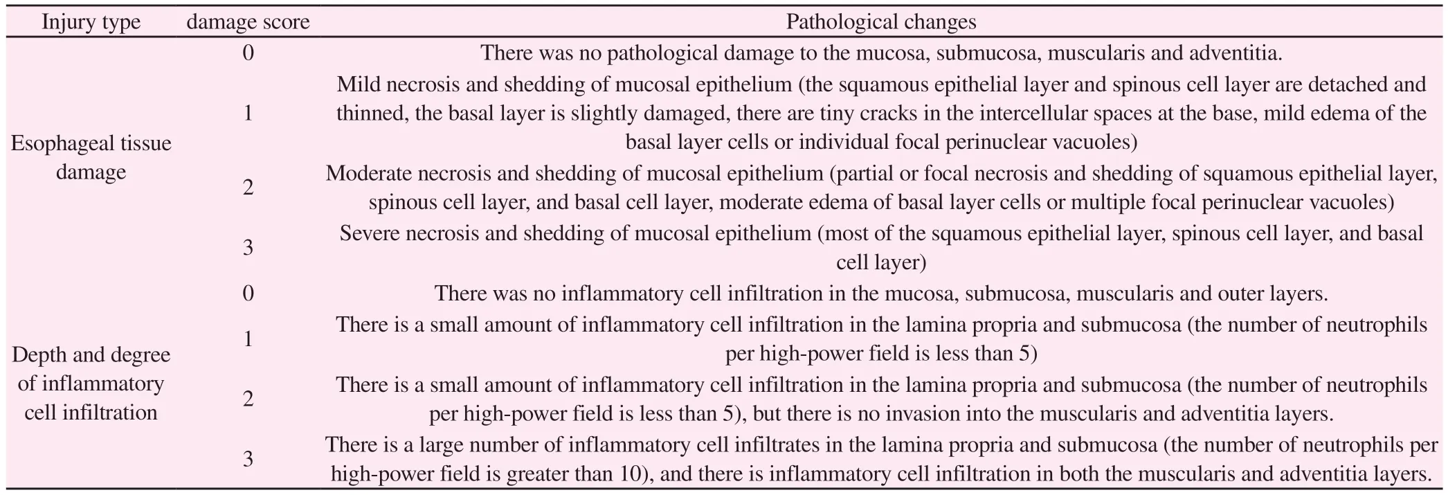
Tab 1 Scoring Table for the Degree of Esophageal Pathological Injury
Western Blotting was used to detect the expression of NF-κB protein in the esophageal tissue of each group.Take the frozen esophageal tissue, add the prepared lysis solution, lyse it on ice for 45 minutes, centrifuge at 4 ℃, 12 000r/min for 15 min, and extract.Supernatant, BCA protein quantification analysis.Prepare the gel,load the sample, and determine the electrophoresis stop time based on the pre-stained marker and NF-κB protein molecular weight.Transfer the membrane with PVDF for 90 min under constant voltage electrophoresis at 100 V, block with 5% skim milk at room temperature for 2 h, add primary antibody NF-κB, and incubate at 4 ℃ overnight.Wash 3 times with TBST, 15 min each time,add secondary antibody and incubate for 2 h, wash 3 times with TBST, 15 min each time.The ECL luminescent solution was used for exposure by chemiluminescence chromogenic method, the gel imaging analysis system was used to detect protein bands, and the software Image J was used to calculate the gray value.
2.7 Statistical methods
The data obtained from the experiment were analyzed and processed using SPSS 25.0 statistical software.After testing, all data conformed to the normal distribution and had homogeneous variances, and were expressed as mean ± standard deviation.The experimental results were compared among multiple groups using one-way analysis of variance, and the LSD test was used for pairwise comparison between groups.P<0.05 was considered as a statistically significant difference.
3.Results
3.1 Changes in body weight and food intake of rats in each group
Judging from the changes in the weight of the rats: the weight of the rats in each group was weighed and recorded every day.The weights of the rats in each group were close before modeling.The rats in the CONTROL group were not irradiated.The weight increased day by day since Day 0, and the weight on the 7th day was (284.3± 6.5) g;the weight of rats in other groups decreased day by day since D0.The weight of the MODEL group on the 7th day was 175.5±4.3 g,which was significantly lower than that of the CONTROL group.The weight of rats in other intervention groups all decreased, but compared with that of the MODEL group.Ratio, the difference was not statistically significant (P>0.05).The body weight of rats in each group before and after intervention is shown in Table 2.The daily weight change curve of rats in each group is shown in Figure 1.
Tab 2 Body weight of rats in each group before and after intervention (unit: g, ±s)
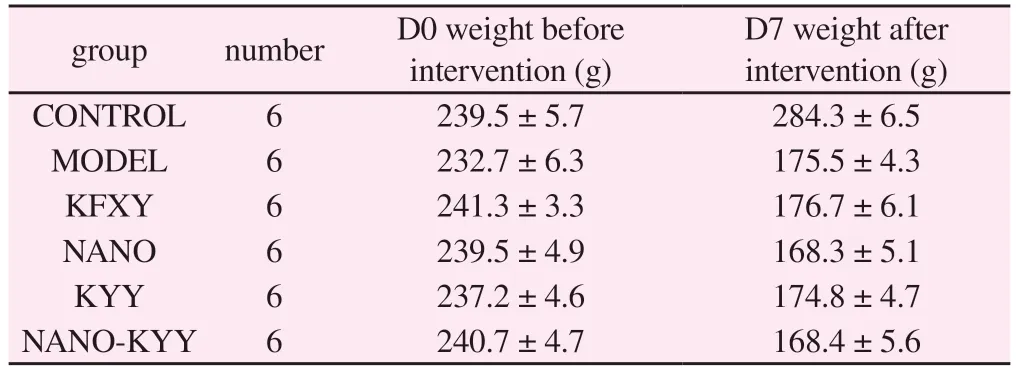
Tab 2 Body weight of rats in each group before and after intervention (unit: g, ±s)
D7 weight after intervention (g)CONTROL 6 239.5 ± 5.7 284.3 ± 6.5 MODEL 6 232.7 ± 6.3 175.5 ± 4.3 KFXY 6 241.3 ± 3.3 176.7 ± 6.1 NANO 6 239.5 ± 4.9 168.3 ± 5.1 KYY 6 237.2 ± 4.6 174.8 ± 4.7 NANO-KYY 6 240.7 ± 4.7 168.4 ± 5.6 group number D0 weight before intervention (g)
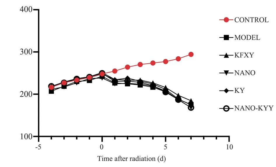
Fig 1 Body weight change curve of rats in each group
Judging from the food intake of the rats: the food intake of the rats in each group before modeling was similar.The rats in the CONTROL group were not irradiated and continued to eat normally since D0.The total food intake in the next 7 d was (192.8±9.4) g;the rats in the other 7 groups had a normal food intake since D0.The food intake from D0 was significantly lower than that before irradiation.The total food intake within 7 d of the MODEL group was (39.3±6.2)g.Compared with the normal group, the weight was significantly lower.The difference was statistically significant.The total food intake of the NANO-KYY group (NANO-KYY) within 7 d was (39.3±6.2)g.The amount was (64.2±4.5) g, which was higher than that of the MODEL group and other intervention groups,and the difference was statistically significant (t=12.65, P<0.05);compared with the MODEL group, the difference between the other intervention groups was not statistically significant (P>0.05 ).The specific values and histograms are shown in Table 3 and Figure 2 below:
Tab 3 Total food intake of rats in each group within 7 days after intervention (unit:±s)

Tab 3 Total food intake of rats in each group within 7 days after intervention (unit:±s)
group number Total food intake 7 days after modeling CONTROL 6 192.8 ± 9.4 MODEL 6 39.3 ± 6.2 KFXY 6 46.0 ± 7.4 NANO 6 42.5 ± 5.9 KYY 6 43.3 ± 7.6 NANO-KYY 6 64.2 ± 4.5
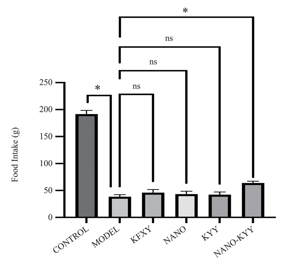
Fig2 Comparison of total food intake within 7 days of prognosis of rats in each group
3.2 Comparison of pathological damage scores of esophageal tissue in rats in each group
The HE staining results of the esophageal tissues of rats in each group are shown in Figure 3.The structure of the esophageal mucosa of rats in the normal group was intact, with no pathological damage to the mucosal layer, submucosal layer, muscular layer and adventitia layer.The basal layer was tightly arranged, and the multi-layer spinous cell layer and granular layer outside the basal layer were densely arranged.The stratum corneum was intact.Exfoliated necrotic cells were seen, with no obvious inflammatory cell infiltration.Compared with the other groups, various degrees of esophageal tissue damage and inflammatory cell infiltration were observed.Partial mucosal loss was seen, mucosal basal cells were arranged in disorder, the spinous cell layer was atrophied, thinned and arranged in disorder, keratinocytes were obviously shed, and a large number of lymphocytes and Granulocytic infiltration.The results of calculating the pathological change scores of rats in each group according to the pathological damage scoring method of radiation-induced esophagitis in rats are shown in Table 4 and Figure 4.No obvious esophageal damage was found in the non-irradiated rats, and the pathological score was 0.33±0.52.After irradiation, the esophageal tissue of the rats in each group was severely damaged,and the pathological scores were significantly higher than those in the CONTROL group; except for the CONTROL group, the nanoulcer solution group (NANO-KYY) had larger The pathological damage score of the mouse esophagus was 2.17±0.75, which was significantly lower than that of other groups, and the difference was statistically significant (t=3.523, P<0.05); there was no statistical difference between the other groups (P>0.05).
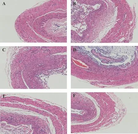
Fig 3 HE staining of rat esophageal tissue in each group (A control; B model; C KFXY; D Nano; E KYY; F Nano-KYY Magnification: ×100)
Tab 4 Pathological damage scores of esophageal tissue in rats in each group ( ±s )

Tab 4 Pathological damage scores of esophageal tissue in rats in each group ( ±s )
group number Pathological damage score CONTROL 6 0.33 ± 0.52 MODEL 6 3.67 ± 1.37 KFXY 6 3.83 ± 0.98 NANO 6 3.50 ± 0.84 KYY 6 4.00 ± 1.41 NANO-KYY 6 2.17 ± 0.75
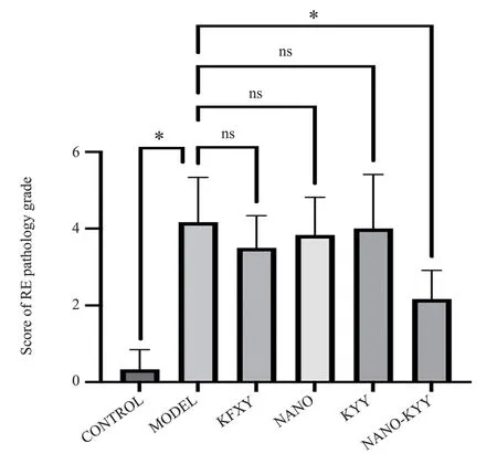
Fig 4 Comparison of pathological damage scores of esophageal tissue in rats in each group
3.3 Secretion of pain-related factors in rats in each group
The ELISA results showed that the expression levels of pain-related factors prostaglandin-2 (PGE-2), substance P (SP), and calcitonin gene-related peptide (CGRP) in the serum of rats after radiation irradiation were (437.2±52.21) ng/L, respectively.(5.84 ± 0.41) ng/L,(29.53 ± 3.91) ng/L, which were significantly higher than the normal group.Simply giving nano-hydralcite matrix could not reduce the level of pain factors.Kangfuxin Liquid, traditional Chinese medicine ulcer liquid, and nano-ulcer liquid could all reduce the levels of three pain factors.The expression of pain factors, PGE-2, SP, and CGRP after nano-ulcer solution intervention were (298.1 ± 71.74) ng/L,(3.23 ± 0.74) ng/L, and (14.62 ± 3.80) ng/L respectively.Among them, CGRP decreased the most, and the difference was statistically significant.Significance (t=8.914, P<0.05).The ELISA results of various pain factors are shown in Table 5 and Figure 5 below:
Tab 5 ELISA test result statistics table (unit: ng/L, ±s)

Tab 5 ELISA test result statistics table (unit: ng/L, ±s)
group number PGE-2 SP CGRP CONTROL 6 241.6 ± 61.87 2.77 ± 0.32 14.67 ± 4.50 MODEL 6 437.2 ± 52.21 5.84 ± 0.41 29.53 ± 3.91 KFXY 6 319.1 ± 78.64 4.20 ± 0.89 19.83 ± 4.17 NANO 6 372.4 ± 76.18 5.30 ± 0.74 24.12 ± 3.79 KYY 6 239.2 ± 39.04 4.44 ± 0.70 20.18 ± 4.08 NANO-KYY 6 298.1 ± 71.74 3.23 ± 0.74 14.62 ± 3.80
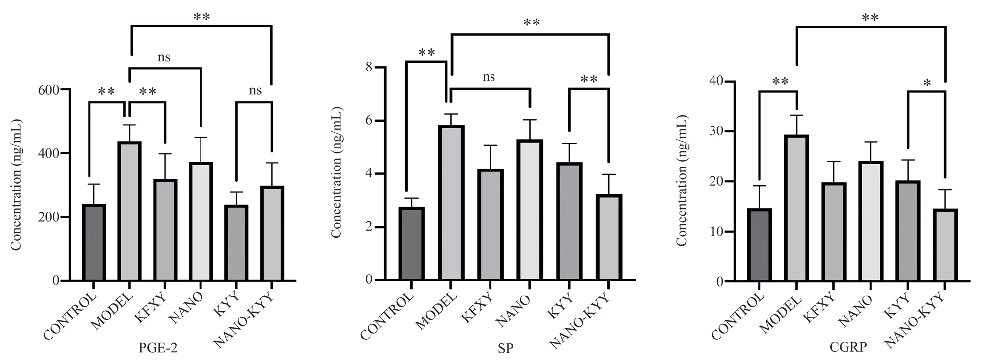
Fig 5 Expression levels of PGE-2, SP, and CGRP
3.4 Expression of inflammation-related proteins in rats in each group
Western Blot results showed that the expression levels of the internal reference GAPDH were consistent in each group, and the WB results were comparable.The Western Blot results of various proteins are shown in Figure 6 below.The relative gray value statistics are shown in Table 6.The histogram analysis statistics are shown in Figure 7.They are as follows.The results showed that radiation exposure will increase the expression of inflammationrelated protein NF-κB in esophageal tissue.The relative gray value of NF-κB protein expression in the MODEL group was 0.46 ±0.08, and the difference was statistically significant compared with CONTROL (t= 6.708, P<0.05); Kangfuxin Liquid, Traditional Chinese Medicine Ulcer Liquid, and Nano Ulcer Liquid can all reduce the expression of NF-κB.After the intervention of Nano Ulcer Liquid, it decreased to 0.10 ± 0.04, and the difference was statistically significant compared with the MODEL group (t =5.369,P<0.05).
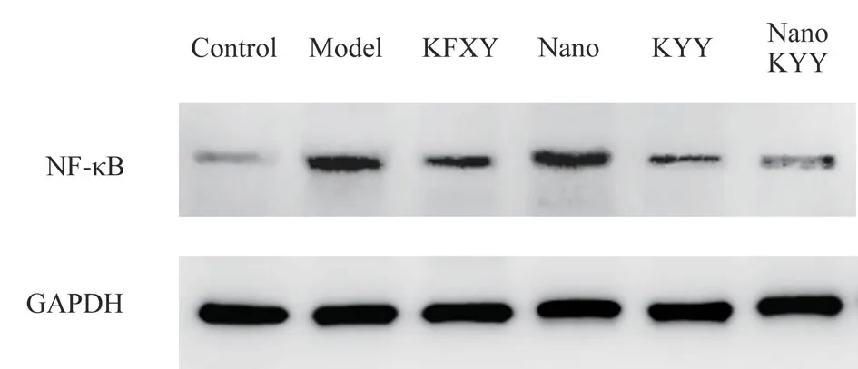
Fig 6 NF-κB protein expression band display

Tab 6 NF-κB protein expression (grayscale relative value)
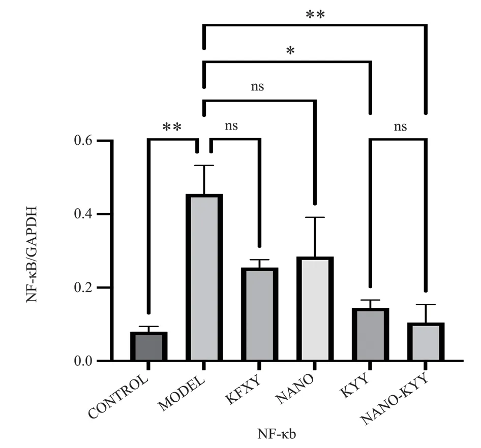
Fig 7 NF-κB protein expression
4.Discussion
Radiation esophagitis is a common complication of radiotherapy for cervical and thoracic tumors.The main clinical manifestations include pain in swallowing, dysphagia, acid regurgitation, and retrosternal burning sensation.In severe cases, esophageal mucosal bleeding, esophageal perforation, esophageal tracheal fistula, and other emergency and severe symptoms may occur.[7], not only brings great pain to patients, but some patients are even forced to interrupt the radiotherapy process due to severe esophagitis, limiting the efficacy of radiotherapy[8].Therefore, early intervention in radiation esophagitis can relieve patients’ swallowing pain, improve nutritional status, ensure the successful completion of radiotherapy plans, improve tumor control rates, and improve patients’ longterm survival rates.Currently, there is a lack of evidence-based treatment methods for the treatment of radiation esophagitis, and the effectiveness of modern medicine in treating radiation esophagitis is limited.
Ulcer fluid is an in-hospital preparation developed by the Department of Oncology of Integrated Traditional Chinese and Western Medicine at the China-Japan Friendship Hospital.It is an empirical medicine for the treatment of acute radiation injury.It can treat radiation-induced skin and mucous membranes such as radiation dermatitis[9], radiation stomatitis[10], and radiation esophagitis[2].Injury diseases have significant effects.The ulcer fluid is composed of raw rhubarb, angelica angelica, angelica root, safflower, lithospermum and other medicines.Raw rhubarb can purge fire and detoxify, cool blood and remove blood stasis;Lithospermum can cool blood and activate blood stasis, clear away heat and toxins in the blood; Safflower can remove blood stasis,activate blood circulation and moisturize blood; Angelica sinensis can activate blood circulation and nourish blood, reduce swelling,relieve pain, and eliminate phlegm.The effect of pus and muscle growth; Angelica dahurica can cure sores, swelling and pain, reduce swelling and drain pus, support sores, and promote muscle growth.All medicines have warm and cool flavors when used together.They can clear away heat, cool blood and detoxify without cooling.They can activate blood circulation, remove blood stasis and relieve pain without drying heat.They can also reduce swelling and pain,discharge pus and promote muscle growth, and together they can detoxify and remove blood stasis.Traditional Chinese medicine ulcer liquid prepared based on the theory of external treatment of traditional Chinese medicine mainly works through skin and mucous membrane absorption.It has achieved significant clinical efficacy in treating radiation dermatitis and stomatitis, but its efficacy in treating radiation esophagitis still needs to be improved.The main reason for this difference may be that the unique anatomical structure and physiological characteristics of the esophagus make it difficult for drugs to adhere to the surface of the esophagus,affecting drug absorption and utilization.In order to solve this problem and improve the adhesion and absorption and utilization of traditional Chinese medicine ulcer liquid, we plan to nano-modify the ulcer liquid.Nanohydrotalcite (nano-LDH) was researched and developed by the State Key Laboratory of Effective Utilization of Chemical Resources, School of Chemistry, Beijing University of Chemical Technology[11].nano-LDH is also called nanoscale layered composite hydroxide.In recent years, the application of nano-LDH in the field of biomedicine has aroused great interest.The synthesis method of nano-LDH materials is simple and relatively low-cost,with high drug loading efficiency and high drug cell membrane permeability[3].Currently, nano-LDH is widely used as a new drug carrier in the fields of antibacterial, anti-tumor and bioimaging.nano-LDH has good biocompatibility, is easily taken up by cells,and is easily degraded under acidic conditions.Based on the material advantages of nano-hydrtalcite, we chose nano-hydrtalcite as a carrier to prepare nano-ulcer fluid.
The inflammatory mechanism of radiation-induced esophageal injury can be divided into direct injury and indirect injury.Direct damage means that radiation acts directly on cellular DNA, causing DNA breakage.Indirect damage refers to the active products indirectly formed by the action of radiation on water, that is, reactive oxygen species (ROS) with high activity and strong oxidizing ability.ROS can serve as second messengers to activate multiple signaling pathways and transcription factors in cells.NF-κB plays an important role in ROS-mediated signaling.NF-κB, as an important pleiotropic nuclear transcription factor, can regulate the expression of a variety of genes[12].When NF-κB is stimulated by ROS, it causes NF-κB to dissociate and transfer to the nucleus in an active form, initiating and regulating the transcription of various inflammatory factors such as tumor necrosis factor (TNF-α)and interleukin 6 (IL-6).[13].Radiation esophagitis is usually accompanied by severe pain on swallowing or retrosternal burning sensation, and is associated with elevated levels of prostaglandin-2(PGE-2), substance P (SP), calcitonin gene-related peptide (CGRP)and other pain factors.Related.In this study, we first conducted a pharmacodynamic study on the nano-ulcer solution, and used NFκB as an inflammation detection indicator; PGE-2, SP, and CGRP as pain-related indicators to study the mechanism.
The results of this study show that after the intervention of nanoulcer solution, the food intake of rats with radiation esophagitis was higher than that of other groups, and the esophageal pathological damage score was lower than that of other groups, indicating that the esophageal inflammation, dysphagia and pain in swallowing were lower in rats in the NANO-KYY group.The other modeling groups are lighter, and the nano-ulcer liquid is more effective in treating radiation esophagitis, which is better than the traditional Chinese medicine ulcer liquid and the commonly used clinical drug for treating radiation damage, Kangfuxin Liquid.However, the food intake and esophageal damage of rats in the NANO group were not significantly different from those in the MODEL group, indicating that the simple nanohydrtalcite matrix has no obvious therapeutic effect on radiation-induced esophagitis in rats.Only when combined with traditional Chinese medicine ulcer liquid to prepare nano-ulcer liquid, the adhesion is enhanced, and the drug Only when the active ingredients stay on the surface of the esophagus for a longer time can the efficacy be improved, reduce inflammation and pain in the esophagus of rats, and promote eating in rats.WB results showed that the NF-κB level in rats increased significantly after modeling,and the NF-κB level decreased significantly after nano-ulcer solution intervention.As an important molecule in the inflammatory response, NF-κB can regulate various inflammatory factors such as TNF-α and IL-6.The activation of NF-κB is the core step of the inflammatory pathway, and the inhibition of NF-κB levels by nano-ulcer fluid also proves for its anti-inflammatory effects.The occurrence of pain symptoms is accompanied by elevated levels of a variety of pain factors.PGE-2 plays an important role in the generation and development of inflammatory pain and hyperalgesia.It can stimulate the peripheral nerves that grow into the esophageal tissue, thereby producing pain[14].Studies have shown that genetic knockout of PGE-2 receptors can significantly reduce acute herpes zoster pain[15]; SP is a polypeptide widely distributed in fine nerve fibers involved in pain transmission, and interacts with neurokinin-1(NK-1) receptor binding to exert physiological effects.By directly or indirectly promoting the release of glutamate, etc., SP participates in pain transmission.After pain occurs, the release of enkephalin plays an analgesic effect[16].SP is a key factor in pain transmission and can be activated to participate in pain transmission to produce pain sensation.Its level is positively related to the degree of pain[17];CGRP is a bioactive peptide released from sensory nerve endings[18] and is widely distributed in the digestive intestines, heart and The vascular system, central and peripheral nervous systems play an important role in the transmission of pain sensation and can promote the occurrence of pain by participating in pain information transmission and central sensitization, mediating neurogenic inflammation and regulating inflammatory responses, and activating glial cells[19].ELISA results showed that after radiation esophagitis modeling, the pain factors of model rats were significantly higher than those of normal rats.Given different intervention methods,pain factors showed different changing trends.The NANO group only used nano-hydrtalcite matrix intervention, and the drug did not contain traditional Chinese medicine ingredients.The levels of the three pain factors were close to those of the MODEL group, so the nano-hydrtalcite ingredients had no therapeutic effect on pain.Kangfuxin Liquid, traditional Chinese medicine ulcer liquid, and nano-ulcer liquid all showed significant inhibitory effects on the expression of three pain factors.The difference in the decrease in PGE-2, SP, and CGRP after nano-ulcer liquid intervention was most obvious compared with the MODEL group.
In summary, nano-ulcer fluid has a therapeutic effect on rats with radiation-induced esophagitis.It can increase the food intake of rats with radiation-induced esophagitis, reduce esophageal tissue damage in rats, and reduce serum pain factor concentrations.The anti-inflammatory effect of nano-ulcer fluid may be related to the reduction of NF-κB inflammatory factor levels are related.
This experiment selected representative inflammatory responserelated factors for detection.In addition, tissue repair is also an important pathological change in radiation-induced esophagitis.Epidermal growth factor and transforming growth factor can be detected in the esophageal tissue of rats with radiation-induced esophagitis in further experiments.and protein expression levels.
Authors’ Contribution
Li Kaixuan: participated in the entire process and wrote the paper;Wang Qin: raised animals, assisted in completing experiments, and participated in writing the paper; Zheng Jiabin: guided experimental operations and paper writing; Teng Feng: guided animal radiation modeling; Jia Liqun: project design, guiding experiments and paper writing.
All authors declare no conflicts of interest.
 Journal of Hainan Medical College2023年16期
- Journal of Hainan Medical College的其它文章
- TCM Intervention on Wnt/β-Catenin signaling pathway in the treatment of diabetic nephropathy
- Research progress on the correlation between intestinal microflora disorder and liver disease
- Research progress on the effects of phthalates on reproductive health of childbearing population and their offspring
- Construction and validation of prognostic model of Cuproptosis-related LncRNA in osteosarcoma
- Effect of glycyrrhetinic acid on Th1/Th2 balance in cough variant asthma mice
- Efficacy and safety of oral Chinese patent medicine combined with sacubitril/valsartan in the treatment of chronic heart failure: A Metaanalysis
