Exploration of the molecular mechanism of Qishen decoction in regulating miR-495/FTO pathway mediated macrophage polarization to improve insulin resistance therapy of type 2 diabetes
SUN Zhi-dong, GAO Jia-wei, YANG Liu-xin, ZHANG Ya-li,3, YUAN Xing-xing,✉
1.Heilongjiang Academy of Traditional Chinese Medicine, Harbin 150006, China
2.Heilongjiang University of Traditional Chinese Medicine, Harbin 150040, China
3.Zhang Yali Minglao Traditional Chinese Medicine Studio, Harbin 150006, China
Keywords:
ABSTRACT Objective: To observe the effect of Qishen decoction on macrophage polarization mediated by miR-495/FTO signaling pathway, and to clarify the molecular mechanism of Qishen decoction in improving insulin resistance in the treatment of type 2 diabetes.Methods: THP-1 was induced to differentiate macrophages with phorbol ester.It was divided into the control group,the model group, the Qishen decoction group, the miR-495 inhibitor group, and the Qishen decoction+miR-495 inhibitor group.Except for the control group, the remaining groups were stimulated with 30 mmol/L glucose to construct a macrophage polarization model, and corresponding drugs were given for intervention.Cells were collected from each group for 24 hours and the content of inflammatory factors (IL-6, IL-1β, IL-4, and IL-10) were detected using enzyme-linked immunosorbent assay.The expression of macrophage polarization marker molecules, miR-495, and FTO were detected by flow cytometry, qPCR, and Western blot to detect.Results: Compared with the control group, there was no significant change in the activity of macrophages in the control serum, Qishen decoction containing serum, and miR-495 inhibitor transfected serum, and the difference was not statistically significant (P>0.05).In addition, compared to the control group, the content of IL-6 and IL-1β, the expression levels of CD68, iNOS, COX-2, miR-495, and the ratio of CD68/CD206, were significantly increased (P<0.01).While the content of IL-4 and IL-10, as well as the expression of CD206,Arg-1, YM-1, and FTO were significantly reduced (P<0.01).Compared with the model group, the QiShen decoction significantly reduced the contents of IL-6 and IL-1β, and the expression levels of CD68, iNOS, COX-2, and miR-495, as well as the ratio of CD68/CD206, while the content of IL-4 and IL-10, as well as the expression of CD206, Arg-1,YM-1, and FTO were significantly increased (P<0.01).Conclusion: Qishen decoction upregulate the expression of FTO to promote M2 type polarization of macrophages, thereby inhibiting inflammation and improving insulin resistance by inhibiting the expression of miR-495.
1.Introduction
Type 2 diabetes (T2DM) is a metabolic disease caused by environmental and genetic factors, which is characterized by relatively reduced insulin secretion and insulin resistance[1].Over the past 30 years, the prevalence of diabetes in China has increased significantly, which has become one of the serious public health problems.T2DM accounts for more than 90% of diabetes in China,which not only brings great harm to patients, but also brings huge economic burden to society[2].
Macrophages are important innate immune cells of the human body.Their functions change when the microenvironment of the body changes, including classic activated M1 type and alternate activated M2 type.Macrophage polarization plays an important role in the occurrence and development of metabolic diseases including obesity, T2DM, non-alcoholic fatty liver disease, and gout[3].Research has confirmed that the role of inflammatory infiltration composed of adipose macrophages in the pathological process of inducing insulin resistance - T2DM cannot be ignored[4].Therefore, regulating the inflammatory microenvironment mediated by macrophage polarization may be an important way to improve insulin resistance.Qishen decoction has been proven to improve insulin resistance induced by a high-fat diet, thereby promoting glucose and lipid metabolism and reducing body mass.However,the specific mechanism is still unclear[5,6].This study induced differentiation of macrophages in THP-1 human monocyte line using phorbol ester, and constructed a macrophage polarization model using high glucose stimulation.By observing the effect of Qishen decoction on macrophage polarization mediated by miR-495/FTO signaling pathway, the molecular mechanism of Qishen decoction improving insulin resistance in the treatment of T2DM was elucidated.
2.Materials and methods
2.1 Experimental animals and cells
20 healthy and clean grade SD rats (male, 6 weeks old, 201~225 grams) were purchased from Beijing Weitong Lihua Co., Ltd.,with an experimental animal production license of SCXK (Beijing)2021-0011.Animals are raised at the Animal Experiment Center of Heilongjiang University of Traditional Chinese Medicine, with a room temperature of 20~25 ℃, circulating light, and free drinking water.The THP-1 (human monocytic leukemia) cell line was purchased from Wuhan Punosai Life Technology Co., Ltd.
2.2 Drugs and reagents
Qishen decoction (15 g raw Astragalus membranaceus, 10 g American ginseng, 10 g Alisma orientalis, 10 g lotus leaf, 10 g raw hawthorn, 10 g salvia miltiorrhiza, 8 g ligustrum lucidum, 8 g Eclipta grandiflora, 5 g Gynostemma pentaphyllum, 5 g Panax notoginseng, and 3 g raw licorice) was purchased from the Chinese Herbal Medicine Bureau of Heilongjiang Academy of Traditional Chinese Medicine.The medicine was boiled and filtered to form a 1 g/mL concentrated solution for future use.Fobo ester was purchased from Biosharp Company in China (product number: BL1127A);RPMI-1640 culture medium and high sugar culture medium were purchased from Wuhan Punosai Life Technology Co., Ltd.(product numbers PM150110B and PM150210B respectively).Both CD86 and CD206 antibodies were purchased from Abcam Company in the UK (ab239075 and ab270647); FTO antibody β- The Actin first antibody, HRP labeled second antibody, ECL chemiluminescence kit, and BCA protein detection kit were purchased from CST Corporation in the United States (product numbers # 31687, # 93473,# 7074, # 6883, and # 7780, respectively).Interleukin-4, IL-6, IL-1β, and IL-10 detection kit were purchased from Jiangsu Enzyme Immunoassay Industry Co., Ltd.(product numbers MM-0051H2,MM-0049H2, MM-0181H2, and MM-0066H2 respectively).Lipo6000 transfection kit was purchased from Shanghai Biyuntian Biotechnology (product number: C0526-10ml); The qPCR kit was purchased from Roche Corporation in the United States (product number: KK4610).
2.3 Methods
2.3.1 Preparation of medicated serum
After one week of adaptive feeding, rats were randomly divided into a control group and model group, with 10 rats in each group.Referring to the previous research method[7], the Qishen decoction group was treated with Qishen decoction concentrated solution(18.8 g/kg) by gavage, while the control group was treated with an equal volume of physiological saline by gavage, once a day for 7 consecutive days.After the last treatment, anesthetized rats were injected intraperitoneally, and blood was collected from the abdominal aorta.Collect serum after 10 min of heart separation at 3 500 rpm, inactivate it in a water bath for 30 min, and filter it with a membrane for later use.
THP-1 was cultured in RPMI-1640 medium (containing 10% FBS,1% penicillin streptomycin double antibody and β-Mercaptoethanol was used for culture, and the cells were prepared in a 37 ℃-incubator containing 5%CO2.The medium was changed once every 2~3 d.When the cells are in the logarithmic growth phase,they are inoculated onto a 6-well plate (1×10⁶/mL).Adding phorbol ester (160 nmol/L) to cells induces THP-1 to differentiate into macrophages.When the cells transition from a suspended state to an adherent state, it indicates successful cell differentiation.
2.3.3 Cell grouping and drug intervention
Divide the differentiated macrophages into 2.5×10⁵/mL density onto 6-well plates, with 900 per well μL.Group according to the plan and provide corresponding drugs for intervention.①Control group: THP-1+normal culture medium (5.6 mmol/L glucose)+100 μL normal serum; ②Model group: THP-1+high glucose medium (30 mmol/L glucose)+100 μL normal serum; ③Qishen decoction group:THP-1+high sugar medium (30 mmol/L glucose)+100 μL medicated serum; ④ MiR-495 inhibitor group: THP-1+high glucose medium(30 mmol/L glucose)+100 μL normal serum +miR-495 inhibitor;⑤ Qishen decoction+miR-495 inhibitor group: THP-1+high sugar medium (30 mmol/L glucose)+100 μL-containing serum+miR-495 inhibitor.Among them, miR-495 inhibitor was synthesized by Shanghai Jima Pharmaceutical Technology Co., Ltd.According to the Lipo6000 kit instructions, miR-495 inhibitor with a final concentration of 200 nmol/L was transfected into macrophages.
2.3.4 CCK8 assay
Divide the differentiated macrophages into 2.5×10⁵/mL density on a 6-well plate, group according to the protocol, and provide corresponding drugs for intervention to detect the toxicity of Qishen decoction containing serum and miR-495 inhibitor transfection to macrophages.① Control group: THP-1+normal culture medium(5.6 mmol/L glucose); ② Control serum group: THP-1+normal culture medium (5.6 mmol/L glucose)+100 μL blank serum; ③Medicated serum group: THP-1+normal culture medium (5.6 mmol/L glucose)+100 μL medicated serum; ④ MiR-495 inhibitor group:THP-1+normal culture medium (5.6 mmol/L glucose)+miR-495 inhibitor.Add 10 without holes μL of CCK8 solution was incubated at 37 ℃ for 4 hours, and the absorbance values (A450) of each well were measured using an enzyme-linked immunosorbent assay(ELISA).
2.3.5 ELISA
Collect each group of macrophage culture medium in 1.3.3 after 48 hours, and use an ELISA kit to detect IL-6, IL-1β, IL-4, and IL-10 in the supernatant of each group of culture medium under the guidance of the kit manual.
2.3.6 Flow cytometry
Turning to ask the Beast what it could all mean, Beauty found that he had disappeared, and in his place stood her long-loved Prince! At the same moment the wheels of a chariot were heard upon the terrace, and two ladies entered the room
Collect macrophages from each group in 1.3.3 after 48 h,centrifuge at 1 000 rpm for 5 min, discard the supernatant, add PBS for resuspension, centrifuge at 1 000 rpm for 5 min, and wash PBS twice before dividing into two groups.Join a group of 200 μL PBS was resuspended and incubated with FITC labeled CD86 antibody;Add another portion to 200 μL After resuspension of PBS, add 1.5 mL of membrane breaker and incubate with FITC labeled CD206 antibody.After incubation for 30 min, the positive expression rates of CD86 and CD206 were detected on the machine after washing with PBS and resuspension.
2.3.7 qPCR
Collect macrophages from each group in 1.3.3 after 48 h, extract total RNA from cells using TRIzol method, and then reverse transcript to synthesize cDNA.Quantitative analysis of mRNA was performed using the qPCR kit method.The primer sequence is as follows: miR-495: F: 5’-GCGAAAAAACATGGTGC-3’,R: 5’-GCAGGGGTCCGAGGTATC-3’; U6: F: 5’-CTCGCTT CGGCAGCACA-3’, R: 5’-AACGCTTCACGAATTTGCGT-3’;FTO: 5’-TTCATGCTGGATGA CCTCAATG-3’, R: 5’-G C C A A C T G A C A G C G T T C TA A G-3’; G A P D H:F: 5’-G C AC C G T C A A G G C T G AG A AC-3’, R: 5’-TGGTGAAGACGCCAGTGGA-3’.The reaction conditions for qPCR are as follows: pre denaturation at 95 ℃ for 3 min; Degenerate at 95 ℃ for 20 seconds, denature at 58 ℃ for 30 seconds, extend at 72 ℃ for 45 seconds, and after 40 cycles, extend at 95 ℃ for 15 seconds, maintaining at 4 ℃.MiR-495 uses U6 as the internal reference, FTO uses GAPDH as the internal reference, and uses 2-ΔΔCtCalculate the relative expression of miR-495 and FTO.
2.3.8 Western blot
Collect macrophages from each group in 1.3.3 after 48 h, extract total protein, and detect protein concentration using BCA reagent kit.After sample loading, electrophoresis, and membrane transfer,seal with skimmed milk at room temperature.Join FTO (1:1 000)and β- actin (1:1 000) primary antibody was incubated overnight at 4 °C, followed by the addition of secondary antibody and continued incubation at room temperature for 2 h.After TBST film washing,ECL was added for development, and the relative expression level of FTO was analyzed after taking photos with a chemiluminescence detection system.
2.3.9 Statistical processing
SPSS 26.0 software was used for data analysis, ANOVA was used for data between multiple groups, and LSD test was used for pairwise comparison between groups.The difference represented by P<0.05 is statistically significant.
3.Results
3.1 Effect of Qishen decoction on Macrophage Activity
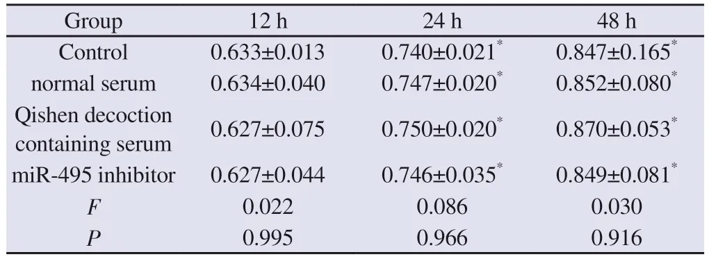
In order to clarify the toxicity of normal serum, Qishen decoction containing serum, and miR-495 inhibitor transfection on macrophages, this study used the CCK8 method to detect the activity of macrophages.Compared with 12 h in this group, the activity of cells significantly increased at 24 and 48 hours, and the difference was statistically significant (P<0.05).There was no statistically significant difference (P>0.05) between the groups during each time.Compared with the control group, there was no significant change in macrophage activity in the blank serum, Qishen decoction containing serum, and miR-495 inhibitor transfected macrophages, and the difference was not statistically significant (P>0.05).See Table 1
Tab 1 Effect of Qishen decoction on macrophage activity(n=3,±s , OD)

Tab 1 Effect of Qishen decoction on macrophage activity(n=3,±s , OD)
Note: Compared with this group for 12 h, *P<0.05.
Group 12 h 24 h 48 h Control 0.633±0.013 0.740±0.021* 0.847±0.165*normal serum 0.634±0.040 0.747±0.020* 0.852±0.080*Qishen decoction containing serum 0.627±0.075 0.750±0.020* 0.870±0.053*miR-495 inhibitor 0.627±0.044 0.746±0.035* 0.849±0.081*F 0.022 0.086 0.030 P 0.995 0.966 0.916
3.2 Effect of Qishen decoction on Inflammatory Factors in Macrophages
Compared with the control group, the content of IL-6 and IL-1β significantly increased in the model group, while the content of IL-4 and IL-10 significantly decreased, with statistically significant differences (P<0.01).Compared with the model group, the content of IL-6 and IL-1β in the supernatant of the Qishen decoction group,miR-495 inhibitor group, and Qishen decoction +miR-495 inhibitor group significantly decreased, and the differences were statistically significant (P<0.01).Compared with the model group, the content of IL-4 and IL-10 in the supernatant of the Qishen decoction group,miR-495 inhibitor group, and Qi Shen Tang+miR-495 inhibitor group significantly increased, and the differences were statistically significant (P<0.05 andP<0.01).See Table 2
Tab 2 Effect of Qishen decoction on inflammatory factors in macrophages(n=3,±s , pg/mL)

Tab 2 Effect of Qishen decoction on inflammatory factors in macrophages(n=3,±s , pg/mL)
Note: Compared with the control group, *P<0.05, **P<0.01.Compared with the model group, ○P<0.05, ○○P<0.01.
Group IL-6 IL-1β IL-4 IL-10 Control 7.93±0.78 18.04±3.35 23.23±0.93 70.07±8.98 Model 24.58±2.15** 57.66±9.69** 8.91±1.26** 28.43±4.19**Qishen decoction 16.48±2.88○○ 40.25±5.63○○ 13.69±1.87○○ 40.12±1.30○○miR-495 inhibitor 17.99±3.29○○ 39.25±6.81○○ 13.10±1.38○ 40.22±2.06○Qishen decoction +miR-495 inhibitor 9.78±1.37○○ 22.67±1.83○○ 19.05±2.63○○ 52.60±2.48○○F 25.662 20.051 31.628 33.981 P<0.001 <0.001 <0.001 <0.001
3.3 Effect of Qishen decoction on Macrophage Phenotype
Compared with the control group, the expression of macrophage CD68 and the ratio of CD68/CD206 in the model group were significantly increased, while the expression of CD206 was significantly reduced, with statistical significance (P<0.01).Compared with the model group, the expression of CD68 and the ratio of CD68/CD206 in macrophages in the Qishen decoction group, miR-495 inhibitor group, and Qishen decoction+miR-495 inhibitor group were significantly reduced, while the expression of CD206 was significantly increased, with statistical significance(P<0.01).See Table 3 and Figure 1
Tab 3 Effect of Qishen decoction on macrophage phenotype(n=3, ±s)

Tab 3 Effect of Qishen decoction on macrophage phenotype(n=3, ±s)
Note: Compared with the control group, **P<0.01.Compared with the model group,○○P<0.01.
Group CD68(%) CD206(%) CD68/CD206 Control 21.26±1.38 24.01±3.16 0.90±0.16 Model 53.53±3.05** 11.20±1.69** 4.84±0.46**Qishen decoction 38.05±3.14○○ 19.64±4.15○○ 2.01±0.52○○miR-495 inhibitor 37.61±5.73○○ 20.85±3.24○○ 1.81±0.27○○Qishen decoction +miR-495 inhibitor 30.49±2.05○○ 29.08±1.98○○ 1.05±0.06○○F 36.219 14.535 65.063 P<0.001 <0.001 <0.001
3.4 Effect of Qishen decoction on the Expression of Macrophage Marker Molecules
Compared with the control group, the expression of iNOS and COX-2 in macrophages of the model group was significantly increased, while the expression of Arg-1 and YM-1 was significantly reduced, with statistical significance (P<0.01).Compared with the model group, the expression of iNOS and COX-2 in macrophages in the Qishen decoction group, miR-495 inhibitor group, and Qishen decoction+miR-495 inhibitor group were significantly reduced, while the expression of Arg-1 and YM-1 were significantly increased, with statistical significance (P<0.01).See Table 4.
3.5 Effect of Qishen decoction on the Expression of MiR-495 and FTO
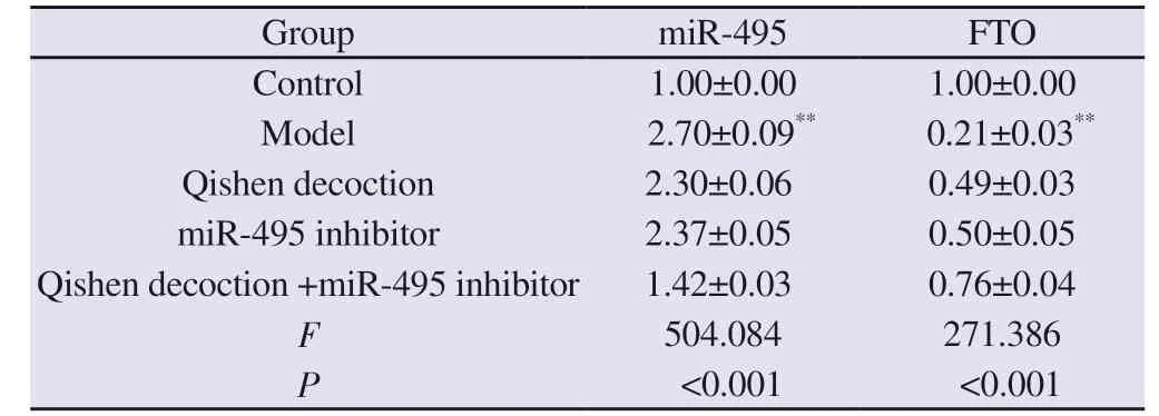
Compared with the control group, the expression of miR-495 in macrophages of the model group was significantly increased, while the expression of FTO was significantly reduced, with statistical significance (P<0.01).Compared with the model group, the expression of miR-495 in macrophages of the Qishen decoction group, miR-495 inhibitor group, and Qishen decoction +miR-495 inhibitor group were significantly reduced, while the expression of FTO was significantly increased, with statistical significance(P<0.01).See Table 5.
Tab 4 Effect of Qishen decoction on the expression of macrophage marker molecules(n=3, ±s)

Tab 4 Effect of Qishen decoction on the expression of macrophage marker molecules(n=3, ±s)
Note: Compared with the control group, **P<0.01.Compared with the model group, ○○P<0.01.
Group iNOS COX-2 Arg-1 YM-1 Control 1.00±0.00 1.00±0.00 1.00±0.00 1.00±0.00 Model 3.14±0.18** 2.80±0.12** 0.25±0.07** 0.34±0.05**Qishen decoction 2.43±0.08○○ 2.41±0.08○○ 0.64±0.07○○ 0.66±0.07○○miR-495 inhibitor 2.45±0.14○○ 2.39±0.13○○ 0.67±0.09○○ 0.68±0.05○○Qishen decoction +miR-495 inhibitor 1.75±0.10○○ 1.45±0.09○○ 0.93±0.05○○ 0.88±0.04○○F 143.029 191.093 64.004 84.613 P<0.001 <0.001 <0.001 <0.001
Tab 5 Effect of Qishen decoction on the expression of miR-495 and FTO(n=3, ±s)

Tab 5 Effect of Qishen decoction on the expression of miR-495 and FTO(n=3, ±s)
Note: Compared with the control group, **P<0.01.Compared with the model group, ○○P<0.01.
Group miR-495 FTO Control 1.00±0.00 1.00±0.00 Model 2.70±0.09** 0.21±0.03**Qishen decoction 2.30±0.06○○ 0.49±0.03○○miR-495 inhibitor 2.37±0.05○○ 0.50±0.05○○Qishen decoction +miR-495 inhibitor 1.42±0.03○○ 0.76±0.04○○F 504.084 271.386 P<0.001 <0.001
3.6 Effect of Qishen decoction on FTO Protein Expression
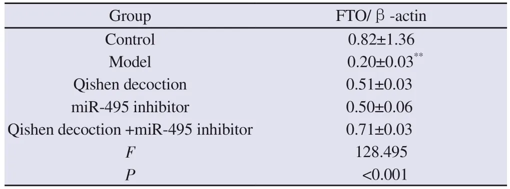
Compared with the control group, the expression of FTO protein in macrophages of the model group was significantly reduced, and the differences were statistically significant (P<0.01).Compared with the model group, the expression of FTO protein in macrophages in the Qishen decoction group, miR-495 inhibitor group, and Qishen decoction +miR-495 inhibitor group were significantly increased,and the differences were statistically significant (P<0.01).See Table 6.
Tab 6 Effect of Qishen decoction on FTO protein expression(n=3,±s )

Tab 6 Effect of Qishen decoction on FTO protein expression(n=3,±s )
Note: Compared with the control group, **P<0.01.Compared with the model group, ○○P<0.01.
Group FTO/β-actin Control 0.82±1.36 Model 0.20±0.03**Qishen decoction 0.51±0.03○○miR-495 inhibitor 0.50±0.06○○Qishen decoction +miR-495 inhibitor 0.71±0.03○○F 128.495 P<0.001

Fig 2 Comparison of FTO protein expression in macrophages of each group
4.Results
Insulin resistance, as an important pathological mechanism of T2DM, has also been proven to play an important role in metabolic syndrome.Obesity can lead to an increase in the size of fat cells,and the increased fat cells cause hypoxia in fat tissue, thus inducing cell dysfunction (such as endoplasmic reticulum stress and oxidative stress reaction) and initiating inflammatory reaction; On the other hand, hypertrophic adipocytes secrete MCP-1 and TNF-α which causing disorder of glucose and lipid metabolism and insulin resistance[8].In addition to adipose tissue macrophages, under the chemotaxis of MCP-1, B cells and T cells, macrophages from monocyte differentiation infiltrate around necrotic adipocytes, which is a sign of chronic inflammation of diabetes[9].
Macrophage function has significant heterogeneity, and its phenotype is dynamically transformed under the influence of local microenvironment.M1 type macrophages are mainly labeled with CD68, iNOS, and COX2, and secrete IL-6 and IL-1 β and TNFα Pro-inflammatory factors play a role in immune monitoring.INOS is an important component of the inflammatory response and participates in the formation of insulin resistance by downregulating the expression level of the insulin signaling molecule Rab8[10].COX2 is an important rate limiting enzyme in the synthesis of prostaglandins, and its expression level is closely related to inflammatory response and tumor formation.Studies have confirmed that COX2 can induce inflammation levels in the pancreatic islets,leading to the formation of pancreatic islets β Impairment of cellular function[11].M2 macrophages express CD206, Arg-1,and YM-1 as main surface marker molecules, and exert antiinflammatory effects by secreting cytokines such as IL-4 and IL-10.The results of this study showed that high glucose induced macrophage differentiation was mainly M1 type and secreted a large amount of IL-6 and IL-1 β.Qishen decoction medicated serum can significantly upregulate the expression of CD206, Arg-1, and YM-1 in macrophages, and downregulate the expression levels of CD68,iNOS, and COX2, thereby promoting the differentiation of M1 into M2 type macrophages and exerting anti-inflammatory effects.
MicroRNAs (miRNAs) are a class of small non-coding RNA in eukaryotes, about 19~25 nucleotides in size.Under the action of Dicer enzyme, the single stranded RNA precursor of 70~90nt hairpin structure is processed to form miRNA, which recognizes and specifically binds to the 3 ‘UTR of mRNA, thereby inhibiting mRNA translation or promoting mRNA degradation at the post transcriptional level and playing a pathological and physiological role.More and more studies have confirmed that miRNA expression affects insulin resistance or regulates pancreatic islets β the function of cells is involved in glucose metabolism[12].In addition,miRNA can also participate in the polarization of macrophages through post transcriptional regulation, and then play a therapeutic role in immune response, insulin resistance and inflammation[13].MiR-495 has been confirmed to be expressed in macrophages and plays a positive role in regulating vascular restenosis, oxidative stress, and macrophage polarization[14-16].FTO is an important downstream target gene of miR-495.As a fat and obesity related gene, FTO has a close relationship with the occurrence and development of metabolic diseases such as diabetes, fatty liver, and obesity.Studies have confirmed that overexpression of FTO can cause weight gain and diabetes,etc.[17].However, the role of FTO in different cells may be opposite.Research has confirmed that FTO participates in the regulation of glucose homeostasis in the body by regulating the expression of glycogenic genes in the liver[18].Current research confirms that the expression level of miR-495 is significantly increased in a high glucose induced macrophage model.Inhibiting the expression of miR-495 can significantly upregulate the expression of FTO, thereby promoting the transformation of M1 type macrophages into M2 type macrophages and exerting antiinflammatory effects.
Qishen decoction is an empirical formula for the clinical treatment of metabolic syndrome, with Astragalus membranaceus and Panax quinquefolium as the main ingredients.Astragalus membranaceus has the functions of tonifying qi and consolidating the surface,while Panax quinquefolium can nourish yin and promote fluid production.When combined, the two have the effects of tonifying qi and nourishing yin, clearing heat and promoting fluid production.Research has confirmed that Astragalus polysaccharides in Astragalus membranaceus can improve insulin resistance in obese rats by upregulating PI3K in skeletal muscle and reducing serum levels of total cholesterol and triglycerides[19].In the formula,Zexie and lotus leaves are used to enter the spleen and kidney meridians, seep dampness and relieve heat.Hawthorn and Salvia miltiorrhiza are also used to enter the liver, spleen, and stomach meridians, strengthening the spleen and dissipating food, promoting qi and blood stasis.The above four drugs are all official medicines.Zhou Yi et al.[20] demonstrated through network pharmacology that lotus leaves can play a coordinated role in treating T2DM through insulin resistance pathways, ErbB signaling pathways, and insulin signaling pathways.The main active component of Salvia miltiorrhiza, tanshinone I, can promote the activation of AKT signaling pathway and improve insulin resistance by inhibiting the expression of PTP1B[21].Ligustrum lucidum and Eclipta grandiflora nourish the liver and kidney, Gynostemma pentaphyllum nourishes deficiency and clears heat, Panax notoginseng promotes blood circulation and dispels blood stasis, and the above four drugs are all adjuvants.Pharmacological studies have confirmed that Gynostemma pentaphyllum and its equivalent components,such as Gynostemma pentaphyllum polysaccharide, Gynostemma pentaphyllum saponin, can regulate the metabolism of glucose and lipid, restore the homeostasis of intestinal flora, improve insulin resistance and antioxidation, and play an important role in the treatment of diabetes.Licorice is used as a detoxifying and medicinal herb to balance its properties.The whole recipe plays the role of tonifying qi, nourishing yin, and dissipating food and blood stasis.In this study, our results confirm that Qishen decoction has significant toxic and side effects on the activity of macrophages,indicating that the anti-inflammatory effect of Qishen Tang is not achieved by inhibiting macrophage activity.Further pharmacological studies have shown that Qishen Tang can significantly increase the proportion of M2 type macrophages and reduce the proportion of M1 type macrophages, thereby reducing IL-6 and IL-1 β Increase the levels of IL-4 and IL-10 and exert anti-inflammatory effects.From the perspective of mechanism, Qishen Tang can significantly inhibit the expression of miR-495 in macrophages, upregulate the expression level of FTO, and thus polarize macrophage M1 towards M2 type.Its pharmacological effect is similar to the trend of miR-495 inhibitors, and the combination and synergistic effect of the two can be achieved.
In summary, based on previous research, we further elucidated the molecular mechanism and targets of Qishen decoction in improving insulin resistance.The research results indicate that Qishen decoction promotes M2 type differentiation of macrophages through the miR-495/FTO pathway, thereby exerting anti-inflammatory mechanisms and improving insulin resistance.
 Journal of Hainan Medical College2023年14期
- Journal of Hainan Medical College的其它文章
- Research progress on key genes of vitamin D signaling pathway
- Research progress on the influence of local hemodynamics on carotid atherosclerosis
- Copy number variation sequencing for diagnosis of cytomegalovirus infection based low‑depth whole‑genome sequencing technology in fetus: Three cases and literature review
- Intervention of Xuduan Zhongzi Formula on spermatogenesis epididymal morphological changes in a mice model of oligospermia
- Expression of miR-9-5p and RHOA in aluminum-induced rat cognitive dysfunction
- miR-483-5p regulates osteoclast generation by targeting Timp2
