Existence Forms of Alizarin in Methanol Solution with Different pH Values
LI Yaqin,HU Yujiang,LIU Hanguang,GAO Chao*,XIONG Jinyan,TIAN Di,HONG Shaoli*,LI Wei*
(1.Hubei Key Laboratory of Biomass Fiber and Eco-dyeing &Finishing,College of Chemistry and Chemical Engineering,Wuhan Textile University,Wuhan 430073,China;2.PetroChina Tarim Oilfield Company,Korla 841000,China)
Abstract:In order to explore the existence forms of Alizarin in methanol solution with different pH values,we designed nine structures,calculated UV-Vis absorption spectra and fluorescence emission spectra by time-dependent density functional theory(TDDFT) method and compared with the experimental spectra at different pH values.By checking the consistency between the simulated and experimental spectra,the reasonable existence forms of Alizarin at different pH values were confirmed.Under the acidic condition,Alizarin exists in the molecular form of a_G_1 and a_G_4;under the basic condition,Alizarin exists in the anion forms of b_G_1,b_G_4,and c_G.On the ground state,a_G_3 is unstable and is transformed to a_G_4;on the excited state,a_E_3 is unstable and is transformed to a_E_1.On the excited state,the proton transfer between 1-position hydroxyl and 9-position carboxyl can be confirmed by fluorescence emission spectra and TDDFT calculations.The UV-Vis absorption spectra and fluorescence emission spectra of Alizarin mainly originate from intra-molecular charge transfer.
Keywords:Alizarin;structure;UV-Vis absorption spectrum;fluorescence emission spectrum;TDDFT
Alizarin(1,2-dihydroxy-9,10-anthraquinone) can be extracted from the roots of madder.As a derivative of anthraquinone,Alizarin is widely used in the dyeing and printing of fiber.Due to the protonation/deprotonation reversible mechanism of the chromophore,Alizarin is applied in the development of wearable textile sensors[1]and pH-responsive film for intelligent packaging[2].Alizarin has a significant bone growth promoting function due to high affinity to calcium,and therefore can effectively reduce bone tumor growth[3].As a potential organic semi-conductors for solar cells,the optical and electronic properties of Alizarin and its functionalized derivatives were investigated[4-5].The investigation of the structure of Alizarin is an interesting topic.Baran[6],Lofrumento[7],de Souza[8],and co-workers studied the structure of Alizarin and Alizarin Red S by SERS and FT-Raman.Jen and co-workers[9]studied the ultrafast intramolecular proton transfer of Alizarin in DMSO solution by means of transient stimulated Raman spectrum.Dworak and co-workers[10]used ultrafast transient absorption spectroscopy to study the photo-induced processes of inorganic-organic CdSe quantum dot-Alizarin hybrid complexes.Nawrocka and co-workers[11]used electro-absorption spectroscopy to study the electronic excited states of Alizarin that adsorbed on TiO2nanoparticles.Tang and co-workers[12]studied the effect of solvent on the excited state intramolecular proton transfer and revealed the formation mechanism of hydrogen bond for Alizarin and its isomers.The complexation and adsorption behaviors of Alizarin were also studied.For example,the complexation of Alizarin and Al(Ⅲ) in methanol solution was investigated by means of UV-Vis spectrum and theoretical calculations[13],the graft of Alizarin on ZnO[14]and Ag nanoparticles[15]were also explored.
The deprotonation of hydroxyl at 1-position or 2-position of Alizarin will take place in basic condition.Therefore,Alizarin exists in different forms under acid-base conditions.It is very difficult to determine the existence forms of Alizarin at different pH values solely by measurement instruments due to the inter-conversion of structures.The combinations of spectral analysis and theoretical calculation can provide a feasible solution.In this paper,we measured the UV-Vis absorption spectra and fluorescence emission spectra of Alizarin in methanol solution with different pH values.Moreover,we designed the structures of Alizarin at different pH values and calculated their UV-Vis absorption spectra and fluorescence emission spectra by time-dependent density functional theory(TDDFT) method.Furthermore,we identified the reasonable structures of Alizarin by the consistency check on the simulated and experimental spectra.
1 Experiment
1.1 UV-Vis absorption spectra and fluorescence emission spectra
Alizarin(AR) was purchased from Sinopharm Chemical Reagent Co.,Ltd..Alizarin was dissolved in methanol and the pH value of solutions were adjusted by NaOH solution and HCl solution.The UV-Vis absorption spectra were recorded on TU-1901 spectrometer(PERSEE,China) and the fluorescence emission spectra were recorded on F-2700 spectrometer(Hitch,Japan).
1.2 Theoretical calculations
Under the acidic and neutral conditions,there are four orientations of hydroxyls at 1-position and 2-position of Alizarin,which are labelled as a_G_1,a_G_2,a_G_3,and a_G_4(in Tab.1).Under basic condition,the deprotonation of one hydroxyl at 1-posotion or 2-position will produce four isomers b_G_1,b_G_2,b_G_3,and b_G_4(in Tab.1).The deprotonations of both hydroxyls at 1-position and 2-position will produce c_G(in Tab.1).The ground state(S0) geometry optimization and vibrational frequency calculations were carried out by DFT method at B3LYP/6-31G(d) level of theory.The vertical excited energies were calculated by TDDFT method at the ground state equilibrium geometry.The B3LYP hybrid functional and 6-311+G(d,p) basis set were selected.The effect of solvent on spectrum was considered by using PCM model[16].The linear-response(L.R.) and state-specific(S.S.) approaches were employed.In order to find the minimum energy point on the excited state potential energy surface(S1),the TDDFT geometry optimization and vibrational frequency calculations

Tab.1 Optimized equilibrium geometries of nine Alizarin structures on ground state and excited state
were carried out along with the equilibrium and linear-response solvation approach.The B3LYP(or CAM-B3LYP) hybrid functional and 6-311+G(d,p) basis set was selected.And then,the state-specific equilibrium solvation of the first excited state at its equilibrium geometry(E1) was calculated by corrected linear-response approach.Finally,the ground state energy with non-equalibrium solvation at the excited state geometry and with the static solvation from the excited state(E2) was calculated by using the corrected linear-response approach.The difference betweenE1andE2gives the vertical emission energy.The UV-Vis absorption spectra and fluorescence emission spectra were simulated by using the Franck-Condon-Herzberg-Teller method.All calculations were carried out on Gaussian16 D01 revision program suite.Multiwfn 3.8[17]and VMD1.9.4 program suite[18]were used to analyze and display graphically the electronic transitions.
2 Results and Discussions
2.1 UV-Vis absorption spectra of Alizarin at different pH values(Fig.1)

Fig.1 UV-Vis absorption spectra of Alizarin in methanol solution with different pH values(a),UV-Vis absorption spectrum of Alizarin at pH≤6 and simulated spectra of a_G_1,a_G_2,and a_G_4(b),UV-Vis absorption spectrum of Alizarin at pH=10 and simulated spectra of b_G_1,b_G_2,and b_G_4(c),and UV-Vis absorption spectrum of Alizarin at pH=13 and simulated spectrum of c_G(d)
As shown in Fig.1,when the pH value is in the range of 1-7,a strong absorption band centered at 430 nm can be observed.When the pH value is equal to 8,the band located at 430 nm is weaken progressively,while a new band centered at 530 nm is observed.When the pH value increases to 10 and 12,the band located at 430 nm is disappeared,while the band located at 530 nm is enhanced steadily.When the pH value increases to 13,a new shoulder band located at 570 nm and 610 nm is observed.
2.2 Simulated UV-Vis absorption spectra of Alizarin(Fig.1)
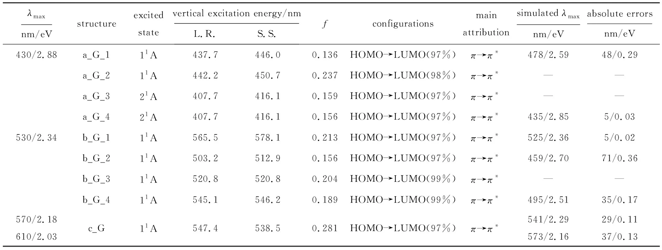
The ground state equilibrium geometries of all nine structures were presented in Tab.1.The vertical excitation energy,oscillator strengths(f),and main configurations were listed in Tab.2.

Tab.2 Vertical excitation energy,oscillator strength(f),configurations,and transition types of nine Alizarin structures on ground state
Inspection of the ground state equilibrium geometries of a_G_1,a_G_2,a_G_3,and a_G_4,we find that a_G_3 is unstable and is transformed to a_G_4.Therefore,a_G_3 has the same absorption spectrum with that of a_G_4.
Under the acidic and neutral conditions(pH≤7),the maximum absorption(λmax) of Alizarin can be observed at 430 nm.The L.R.excitation energies of a_G_1,a_G_2,a_G_3,and a_G_4 are 437.7 nm,442.2 nm,407.7 nm,and 407.7 nm,respectively,and their S.S.excitation energies are 446.0 nm,450.7 nm,416.1 nm,and 416.1 nm,respectively.The S.S.excitation energies are lower than L.R.excitation energies.As for a_G_2,due to the proton transfer on the excited state,there are different structures for the ground state and the excited state.The Franck-Condon-Herzberg-Teller method cannot provide a reasonable absorption spectrum.The simulated UV-Vis absorption spectra of a_G_1 and a_G_4 show that theλmaxof a_G_1 and a_G_4 are 478 nm and 435 nm,respectively.The absolute errors onλmaxare 48 nm(0.29 eV) and 5 nm(0.03 eV),respectively.These small errors reveal that the Alizarin exists in the form of a_G_1 and a_G_4.TDDFT calculations show this absorption band dominatingly originates from the excitation of HOMO→LUMO and has the characteristic ofπ→π*transition.
As the pH value is equal to 8,the deprotonation of hydroxyl at 1-position or 2-position of a fraction of Alizarins will produce an anion with a negative charge,but most of the Alizarins exist in the form of molecule.Therefore,two main absorption bands can be observed at 430 nm and 530 nm.According to the analyses above,the 430 nm band arises from the absorption of Alizarin molecule,and the 530 nm band may arise from the absorption of b_G_1,b_G_2,b_G_3,and b_G_4.The L.R.excitation energies of b_G_1,b_G_2,b_G_3,and b_G_4 are 565.5 nm,503.2 nm,520.8 nm,and 545.1 nm,respectively,and the S.S.excitation energies are 578.1 nm,512.9 nm,520.8 nm,and 546.2 nm,respectively.The simulatedλmaxof b_G_1,b_G_2,and b_G_4 are 525 nm,459 nm,and 495 nm,and the absolute errors onλmaxare 5 nm(0.03 eV),71 nm(0.36 eV),and 35 nm(0.17 eV).The good agreements onλmaxindicate that there are b_G_1 and b_G_4 in the system.TDDFT calculations show that this absorption band mainly originates from HOMO→LUMO transition and has the characteristic ofπ→π*transition.In summary,as the pH value is equal to 8,there are a_G_1,a_G_4,b_G_1,and b_G_4 in the system.When the pH value increases to 10 and 12,a sole absorption band can be observed at 530 nm,it can be inferred that there are b_G_1 and b_G_4 in the system.
As the pH value increases to 13,a shoulder band whose peaks located at 570 nm and 610 nm is observed.Under the strong basic condition,the deprotonation of two hydroxyls at 1-position and 2-position will produce c_G.The Franck-Condon-Herzberg-Teller method well reproduces the UV-Vis absorption spectrum of c_G.The simulatedλmaxof c_G are 541 nm and 573 nm,the absolute errors onλmaxare 29 nm(0.11 eV) and 37 nm(0.13 eV).TDDFT calculations reveal that this band originates from the HOMO→LUMO configurations,and the orbital component analysis manifests that this UV-Vis absorption band originates fromπ→π*electronic transition.
2.3 Fluorescence emission spectra of Alizarin at different pH values(Fig.2)

Fig.2 Fluorescence emission spectra of Alizarin in methanol solution with different pH values(a),fluorescence emission spectrum of Alizarin at pH=6 and simulated spectra of a_E_1,a_E_2,and a_E_4(b),fluorescence emission spectrum of Alizarin at pH=10 and simulated spectra of b_E_1,b_E_2,and b_E_4(c),and fluorescence emission spectrum of Alizarin at pH=13 and simulated spectrum of c_E(d)
As shown in Fig.2,under the acidic condition(pH=6),the fluorescence emission bands of Alizarin(λem) can be observed at 531 nm and 582 nm;under the near neutral condition(pH=8),a single emission band located at 526 nm can be observed;under the weak basic condition(pH=10),a bathochromic shift can be observed and theλemis shifted to 628 nm;under the strong basic condition(pH=13),the emission band continues shifting towards long wavelength to 649 nm.
2.4 Simulated fluorescence emission spectra of Alizarin(Fig.2)
The excited state equilibrium geometries of all nine structures of Alizarin were presented in Tab.1.Comparing the equilibrium geometries of ground state and excited state,some interesting finds are obtained.a_E_3 is unstable on the excited state and is transformed to a_E_1.Under the radiation excitation,the proton transfer between 1-position hydroxyl and 9-position carbonyl of Alizarin will take place[9].Comparing the geometries of a_G_1 with that of a_E_1,a strong tendency on proton transfer is observed.The O1-H is elongated from 0.991 Å to 1.056 Å,and the O2-H is shorten from 1.687 Å to 1.462 Å,respectively.A complete proton transfer is observed on a_E_2,the O1-H is elongated from 0.986 Å to 1.594 Å,and the O2-H is shorten from 1.673 Å to 1.006 Å.A strong tendency on proton transfer is also observed on b_G_1.Comparing the geometries of b_G_1 with that of b_E_1,the O1-H is elongated from 0.993 Å to 1.028 Å and the O2-H is shorten from 1.635 Å to 1.504 Å.
The vertical emission energies,oscillator strength (f),and main configurations of the first singlet excited state were presented in Tab.3.The emissions spectra were simulated by using the Franck-Condon-Herzberg-Teller method and presented in Fig.2 together with experimental spectra.

Tab.3 Vertical emission energy,oscillator strength(f),configurations,and simulated λem of nine Alizarin structure on excited state
To be specific,under the acidic condition(pH=6),two main emission bands located at 531 nm and 582 nm can be observed.The vertical emission energy of a_E_1,a_E_2,a_E_3,and a_E_4 are 612.4 nm,551.0 nm,645.1 nm,and 555.7 nm,respectively.As
for a_E_2,due to the proton transfer,the ground state structure is different from the excited state structure.The Franck-Condon-Herzberg-Teller method does not provide a reasonable emission spectrum.The simulatedλemof a_E_1 are 532 nm and 567 nm and of a_E_4 are 474 nm and 503 nm,respectively(Fig.2b).The 567 nm band of a_E_1 can correspond to the bandat 581 nm,and 503 nm band of a_E_4 can correspond to the band at 531 nm.The absolute errors are 15 nm(0.06 eV) and 28 nm(0.13 eV).A good agreement on theλemshows that under the acidic condition,there are a_E_1 and a_E_4 in the system.TDDFT calculations reveal that these two emission bands both origin from HOMO→LUMO(99%) configurations and have the characteristic ofπ→π*transition.Under the near neutral condition(pH=8),there is only one fluorescence emission band observed at 526 nm.The fluorescence emission is shifted from 531 nm to 526 nm due to the different acidic and basic conditions.According to the analyses above,the band at 526 nm corresponds to the fluorescence emission of a_E_4 and the absolute error is 23 nm(0.11 eV).
Under the weak basic condition(pH=10),a strong band is observed at 628 nm.It is possibly originated from the fluorescence emission of Alizarin anion.The calculated vertical emission energy of b_E_1,b_E_2,b_E_3,and b_E_4 are 660.4 nm,594.1 nm,543.1 nm,and 597.2 nm,respectively.The simulatedλemof b_E_1,b_E_2,and b_E_4 are 617 nm,532 nm,and 581 nm,and the absolute errors onλemare 11 nm(0.04 eV),96 nm(0.36 eV),and 47 nm(0.17 eV),respectively.The simulatedλemof b_E_1 and b_E_4 is close to the experimental value,therefore,the band located at 628 nm originates from the emission of b_E_1 and b_E_4.TDDFT calculations reveal that this emission mainly originates from the HOMO→LUMO configuration,and the oscillator strength(f) is 0.299 or 0.312 for b_E_1 and b_E_4,respectively.
Under the strong basic condition(pH=13),the deprotonation of two hydroxyls at 1-position and 2-position produce c_G.Under radiation excitation,c_G is excited to c_E.A sole emission band at 649 nm is observed(Fig.2d).The calculated vertical emission energy of c_E is 593.3 nm.The simulatedλemof b_E_1 is 622 nm,and the absolute error is 27 nm(0.08 eV).A good agreement on theλemreveals that the band located at 649 nm originates from the emission of c_E.TDDFT calculations show this emission mainly originates from the HOMO→LUMO(98%) configuration,and the oscillator strength(f) is 0.493.
3 Conclusions
On the ground state,a_G_3 is unstable and is transformed to a_G_4;on the excited state,a_E_3 is unstable and is transformed to a_E_1.By comparing the simulated UV-Vis absorption and fluorescence emission spectra with experimental spectra of nine Alizarin structures,the reasonable existence forms of Alizarin are confirmed.Under the acidic condition,Alizarin exists in the form of a_G_1 and a_G_4;under the weak basic condition,Alizarin exists in the form of b_G_1 and b_G_4;under the strong basic condition,Alizarin exists in the form of c_G.On the excited state,the proton transfer between 1-position hydroxyl and 9-positon carbonyl can be observed by fluorescence emission spectra and TDDFT calculations.The UV-Vis absorption spectra and fluorescence emission spectra of Alizarin mainly originate from intra-molecular charge transfer.
Acknowledgements:
This work is supported by the National Natural Science Foundation of China(22204123).

