Effects of different moxibustion time on knee cartilage morphology and the expression of TNF-α and IL-10 in rats with knee osteoarthritis
LI Qing (李情), XUE Pingju (薛平聚), ZHANG Xiaoqin (张小琴), JIA Yejuan (贾叶娟), XU Jing (徐晶), CHEN Zilong (陈子龙),JIA Chunsheng (贾春生), XING Haijiao (邢海娇)
Hebei University of Chinese Medicine, Shijiazhuang 050091, China
Abstract
Keywords: Moxibustion Therapy; Mild Moxibustion; Osteoarthritis, Knee; Cartilage; Inflammation; Moxibustion Dosage;Dose Response Relationship, Acupuncture-moxibustion; Rats
Knee osteoarthritis (KOA) is the most common osteoarthritis type, and the main symptoms include pain, numbness, adverse knee joint flexion and extension, and the main pathological features of joint space narrowing, subchondral osteosclerosis,subchondral cystic degeneration, and osteophyte formation at joint margin[1].Epidemiological investigation and analysis have shown an 18%prevalence rate of KOA with a higher prevalence in females than males.KOA has become one chronic disease seriously affecting the quality of life in middle-aged and elderly people in China[2].For KOA, the articular cartilage is the main pathological region; the apoptosis and functional changes of chondrocytes, the destruction of extracellular matrix in the articular cartilage are the main causes[3], and cartilage inflammation is one important part of pathogenesis[4].Moxibustion, as an important part of acupuncturemoxibustion treatment, has the function of warming meridians and promoting local blood circulation.Studies have proven that moxibustion effectively repairs knee cartilage and improves inflammatory symptoms of cartilage in KOA patients[5-7], but the relationship between moxibustion dosage and curative effect is not clear.Therefore, this study explored the dosage-effect relationship of moxibustion in KOA treatment so as to provide a reference for moxibustion in treating KOA.
1 Materials and Methods
1.1 Laboratory animals and grouping
The same batch of 50 healthy specific-pathogen-free male Wistar rats, aged 7 weeks, with a body mass of(250±30) g, were provided by Beijing Vital River Laboratory Animal Technology Co., Ltd., China, with the production license number of [SCXK (Jing) 2016-0006].The experiment was carried out in the Experimental Center of Hebei University of Chinese Medicine.All rats were free to eat and drink with the same animal feed during the whole experiment.The animal feed and padding were provided by Beijing Vital River Laboratory Animal Technology Co., Ltd., China.Five rats in each cage were kept at (24±2) ℃, (55%±5%) relative humidity,12 h/12 h light/dark cycle.After one week of adaptive feeding, the 50 Wistar rats were randomly divided into 5 groups: a blank group, a model group, a 15-minutemoxibustion group, a 30-minute-moxibustion group,and a 60-minute-moxibustion group, with 10 rats in each group.This experiment strictly complied with the requirements of theGuiding Opinions on Treating Laboratory Animalsissued by the Ministry of Science and Technology of the People’s Republic of China and was reviewed and approved by the Laboratory Animal Ethics Committee of Hebei University of Chinese Medicine (Approval No.DWLL2019014).
1.2 Main reagents and instruments
Moxa sticks of 7 mm in diameter and 110 mm in length (Nanyang Wanbei Moxa Factory, China); sodium iodoacetate (Item No.I2512, Sigma, USA); tumor necrosis factor (TNF)-α enzyme-linked immunosorbent assay (ELISA) kit (Item No.88-7340, Invitrogen, USA);interleukin (IL)-10 ELISA kit (Item No.EK310/2-01,UNOCI, China).
ME203E/02 electronic balance (Shanghai Mettler Toledo Instrument Co., Ltd., China); D3024R desktop high-speed refrigeration centrifuge (Dalong Xingchuang Experimental Instrument Co., Ltd., China); Epoch microplate reader (BioTeK, USA); optical microscope(Leica, Germany).
1.3 Modeling method
The model was induced by sodium iodoacetate[6].Rats in all groups except for the blank group were fed adaptively for one week, and then anesthetized by intraperitoneal injection of 3% pentobarbital sodium[1 mL/(kg·bw)].The knee joints of both hind limbs were shaved, sterilized with iodophor, and flexed by 90°.The sodium iodoacetate saline solution (50 μL) was injected into the right posterior knee joint cavity through the infrapatellar ligament.In the same way, 50 μL of normal saline was injected into the left posterior knee joint cavity, then the knee joint was stretched back and forth 5 times and the rat was put back into the cage.Four weeks later, the knee joint diameter was measured with a vernier caliper.A 20% higher swelling degree of the right knee joint greater than the left knee joint indicated successful modeling.
1.4 Intervention methods
The 15-minute-moxibustion group, the 30-minutemoxibustion group, and the 60-minute-moxibustion group were all treated with mild moxibustion intervention at Neixiyan (EX-LE4) and Dubi (ST35) points near the patella.
Methods: The rats were fixed on the rat rack to expose the treatment position of the right hind limb knee joint, which was shaved and disinfected routinely.The moxibustion frame was fixed 2-3 cm above the patella.Fully ignited the moxa sticks and made sure that the rats had no obvious struggle.Treatment with moxibustion lasted for 15 min in the 15-minutemoxibustion group, 30 min in the 30-minutemoxibustion group, and 60 min in the 60-minutemoxibustion group.After each intervention, an infrared thermometer was used to measure the temperature at Neixiyan (EX-LE4) and Dubi (ST35), and an increase in the temperature by (1.5±0.5) ℃ indicated an effective intervention.
Rats in the blank group and the model group were only fixed on the rat rack for 30 min without moxibustion intervention.
All groups were intervened between 9:00 a.m.and 12:00 a.m.every Monday, Wednesday, and Friday,3 times a week for 4 consecutive weeks, for a total of 12 interventions.
1.5 Detection indicators and methods
1.5.1 General observation
The changes in body mass and knee joint diameter of experimental rats were observed dynamically.After intervention, the body mass and knee joint diameter of rats in each group were measured every week.The body mass of rats was measured by the electronic scale,and the knee joint diameter was measured by a vernier caliper.The same person performed the measurement each time to reduce the error.
1.5.2 Morphological observation of cartilage tissue
Macroscopic observation of cartilage: After the intervention, all rats were anesthetized by intraperitoneal injection of 3% pentobarbital sodium solution [1 mL/(kg·bw)], and the articular cavity was exposed.Macroscopic observation of the surface color,transparency, and cartilage surface smoothness in the femoral condyle was performed.
Hematoxylin-eosin (HE) staining: After macroscopic observation of cartilage, femoral condyle cartilage specimens of rat knee joint were cut and fixed in 40 g/L paraformaldehyde fixation solution.After 48 h, the specimens were embedded in paraffin and cut into sections of 5 μm in thickness.After oven dry, the sections were dewaxed in xylene for 10 min; then dehydrated for 5 min in 100%, 90%, 80%, and 70%ethanol solutions, respectively; washed with distilled water for 5 min; then soaked in hematoxylin solution for 5 min; washed with distilled water for 1 min; separated with 5% acetic acid for 1 min; washed with distilled water for 1 min; turned blue in alkaline water for 20 s;washed with distilled water for 1 min; dehydrated with ethanol, and sealed with neutral gum.The smooth degree and thickness of cartilage surface, the density and distribution of cells, and the structure level of cartilage were observed under the light microscope.
Toluidine blue staining and Mankin score: After macroscopic observation of cartilage, the cartilage surface of femoral condyle in the knee joint was cut for toluidine blue staining and Mankin score evaluation.Sample collection, paraffin embedding, section, and deparaffinage methods were the same as in HE staining.The specimen sections were stained in toluidine blue for 10 min, washed with distilled water for 1 min, color separated in glacial acetic acid solution, dehydrated for 1 min in 100%, 90%, 80%, and 70% ethanol solutions,respectively, and then mounted with neutral gum.Three visual fields were selected for each slide and scored according to the Mankin scoring for the average calculation[8].See Table 1 for details.

Table 1 Mankin score
1.5.3 Inflammatory mediator detection
Serum TNF-α and IL-10 concentrations were detected by ELISA.After anesthesia, the abdominal cavity was opened to expose the abdominal aorta, and blood samples were collected by disposable blood collection vessels.The samples were centrifuged at room temperature and 2 500 r/min for 15 min in batches.The supernatant was transferred into cryopreservation tubes with a pipette and stored in a refrigerator at-20 ℃.The serum samples were thawed at room temperature, and all the required reagents were prepared.Washed the plate with washing buffer solution 3 times, added standard diluent (R1) into blank wells, added standard products with different concentrations into other labeled wells, reacted at 37 ℃ for 2 h, and washed the plate 3 times.Added biotinylated antibody solution into each well, reacted at 37 ℃ for 1 h, and washed the plate 3 times.The reaction was carried out at 37 ℃ for 30 min after the Streptavidin-HRP working solution was added, and the plate was washed 3 times.Added TMB substrate and incubated at 37 ℃ for 15 min.Added the termination solution and measured the optical density (OD) value with a microplate reader at 450 nm wavelength 3 times each well to calculate the average value.
1.6 Statistical processing
All data were analyzed by the SPSS version 23.0 software.Measurement data all conformed to normal distribution and were expressed as mean ± standard deviation.One-way analysis of variance was used for comparisons among multiple groups.The least significant differencet-test was used for pairwise comparisons between groups.Repeated measurement analysis of variance was used to compare multiple observation points of the same indicator.P<0.05 indicated that the difference was statistically significant.
2 Results
2.1 General observation
2.1.1 Changes in body mass
Body mass changes of rats in each group are shown in Table 2.
Before intervention, there was no statistical difference in the body mass among the groups (P>0.05),suggesting comparability.The interaction between group and time had statistical significance on rat body mass (P<0.05).
After 1-week intervention, there was no significant difference in the body mass among the groups (P>0.05).After 2, 3, and 4 weeks of interventions, the body mass of the model group was increased compared with that of the blank group at the same time point (P<0.05).Compared with the model group at the same time point,the body mass of the 15-minute-moxibustion group had no significant change (P>0.05), while the body mass of the 30-minute-moxibustion group and the 60-minutemoxibustion group was decreased (P<0.05).There was no statistical difference between the 30-minutemoxibustion group and the 60-minute-moxibustion group (P>0.05).
Compared among different time points of the same group, the body mass of rats showed an upward trend in each group (P<0.05) and reached the highest after 4-week intervention (P<0.05).
Table 2 Changes in rat average body mass Unit: g

Table 2 Changes in rat average body mass Unit: g
Note: Compared with the blank group at the same time point, 1) P<0.05; compared with the model group at the same time point, 2) P<0.05;compared with the 15-minute-moxibustion group at the same time point, 3) P<0.05; compared with the same group after 1-week intervention,4) P<0.05; compared with the same group after 2-week intervention, 5) P<0.05; compared with the same group after 3-week intervention,6) P<0.05.
Group n1-week intervention 2-week intervention 3-week intervention 4-week intervention Blank 10 251.50±26.27 282.78±19.704)366.63±27.054)5)414.11±19.504)5)6)Model 10 238.16±22.90 336.85±24.861)4)415.52±35.001)4)5)460.25±17.641)4)5)6)15-minute-moxibustion 10 229.13±14.87 328.97±20.754)414.36±22.964)5)452.85±17.664)5)6)30-minute-moxibustion 10 251.20±22.17 293.63±11.602)3)4)372.50±21.222)3)4)5)427.96±17.412)3)4)5)6)60-minute-moxibustion 10 247.77±21.37 308.78±11.822)3)4)383.76±21.252)3)4)5)431.63±25.522)3)4)5)6)Intervention F=11.837, P<0.001 Time F=937.080, P<0.001 Intervention * Time interaction F=5.901, P<0.001
2.1.2 Changes in knee joint diameter
Changes in knee joint diameter in each group are shown in Table 3.
Before intervention, there was no significant difference in the knee joint diameter among the groups(P>0.05), suggesting comparability.The rat knee joint diameter was significantly affected by the group and time interaction (P<0.05).
Compared with the blank group at the same time point, the knee joint diameter in the model group was increased significantly (P<0.05).After 1-week or 2-week intervention, the 15-minute-moxibustion group, the 30-minute-moxibustion group, and the 60-minutemoxibustion group had no statistical differences compared with the model group at the same time point(P>0.05).After 3-week or 4-week intervention, there was no statistical difference between the 15-minutemoxibustion group and the model group at the same time point (P>0.05); the knee joint diameter in the 30-minute-moxibustion group and the 60-minutemoxibustion group was significantly smaller than that in the model group at the same time point (P<0.05); there was no statistical difference between the 30-minutemoxibustion group and the 60-minute-moxibustion group at each time point (P>0.05).
Comparison of the same group among different time points showed that the knee joint diameter displayed an upward trend in all groups (P>0.05) and reached the highest level after 4-week intervention (P<0.05).
Table 3 Changes in the mean knee joint diameter in rats Unit: mm

Table 3 Changes in the mean knee joint diameter in rats Unit: mm
Note: Compared with the blank group at the same time point, 1) P<0.05; compared with the model group at the same time point, 2) P<0.05;compared with the 15-minute-moxibustion group at the same time point, 3) P<0.05; compared with the same group after 1-week intervention,4) P<0.05; compared with the same group after 2-week intervention, 5) P<0.05; compared with the same group after 3-week intervention,6) P<0.05.
Group n1-week intervention 2-week intervention 3-week intervention 4-week intervention Blank 10 10.63±0.60 10.84±0.594)11.08±0.564)5)11.22±0.524)5)6)Model 10 11.37±0.411)12.04±0.451)4)13.09±0.711)4)5)13.20±0.701)4)5)6)15-minute-moxibustion 10 11.52±0.73 12.07±0.324)12.86±0.554)5)12.98±0.564)5)6)30-minute-moxibustion 10 11.80±0.57 11.99±0.524)12.17±0.492)3)4)5)12.31±0.452)3)4)5)6)60-minute-moxibustion 10 11.75±0.50 11.86±0.514)12.19±0.582)3)4)5)12.30±0.562)3)4)5)6)Intervention F=17.639, P<0.001 Time F=130.553, P<0.001 Intervention * Time interaction F=6.715, P<0.001
2.2 Morphological observation of cartilage
2.2.1 Macroscopic observation of articular cartilage
The femoral cartilage surface was translucent milky white with faintly visible even reddish subchondral bone, and was smooth, bright, without cracks, decay spots or softening focus; the joint edge was smooth and regular without osteophyte in the blank group.
The femoral articular cartilage surface was light yellow; the articular surface was rough with ulceration and weak luster; the cartilage defect depth was different with many decay spots and serious cartilage damage; the subchondral bone was exposed; the joint edge was not smooth with obvious osteophyte formation in the model group.
The appearance of femoral cartilage in the 15-minute-moxibustion group was slightly yellow compared with that in the blank group.The joint surface was rough with different degrees of cartilage damage without ulcer.The edge of the joint was slightly irregular, with slight osteophyte formation.In the 30-minute-moxibustion group, the articular surface of femur was smooth without cracks or softening foci; the joint edge was regular and neat without osteophyte.The articular surface of femur was smooth but darker in color than in the blank group without cracks, and the joint edge was regular and neat without obvious osteophyte formation at the joint edge in the 60-minute-moxibustion group (Figure 1).
2.2.2 Observation of articular cartilage HE staining
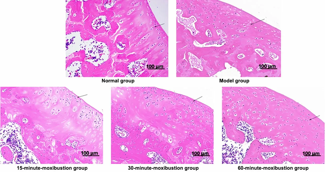
The chondrocytes showed clear structure, neat arrangement, and uniform staining, without obvious cell clusters in the blank group.Compared with the blank group, chondrocytes in the model group were disordered with occasional chondrocyte clusters and nuclear necrosis.The cartilage structure and the cell morphology and arrangement of the 15-minutemoxibustion group, the 30-minute-moxibustion group,and the 60-minute-moxibustion group were similar to those of the blank group.Compared with the 15-minute-moxibustion group, the 30-minutemoxibustion group, and the 60-minute-moxibustion group had clearer cell structure, more neat arrangement, and more uniform staining (Figure 2).
2.2.3 Observation of articular cartilage by toluidine blue staining
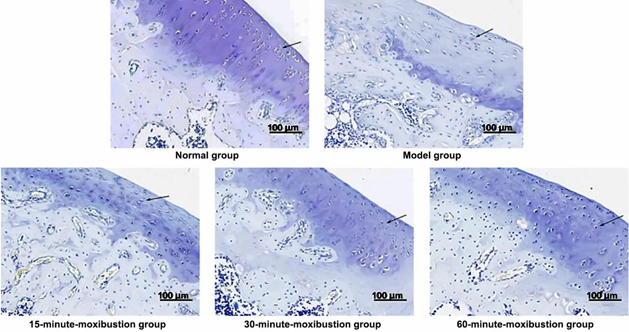
In the blank group, the cartilage was blue-purple with a darker color, uniform staining, and regular chondrocyte arrangement.The staining color of each part in the model group was light blue with a lighter color, uneven staining, and larger chondrocytes than in the blank group, and the Mankin score was higher(P<0.05).Compared with the model group, the 15-minute-moxibustion group, the 30-minutemoxibustion group, and the 60-minute-moxibustion group were stained deeply and evenly, with decreased Mankin scores (P<0.05).Compared with the 15-minutemoxibustion group, the staining was darker, close to that in the blank group, and the Mankin score was decreased significantly in the 30-minute-moxibustion group and the 60-minute-moxibustion group (P<0.05).See Figure 3 and Table 4.
2.3 Detection of inflammatory mediators
2.3.1 Changes in TNF-α expression in serum
Compared with the blank group, the serum TNF-α level in the model group was increased significantly(P<0.05).Compared with the model group, the serum TNF-α level in the 15-minute-moxibustion group, the 30-minute-moxibustion group, and the 60-minutemoxibustion group was decreased significantly (P<0.05).Compared with the 15-minute-moxibustion group, the serum TNF-α level in the 30-minute-moxibustion group and the 60-minute-moxibustion group was decreased significantly (P<0.05).There was no significant difference in serum TNF-α between the 30-minutemoxibustion group and the 60-minute-moxibustion group (P>0.05).See Table 5 for details.
2.3.2 Changes in IL-10 expression in serum
Compared with the blank group, the serum IL-10 level in the model group decreased significantly(P<0.05).There was no significant difference between the 15-minute-moxibustion group and the model group(P>0.05); the serum IL-10 level in the 30-minutemoxibustion group and the 60-minute-moxibustion group was significantly higher than that in the model group (P<0.05).There was no significant difference in the serum IL-10 between the 30-minute-moxibustion group and the 60-minute-moxibustion group (P>0.05).See Table 5 for details.

Figure 1 Macroscopic observation of femur articular cartilage surface in each group

Figure 2 Hematoxylin-eosin staining of rat articular cartilage in each group (×200)

Figure 3 Rat articular cartilage observation by toluidine blue staining in each group (×200)
Table 4 Comparison of the Mankin scoreUnit: point

Table 4 Comparison of the Mankin scoreUnit: point
Note: Compared with the blank group, 1) P<0.05; compared with the model group, 2) P<0.05; compared with the 15-minute-moxibustion group,3) P<0.05.
Group nScore Blank 10 1.4±0.8 Model 10 12.2±1.61)15-minute-moxibustion 10 9.8±1.32)30-minute-moxibustion 10 7.3±1.52)3)60-minute-moxibustion 10 7.9±1.62)3)F-value 81.960P-value <0.001
Table 5 Comparison of the inflammatory factor expression levels Unit: pg/mL

Table 5 Comparison of the inflammatory factor expression levels Unit: pg/mL
Note: TNF-α=Tumor necrosis factor-α; IL-10=Interleukin-10; compared with the blank group, 1) P<0.05; compared with the model group, 2)P<0.05; compared with the 15-minute-moxibustion group, 3) P<0.05.
Group nTNF-α IL-10 Blank 10 0.060±0.007 0.063±0.003 Model 10 0.086±0.0081)0.040±0.0031)15-minute-moxibustion 10 0.077±0.0052)0.041±0.005 30-minute-moxibustion 10 0.065±0.0062)3)0.049±0.0032)3)60-minute-moxibustion 10 0.069±0.0072)3)0.051±0.0032)3)F-value 24.203 67.636P-value <0.001 <0.001
3 Discussion
The pathogenesis of KOA has not been fully elucidated.Modern medicine has explored its pathogenesis based on molecular protein[9],biochemical metabolism[10], and histomorphology[11].Cartilage inflammation is an important part of the pathogenesis in KOA.The occurrence and development of KOA depend on whether the interaction between pro-inflammatory factors and anti-inflammatory factors is in a balanced state.A large number of proinflammatory substances are involved in the occurrence and development of KOA[12], such as complements,macrophages, mast cells, cytokines, chemokines,growth factors, and adipokines.Pro-inflammatory substances promote the synthesis and release of chondrocyte protease, the degradation of extracellular matrix, and the production of other pro-inflammatory substances, thus inducing the aging and apoptosis of chondrocytes.Among the cytokines, TNF-α is considered the leading pro-inflammatory factor that triggers inflammation and destroys articular cartilage[13].TNF-α is an inflammatory activity indicator in knee cartilage.It regulates chondrocyte apoptosis by promoting the production and secretion of cartilage matrix metalloproteinases and participates in multiple signaling pathways to regulate inflammatory factors[14-18].IL-10, an important anti-inflammatory factor, is negatively correlated with inflammation degree and inhibits inflammation development mainly by inhibiting the activity of pro-inflammatory factors[19].Therefore, in this study, the serum TNF-α and IL-10 levels in rats were used to measure the development of cartilage inflammation.
Chinese medicine believes that “pathogenic factors cannot invade when the healthy Qi exists”, the balance between healthy Qi and pathogenic Qi is the key to the healthy operation of human body, and moxibustion has the therapeutic effect of reinforcing healthy Qi to eliminate pathogenic factors.Studies have shown that moxibustion is more effective in treating KOA than filiform needle acupuncture and electroacupuncture[20],since moxibustion can inhibit inflammatory reaction[21],has the effects of warming viscera and unblocking meridians to help Yang and invigorate Qi[22-23], and can stimulate skin temperature receptors[24-26], promoting local microcirculation and increasing blood flow speed[27]to achieve the therapeutic purpose of strengthening healthy Qi and exorcising pathogenic factors.The dosage of moxibustion is an important factor affecting the therapeutic effect of moxibustion.Only the appropriate moxibustion dosage will achieve the best therapeutic effect[28].Yi Zong Jin Jian(Golden Mirror of the Medical Tradition) mentions that the function of Qi and blood in the body can be fully mobilized only when the moxibustion dosage reaches a certain level, thus effectively treating diseases.The curative effect cannot be guaranteed if the moxibustion dosage is insufficient.The moxibustion dosage can be controlled by size, cone numbers, and moxibustion time or frequency[29-31].In this study, the dosage-effect relationship in KOA treatment with moxibustion was explored by controlling the moxibustion dosage by changing the moxibustion time.Adequate moxibustion inhibits the secretion of serum inflammatory factors and alleviates inflammatory reactions in rats[32].
The results of this study showed that the average body mass of rats changed differently with different moxibustion dosages after 2-week intervention.The average body mass of the model group rats was larger than that of the blank group, while the average body mass of the 15-minute moxibustion group, the 30-minute moxibustion group, and the 60-minute moxibustion group was smaller than that of the model group.Because the body mass of rats is inversely proportional to the activity of rats, a greater body mass indicates less activity in rats, showing that the activity restriction is more serious; therefore, moxibustion intervention improves the knee joint activity restriction of rats.
The rat knee joint diameter indicates the inflammation severity of rat knee joint.A larger diameter indicates a greater swelling degree and more serious inflammatory reactions.Compared with the blank group, the knee joint diameter was obviously larger, the joint was swollen and seriously deformed,the surface of articular cartilage was rough and thinner,and even subchondral bone could be seen in the model group.HE staining and toluidine blue staining showed significantly disordered chondrocyte structure,increased Mankin score and inflammation-related factors, and decreased anti-inflammatory factors.Compared with the model group, after moxibustion intervention, the diameter became smaller, the swelling and deformation were decreased in the knee joint; the articular cartilage surface became smooth, and the damage degree was decreased.HE staining and toluidine blue staining showed that the structure of chondrocytes tended to be neat, the Mankin score was decreased significantly, inflammation-related inflammatory factors were decreased significantly, and the anti-inflammatory factors were increased significantly.Regarding the three moxibustion intervention groups, the articular cartilage surface was more smooth, HE staining and toluidine blue staining showed that the structure of chondrocytes was more orderly, the Mankin score was more obviously decreased, the expression level of the inflammationrelated inflammatory factor was lower, and the expression level of the anti-inflammatory factor was higher in the 30-minute-moxibustion group and the 60-minute-moxibustion group than in the 15-minutemoxibustion group; there was no significant difference between the 30-minute-moxibustion group and the 60-minute-moxibustion group.The above results showed that moxibustion intervention effectively improved the cartilage morphology of KOA, and the moxibustion time was the key influencing factor;moxibustion for a longer time could achieve better intervention effects; however, the intervention effect will not be affected by time after a certain period of time.This study has provided a reference for the duration selection in clinical moxibustion treatment of KOA.
Conflict of Interest
The authors declare that there is no potential conflict of interest in this article.
Acknowledgments
This work was supported by the Scientific Research Project of Hebei Provincial Administration of Traditional Chinese Medicine (河北省中医药管理局科研计划项目,No.2020135).
Statement of Human and Animal Rights
The treatment of animals conformed to the ethical criteria in this experiment.
Received: 8 March 2022/Accepted: 13 July 2022
 Journal of Acupuncture and Tuina Science2023年3期
- Journal of Acupuncture and Tuina Science的其它文章
- Journal of Acupuncture and Tuina ScienceInstructions for Authors
- Editorial Members of Journal of Acupuncture and Tuina Science
- Acupuncture intervening depressive disorder:research progress in its neurobiological mechanism
- Efficacy and safety of acupuncture-moxibustion for cerebral palsy-induced speech impairment:a systematic review and meta-analysis
- Clinical observation of acupuncture treatment for children with accommodative myopia
- Clinical observation of Tuina combined with Bu Zhong Yi Qi Tang in the treatment of rectocele
