A case of primary ovarian carcinoid and the review of the literature
Zhi-Ying Xia,Pei-Fang Chen,Lu-Shan Chen,Xiu-Shan Feng*
1Department of Gynecology & Obstetrics, Fuding Hospital, Fuding 355200, China.2Department of Gynecology & Obstetrics, Fujian Medical University Union Hospital,Fuzhou 350001,China.3Department of Pathology,Fujian Medical University Union Hospital,Fuzhou 350001,China.
Abstract Objective: To investigate the clinicopathologic features of primary ovarian carcinoid.Methods: A case of primary ovarian carcinoid of the ovary were reported.Clinicopathological analysis was performed.Results: The case was just an island-like carcinoid.The tumor cells were round or polygonal, arranged into island-like or pseudo chrysanthemum clusters, and got bilateral tubal and oophorectomy.No evidence of recurrence was consulted in 28 months of follow-up.Conclusion:Primary ovarian carcinoid is rare in clinic.The diagnosis should be differentiated from granulosa cell tumor and sertolithoid cell tumor, and metastatic carcinoid should be excluded.As a low-grade malignant tumor, most ovarian carcinoids have a real good prognosis.
Keywords:ovarian tumor; carcinoid; pathology; immunohistochemistry
Case report
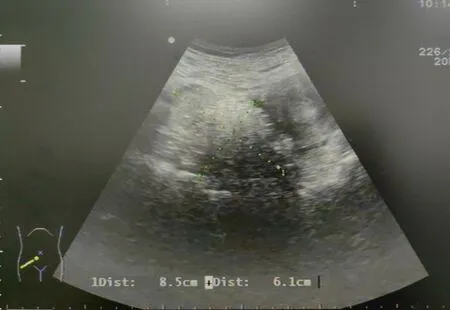
A 57-year-old woman,G2P2,has been postmenopausal for 3 years and has been admitted to the hospital for more than 5 years with a right adnexal mass.The patient had regular menstruation, had been menopausal for 3 years, and had no discomfort such as abdominal pain and vaginal bleeding.Gynecological physical examination found mass in the adnexal area, and gynecological ultrasound suggested uneven echo mass in the right adnexal area.So ovarian teratoma was considered.The size is approximately 8.5 cm × 6.1 cm (Figure 1).After that, gynecologic ultrasound was repeated every year, and no significant enlargement of the mass in the right adnexa was observed.After 5 years of follow-up, the adnexal mass still existed, and color ultrasound suggested that the size of the mass was about 9.6 cm * 8.6 cm *7.1 cm, requiring surgical treatment.
The patient had a history of multiple uterine fibroids and thyroid nodules.Blood samples took one year before admission showed an increase in CA199 (105 U/L).There was not any abnormal vaginal discharge or bleeding after menopause.Gynecological examination found that the right adnexal area can touch a mass, the size of about 10 cm× 8 cm,cystic, boundary clear, poor motion, no tenderness.
After admission, relevant auxiliary examinations were improved and conjunctions were eliminated,and the patient underwent bilateral appendectomy for single-hole laparoscopy.Intraoperative exploration showed that the uterus was slightly enlarged with a smooth surface,and the cystic enlargement of the right ovary was about 8 cm *7 cm*5 cm,which adhered to the right posterior wall of the uterus and local intestinal duct.Left ovarian atrophy, left hydrosalpinx.The right adnexa was removed and frozen.Pathology showed cystic mature teratoma.Due to leave hydrosalpinx, the left appendage was removed with the consent of the patient and her family.
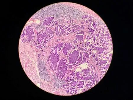
The patient recovered well after surgery and was discharged 3 days after surgery.Postoperative pathology: the tissue submitted for examination was widely sampled, and it was cystic mature teratoma of ovary(Figure 2)with island-like carcinoid(Figure 3).The carcinoid was 0.2 cm in diameter, and no mitotic image was observed.Immunohistochemical results: carcinoid CK, CgA, CD56, syn positive,Ki67 <1%; S100, AR, GCDFP15 and GFAP were negative.

Figure 1 Gynecological ultrasound scan results

Figure 2 Mature teratoma area (×100)

Figure 3 Carcinoid (×100)
Gynecological examination, gynecological color ultrasound and female tumor markers were followed up closely after the operation.At present,after 28 months of postoperative follow-up,the patient was in good condition with no signs of recurrence.
Discussion
Clinical characteristics of ovarian island-like carcinoid
Carcinoid cancer occurs more often in pre-or postmenopausal women with an average age of 53.Carcinoid originates from the primitive embryonic gut,is argyrophilic,can secrete polypeptide hormones,and can also lead to carcinoid syndrome.Carcinoids are more common in the gastrointestinal tract, followed by the lungs, bronchus and biliary ducts.Carcinoid cancers originating in the ovary are very rare and are found in ovarian malignancy, about 0.1% [1].Primary ovarian carcinoid tumors are usually unilateral, confined to the ovary, and histologically difficult to distinguish from metastatic cancers.Its appearance is similar to that of carcinoids found anywhere else (e.g.,gastrointestinal tract, respiratory tract).It is composed of nests and cords of cells (identical cells without atypia nuclei) and a network of fine blood vessels with endocrine characteristics and relatively low malignancy.Patients with ovarian carcinoid cancer often have no obvious clinical symptoms.Most of them are found accidentally or during physical examination, and ultrasound suggests ovarian mass.Carcinoid can produce 5-hydroxytryptamine, histamine, dopamine and other endocrine substances, these substances acting on different organs can cause skin flushing, abdominal pain, diarrhea, peripheral vascular dysfunction, heart damage syndrome, clinically known as carcinoid syndrome.It has been reported that the appearance of carcinoid syndrome is related to tumor size, and the larger the tumor size, the more likely the carcinoid syndrome will occur.It also believed that it is related to tumor type.1/3 patients with island-like carcinoid show symptoms and signs of the carcinoid syndrome, and most of them disappear after tumor resection [1-3].Usually, these tumors are asymptomatic or associated with non-specific symptoms,such as diffuse abdominal pain, but in some cases, they can express clinical signs of carcinoid syndrome induced by serotonin, chronic constipation, exercise, or hyperandrogenemia induced by peptide YY(intestinal inhibitors) [4, 5].
The cause of ovarian carcinoid is not clear at present.The literature reports that ovarian teratoma of monoblastic layer is easy to develop into ovarian carcinoid [6].The histological types of primary ovarian carcinoid can be divided into an island type, beam type, goiter type,mucous type and mixed type,and the rare clear cell type has also been reported in recent years [7-10].Island-like carcinoid accounts for nearly half of the primary ovarian carcinoid.Its morphology is similar to that of mid-gut carcinoid.The tumor cells are round or polygonal,arranged into island-like or pseudo chrysanthemum clusters, and red particles can be seen in the cytoplasm.Beam type carcinoid is a carcinoid similar to the hindgut or foregut.The cells are mostly columnar, single or multilayer cancer cells are arranged in a ribbon,and the longitudinal axis of the tumor cells is perpendicular to the longitudinal axis of the beam and cord structure.Goiter carcinoid is composed of carcinoid and thyroid follicle.There is thyroid glia stained with homogeneous powder in the follicle cavity.There is transitional interlacing between carcinoid and goiter.Myxoid carcinoids are rare in the ovary, and are mostly composed of myxoid cells, such as columnar and goblet cells, with red colored neuroendocrine granules.The mixed structure of several types can be seen in the same tumor, and the mixed carcinoid is mostly composed of mixed island type and beam type.
According to literature reports, more than 85% of primary ovarian carcinoids are associated with other teratoma components, and only 10%-15%of pure carcinoids.The origin of ovarian carcinoid has been controversial.In particular, it is suggested that the carcinoid component of goiter carcinoid originates from thyroid parafollicular cells, while it is also suggested that it mimics endodermal meta intestinal differentiation.In the pathological and genetic classification of breast and female genital organ tumors, WHO (2003) classified ovarian carcinoid into monoblastic teratoma and mature teratoma associated with dermoid cysts of germ cell tumors, which were defined as ovarian tumors containing a large number of well-differentiated neuroendocrine cells and mostly similar to gastrointestinal carcinoid types.The 2014 WHO classification of female reproductive tumors does not categorize them separately, but in mesodermal teratoma and somatic tumors originating from epithelioid cysts.Existing studies have shown that ovarian carcinoid,regardless of histological type, expresses calcitonin as well as other peptide hormone proteins.This fully demonstrates the “teratoma”nature of multisite tissue differentiation in ovarian carcinoid.
Differential diagnosis of ovarian insular carcinoid
Primary ovarian carcinoid should be distinguished from the following diseases: 1.Ovarian granulosa cell tumor.In this tumor, the tumor cells were round or ovoid with common nuclear sulci, and its characteristic Call-Exner bodies were similar to the acinus in the island-type carcinoid, but the carcinoid chromatin was delicate, had no nuclear sulci and no luteinization change.Immunohistochemistry could help to distinguish the two.Granular cell tumor expressed α inhibin and CD 99, etc.However, carcinoid cancer expressed neuroendocrine markers such as CgA and Syn.2.Sertoli cell tumors.Also known as male blastoma, beam and cord structure is easily confused with beam type carcinoid.However, in addition to the beam structure, tubular structure is common in this tumor, and can be seen in the shape of"sunflower seed"tumor cells,often accompanied by the clinical expression of increased estrogen.Now.Immunohistochemistry can help differentiate between the two types by supporting the expression of alpha-inhibin, CD99 or/and calretinin in cell tumors without the expression of neuroendocrine markers.3.Malignant goiter of ovary.That is, a malignant transformation of the ovarian thyroid gland into thyroid carcinoma, which may present as follicular or papillary carcinoma, lacking the girdle and island-like structure of carcinoid.Immunohistochemical expression of TG,TTF1,but not CgA,Syn and other neuroendocrine markers.4.Metastatic carcinoid.Primary ovarian carcinoid is usually unilateral, and other teratoma components can be found by extensive sampling, while metastatic carcinoid often accumulates bilateral ovaries, mostly nodules, and metastases of other organs can be found, and primary lesions can be found.
Treatment and prognosis of ovarian insular carcinoid
It is generally believed that primary ovarian carcinoid is a kind of tumor with low malignant degree and slow growth, so surgery should be the first treatment [11, 12].In addition to causing primary symptoms, carcinoids can also cause metastatic diseases such as heart and liver disease, and early diagnosis and treatment are critical to achieving good outcomes [13, 14].For patients without complications, the current view is to use double salpingo-ovary and hysterectomy, while for clinical stage I young patients with fertility requirements,only the affected side of the annex can be removed.The current view is that the tumor is not sensitive to radiotherapy or chemotherapy.Most primary ovarian carcinoids have a good prognosis, and their cure rate and survival time are related to the stage.However, when the tumor has large atypia, prolonged mitosis,or neoplastic necrosis,the prognosis is usually poor.Radical operation was performed to treat ovarian localized tumor, and the effect was good.However, given its virulent potential, the patient should continue to be closely monitored [15].
Conclusion
Most ovarian carcinoids are cystic and solid masses with a maximum diameter of about 10 cm.They are almost always unilateral, but up to 15% of cases present with a mature cystic teratoma or mucinous tumor in the controlling side of the ovary.The prognosis of ovarian carcinoid carcinoma is good.If adolescent patients are willing to preserve fertility function, fertility conservation therapy can be performed.Close monitoring and regular follow-up are mandatory after surgery.Long-term follow-up of at least 5 years is recommended.

