New seco-anthraquinone glucoside from the roots of Rumex crispus
Yong-Xiang Li, Na Li, Jing-Juan Li, Man Zhang, Hong-Tao Zhu, Dong Wang and Ying-Jun Zhang,3*
Abstract A new seco-anthraquinone, crispuside A (1), and three new 3,4-dihydronaphthalen-1(2H)-ones, napthalenones A-C (2-4), were isolated from the roots of Rumex crispus L., along with 10 known anthraquinones (6—14) and naphthalenone (5).Their structures were fully determined by extensive spectroscopic analyses, including ECD, and X-ray crystallography in case of compound 5, whose absolute configuration was determined for the first time.The isolates 1, 6-14 were evaluated for their anti-inflammatory and anti-fungal activity against three skin fungi, e.g.,Epidermophyton floccosum, Trichophyton rubrum, and Microsporum gypseum.Most of the isolates showed weak antifungal and anti-inflammatory activity.Only compound 9 exhibited obvious anti-fungal activity against E.floccosum(MIC50 = 2.467 ± 0.03 μM) and M.gypseum (MIC50 = 4.673 ± 0.077 μM), while the MIC50 values of the positive control terbinafine were 1.287 ± 0.012 and 0.077 ± 0.00258 μM, respectively.The results indicated that simple emodin type anthraquinone is more potential against skin fungi than its oxyglucosyl, C-glucosyl and glycosylated seco analogues.
Keywords: Polygonaceae, Rumex crispus L., Anthranoids, Anti-fungal activity
1 Introduction
The genusRumexwith more than 200 species distributing widely in the world is the second largest genus in the family Polygonaceae, in which some species displayed nutritional and medicinal properties.For example,Rumex patientiaL.was reported having abundant amino acid and cellulose [1].A series of anthranoids, tannins,naphthalenes, and flavonoids were identified as the major chemical compositions fromRumexspecies [2-5].
Rumex crispusL., a perennial herbaceous species with stout and straight root system, is widely distributed in China, Korea, Kazakhstan, Russia, Japan, Europe, and North America [6, 7].It has been used medicinally for treating jaundice and related liver diseases, stomachache, neckache, low blood pressure, pneumonia, wound healing, and rheumatism [8-10].The crude extract was reported to possess anti-inflammatory, antimicrobial,antioxidant, and anti-diabetic properties [11-14].However, the chemical compositions are so far not wellknown.Our detailed phytochemical study on the roots ofR.crispusled to the isolation of four new compounds including a seco-anthraquinone glucoside (1) and three naphtholones (2-4), along with 10 known anthraquinones (6-14) and napthalenone (5) (Fig.1).Most of the isolates, 1 and 6-14, were evaluated for their anti-fungal activity against three skin fungi (Epidermophytonfloccosum,Trichophyton rubrum,Microsporum gypseum), and the anti-inflammatory properties.Herein,we report the study.
2 Result and discussion
The air-dried roots ofR.crispuswere crushed into small grains and extracted with 90% aqueous MeOH.After removal of the organic solvent, the crude extract was suspended into water and fractionated with ethyl acetate.The ethyl acetate fraction was further applied to repeated column chromatography (CC) over Sephadex LH-20, macroporous resin D101, silica gel, and RP-18, followed with semipreparative HPLC, yielded 14 compounds.Among which, one seco-anthraquinone glucoside (1) and three naphtholones (2-4) are new compounds.Ten known compounds were identified as 3-acetyl-2-methyl-1,4,5-trihydroxy-2,3-epoxynaphthoquinol (5) [15], nepalensides A (6) and B (7) [16], polyanthraquinoside A (8) [17], emodin (9) [18], physcion(10) [19], chrysophanol (11) [20], emodin-1-O-β-Dglucopyranoside (12) [21], 6-methoxyl-10-hydroxyaloin B (13) [22], (10R)-3-methyl-1,8,10-trihydroxy-10-Dglucopyranosyl-9(10H)-anthracenone (14) [23] (Fig.1),respectively, by comparison of their spectroscopic data with literature values.Seven compounds, 5-8 and 12-14, were isolated fromR.crispusfor the first time.

Fig.1 Compounds 1-14 isolated from the roots of Rumex crispus L.
2.1 Structural identification of compounds
Compound 1 was obtained as yellowish amorphous powder.Its molecular formula, C21H22O11, was determined by negative HRESI-TOF-MS (m/z449.1094 [M-H]-,calcd.for C21H21O11, 449.1089).In the13C NMR spectrum of 1 (Table 1), 14 carbon signals due to one ketone(δC203.3), one carboxyl (δC170.8) and two benzene ring (δC106.0-165.0, 12 × C) were observed, assignable to a carboxylated benzophenone.In addition, 6 carbon signals atδC101.3 (C-1″), 74.7 (C-2″), 78.2 (C-3″), 71.7(C-4″), 77.8 (C-5″), and 62.5 (C-6″) from a glucosyl moiety and a methyl signal atδC21.4 were also observed.In the1H NMR spectrum of 1, three characteristic proton resonances atδH6.61 (1H, d,J= 8.3 Hz, H-4), 7.36 (1H,t,J= 8.3 Hz, H-5) and 6.56 (1H, d,J= 8.3 Hz, H-6) suggested the existence of a 1,2,3-trisubstituted benzene ring [24].Moreover, two aromatic singlet resonances atδH6.90 (1H, s, H-3′) and 7.34 (1H, br s, H-5′)] and a 3-H singlet signal atδH2.38 (3H, s) due to a methyl group were observed, together with one anomeric proton atδH4.85 (1H, d,J= 7.8, H-1″) and a set of proton signals in the range betweenδH2.3-3.8, disclosing the existence ofa sugar moiety.TheJ1″-2″coupling constant of anomeric proton (7.8 Hz) revealed the glucosyl anomeric center to beβconfiguration.The1H and13C NMR data of 1 were closely related to those of 6 [16].However, instead of a symmetric 1,2,3-trisubstituted benzene ring in 6, an unsymmetric 1,2,3-trisubstituted benzene ring appeared in 1.This was confirmed by the HMBC correlations of H-4 (δH6.61) with C-1 (δC114.9), C-2 (δC165.0), C-3(δC160.0) and C-6 (δC106.0), and H-6 (δH6.56) with C-1(δC114.9), C-4 (δC112.4) and C-7 (δC203.3).Moreover,the HMBC correlations from methyl proton atδH2.38 to C-3′ (δC121.8), C-4′ (δC140.8), and C-5′ (δC122.8),from H-3′ (δH6.90) to C-5′ and C-6′ (δC132.3), and from H-5′ (δH7.34) to C-3′, C-6′ and C-7′ (δC170.8) (Fig.2)confirmed the structure of 1.Therefore, the structure of compound 1 was established as shown in Fig.1 and named as crispuside A.
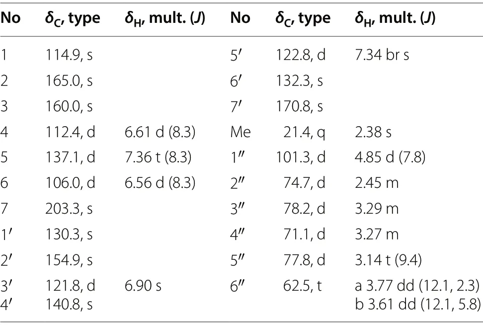
Table 1 1H (600 MHz) and.13C (150 MHz) NMR data of 1 in CD3OD (δ in ppm, J in Hz)
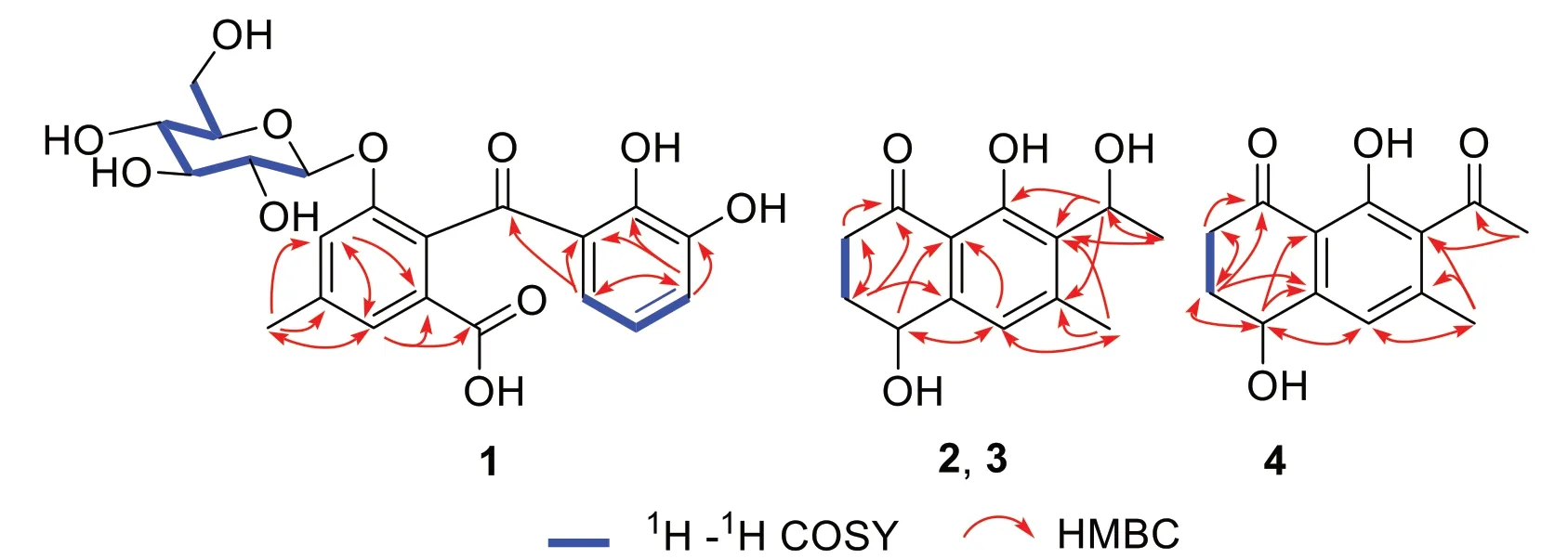
Fig.2 Key 1H-1H COSY and HMBC correlations of 1, 2, 3 and 4
Compound 1 is a new anthraquinone-related compound, whose formation mechanism might be similar to that of desmethylsulochrin, which was established by the ring-opening process of questin catalyzed by GedF (Geodin synthesis protein F) and GedK [25].InR.crispus,compound 1 maybe formed from reduction firstly and then ring-opening of ziganein-1-O-β-glucopyranoside catalyzed by GedF and GedK respectively (Fig.3).

Fig.3 Possible formation of compound 1
Compounds 2 and 3, obtained as colorless powder, are a pair of enantiomers possessing the same molecular formula C13H16O4, as deduced by the negative HRESIMS(m/z235.0979 [M-H]-calcd C13H15O4, 235.0976).The1H,13C NMR, and HSQC spectral data of 2 and 3 revealed the existence of two methyls [δH2.48 (3H, s,H-11),δC21.1;δH1.52 (3H, d,J= 6.7 Hz, H-10),δC21.1],two methylenes [δH2.88 (1H, m, H-2a), 2.67 (1H, m,H-2b),δC35.9;δH2.27 (1H, m, H-3a), 2.08 (1H, m, H-3b),δC32.6], one aromatic methine [δH6.89 (1H, s, H-5),δC121.9], two oxymethines [δH(1H, 4.78, dd,J= 8.0, 3.8 Hz,H-4),δC68.1;δH5.34 (1H, q,J= 6.7 Hz, H-9),δC65.6],one carbonyl group (δC206.1, C-1), and a set of quaternary aromatic carbons (δC146.4, 147.2, 130.9, 161.5,114.6).These spectroscopic features were similar to those of (4S,9S)-9-hydroxy-O-methylasparvenone (4S,9S-HM)[26], whose molecular weight was 252 Da, 16 Da more than those of 2 and 3.However, the chemical shifts of C-6 and C-11 in 2 and 3 were obviously different with those of 4S,9S-HM, indicating that the substituent at C-6 in 2 and 3 was methyl group, instead of a methoxyl group in 4S,9S-HM.Moreover, the HMBC correlations of H-11 (δH2.48) with C-5 (δC121.9), C-6 (δC147.2)and C-7 (δC130.9) confirmed the substitution of methyl at C-6 position.The1H-1H COSY correlations between H-2 and H-3, and the key HMBC correlations from H-2 to C-1/C-3, from H-3 to C-1/C-2, from H-4 to C-5/C-8a, from H-11 to C-5/C-6/C-7, from H-5 to C-8a, from H-10 to C-7/C-9, from H-9 to C-6/C-7/C-8 determined the planar structures of 2 and 3 as 4,8-dihydroxy-7-(1-hydroxyethyl)-6-methyl-3,4-dihydronaphthalen-1(2H)-one.Comparison of the experimental ECD spectrum of 2 with the calculated ECD (Fig.4) of the four stereoisomers, (4S,9S)-, (4R,9R)-, (4S,9R)- and (4R,9S)- HM [26]showed the ECD of 2 was more comparable with the computationally derived data for (4S,9S)- and (4S,9R)-with positive Cotton effects (CEs) at 248 and 214 nm and negative CE at 280 nm.However, the CE amplitudes of 2 were notably closer to those of the (4S,9S)- diastereomer, thus favoring the (4S,9S) absolute configuration for 2.Therefore, compound 2 was proposed as (4S,9S)-4,8-dihydroxy-7-(1-hydroxyethyl)-6-methyl-3,4-dihydronaphthalen-1(2H)-one.Similarly, the absolute configuration of 3 was deduced as (4R,9R)-4,8-dihydroxy-7-(1-hydroxyethyl)-6-methyl-3,4-dihydro-naphthalen-1(2H)-one.The structures of 2 and 3 were established as shown and named as naphthalenones A (2) and B (3),respectively.
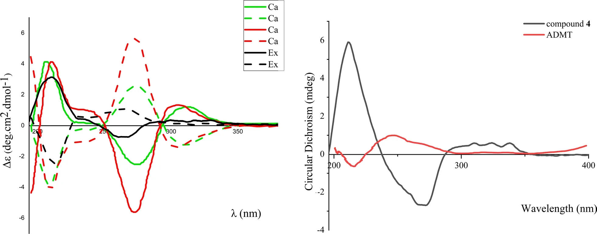
Fig.4 Experimental ECD curves of 2, 3, 4 and ADMT, and calculated ECD curves of four stereoisomers of HM
Compound 4 was isolated as white powder.Its molecular formula was assigned to be C13H14O4, as deduced from the HRESIMS (m/z233.0814 [M-H]-, calcd.C13H13O4, 233.0814).The UV spectrum showed absorption bands atλmax286 nm.The 1D and 2D NMR data of 4 (Table 2) indicated it shared the same 3,4-dihydronaphthalen-1(2H)-one skeleton with 2.The molecular weight of 4 is 2 Da less than that of 2.Comparing the DEPT and13C NMR data of 4 with those of 2 indicated that,a ketone group (δC206.5) appeared in 4, instead of an oxymethine C-9 (δC65.6) in 2.This was further verified by the HMBC correlation of H-10 with C-9 in the HMBC spectrum of 4.Comparing with the ECD curve of the known 7-acetyl-4R,8-dihydroxy-6-methyl-1-tetralone(ADMT) [27], compound 4 showed a negative CE at 260-270 nm in ECD spectrum, which is in contrast to ADMT(Fig.4).Therefore, the structure of 4 was determined to be 7-acetyl-4S,8-dihydroxy-6-methyl-1-tetralone and named as naphthalenone C.
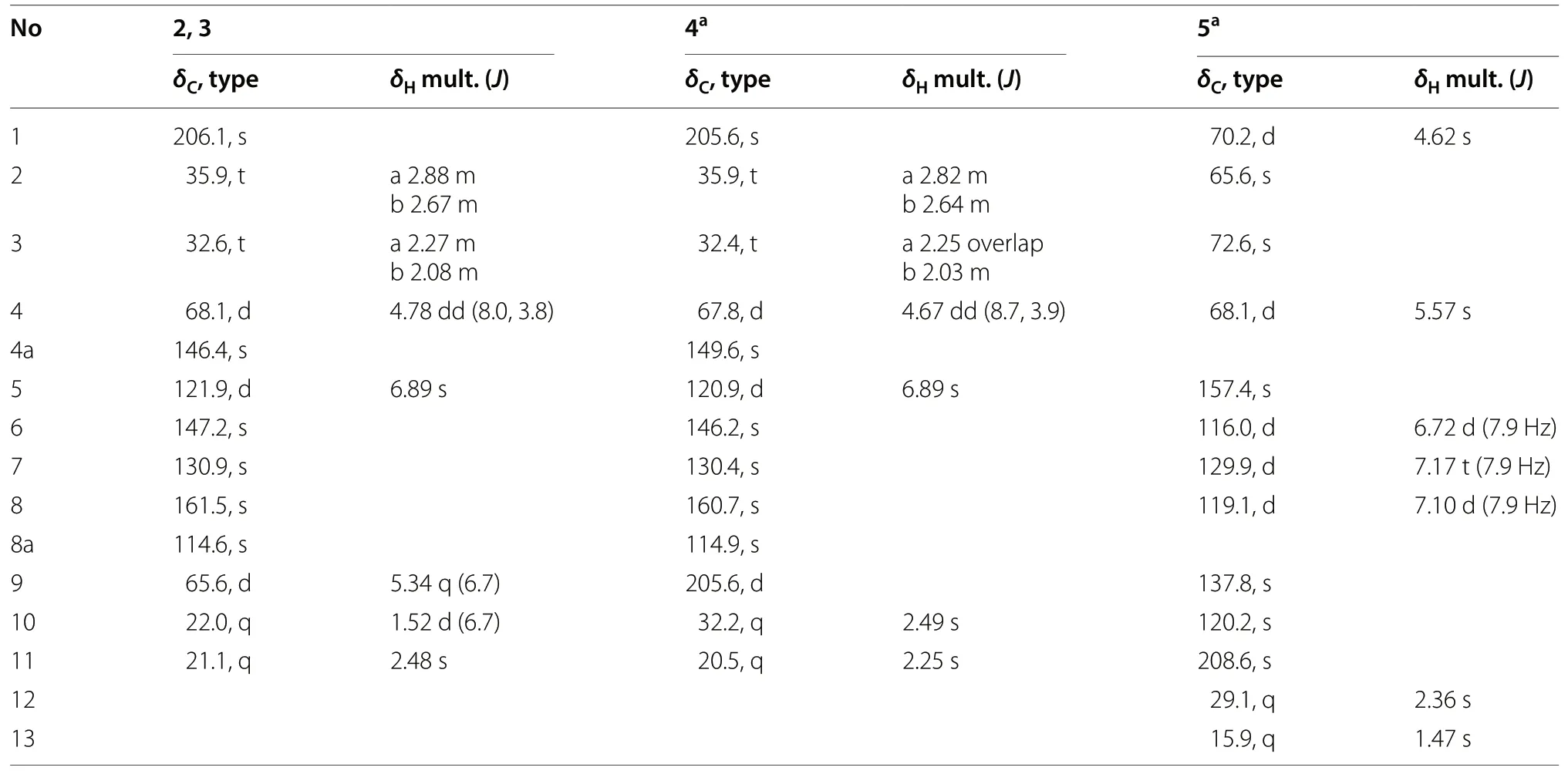
Table 2 1H (600 MHz) and13C (150 MHz) NMR data of 2, 3, 4 and 5 in CD3OD (δ in ppm, J in Hz)
Compound 5 was isolated as colorless needle crystal with a molecular formula of C13H14O5, as determined by the ESI-MS (negative ion mode)m/z249 [M-H]-, and13C NMR and DEPT spectroscopic data.The13C NMR spectrum of 5 showed one carbonyl (δC208.6), six aromatic carbon signals (δC157.4, 116.0, 129.9, 119.1, 137.8,120.2) with three methines and one oxygenated quarternary carbon, and sixsp3carbon signals (δC70.2, 65.6,72.6, 68.1, 29.1, 15.9) assignable to two methyls, two oxymethines and two oxy quarternary carbons.The1H NMR spectrum showed three characteristic proton resonances atδH6.72 (1H, d,J= 7.9 Hz, H-6), 7.17 (1H, t,J= 7.9 Hz, H-7), 7.10 (1H, d,J= 7.9 Hz, H-8) suggesting the existence of a 5,9,10-trisubstituted benzene ring.In addition, two methyls (δH2.36, s, H-12; 1.47, s, CH3-2)and two oxymethines (6.72, d,J= 7.9 Hz, H-6; 5.57, s,H-4) signals were observed.The above data indicated that 5 was a naphthoquinol derivative, whose epoxide ring was inferred by chemical shifts (δC65.6, C-2;δC72.6,C-3) and seven degrees of unsaturation.The detailed analyses of the 2D NMR (1H-1H COSY, HMQC, HMBC)spectra (Fig.5) deduced the planar structure of 5, which is the same as the reported 3-acetyl-2-methyl-1,4,5-trihydroxy-2,3-epoxynaphthoquinol without determination of the absolute configuration [15].In the present study,fine crystal from acetone was obtained and the absolute configuration of 5 was determined by single crystal X-ray diffraction (CDCC Number: 2158643) (Fig.5).The result confirmed the planar structure of 5, and revealed unambiguously the absolute configuration as 1R, 2R, 3S, 4S.
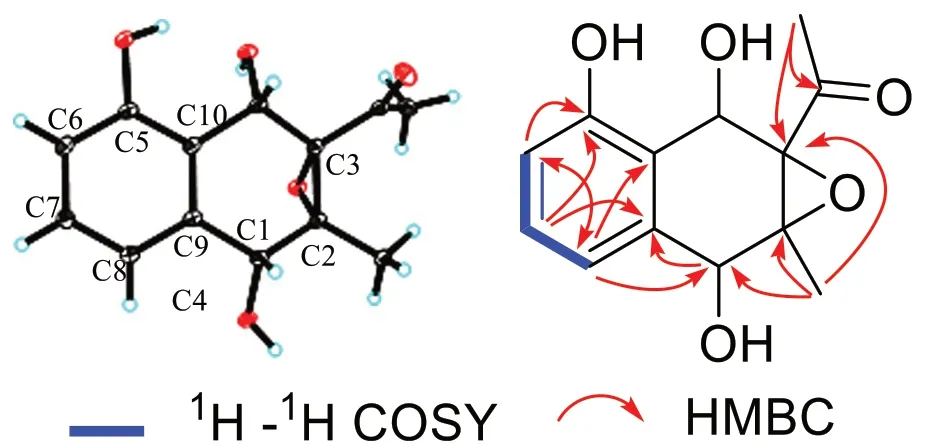
Fig.5 1H-1H COSY, HMBC spectra and X-ray crystallographic structure of 5
2.2 Anti-fungal and anti-inflammatory inhibitory activity
Compounds 1 and 6-14 were evaluated for their inhibitory effects against three skin fungi (Epidermophyton floccosum,Trichophyton rubrum,Microsporum gypseum)at a concentration of 100 μM (Table S1), as previously described [28], with terbinafine as positive control.Most of them displayed only weak antifungal activity against the three skin fungi, while compound 9 showed obvious antifungal activities againstE.floccosumandM.gypseumwith MIC50values of 2.467 ± 0.03 μM and 4.673 ± 0.077 μM, respectively.In case of the antifungal effects againstE.floccosumandM.gypseum,simple emodin type anthraquinone 9 showed the strongest inhibition, followed by oxyglucoside anthraquinone (12)orC-glucoside oxanthrones (13, 14), and finally glycosylated seco analogues (1, 6-8).These compounds (1,6-14) were also evaluated for their anti-inflammatory activity by using LPS to induce the production of iNOS from mouse monocyte macrophage RAW264.7, with L-NMMA as a positive control.The inhibition rate was shown in Table S2, all 10 anthraquinones showed NO inhibitory activity at a concentration of 50 μM, and the order of their inhibition rates is as follows: 9 > 11 > 12 >13 > 14 > 8 > 10 > 1 > 6 > 7.Compound 9 had the strongest anti-inflammatory effect, followed by oxyglucoside anthraquinone,C-glucoside oxanthrones and finally seco-anthraquinone glucosides.It is noted that the antiinflammatory and anti-fungal potential of emodin (9)decreased when it become glycosides or seco-anthraquinone cleavaged between C-10 and C-4a.
3 Experimental
3.1 General experimental procedures
UV spectra were given on a UV-2410PC Shimadzu spectrometer.One and two-dimensional NMR spectra were determined on acetone-d6and methanol-d4with Bruker Ascend-600 and AV-800 spectrometers.Chemical shifts(δ) were recorded in (parts per million, ppm) scale with TMS (Bruker, Zurich, Switzerland) as an internal standard.Coupling constants were expressed in hertz (Hz).ESI mass spectra were measured on a VG Auto Spec300 spectrometer.High-resolution electro-spray ionization mass (HRESIMS) spectra were performed on an API QSTAR Pular-1 spectrometer.Semi-preparative HPLC was performed on a Hanbon Sci & Tech with Capcell Pak Phenyl (250 mm × 10 mm, 5 μm) and Thermo Hypeersil GOLD aQ (250 mm × 9.4 mm × 5 μm) columns.Analytical HPLC was performed on a Waters 2695 Series HPLC system equipped with a reverse-phase ZORBAX SB-C-18 column (4.6 mm × 150 mm, 5 μm, Agilent Corporation, USA).Column chromatography (CC) was carried out using Sephadex LH-20 (25-100 μm, Pharmacia Fine Chemical Co., Ltd., Uppsala, Sweden), 75-100 μm MCI-gel CHP20P (Mitsubishi Chemical Co.Ltd., Tokyo,Japan), silica gel (100-200 mesh, Qingdao Marine Chemical, Inc., Qingdao, China) and macro-porous absorption resin (D101, Donghong Chemical Co., Ltd., People’s Republic of China).Acetonitrile (chromatographic grade)were purchased from XinLanJing (Pennsylvania, USA).Mouse mononuclear macrophage RAW264.7 was purchased from the Shanghai Cell Bank of the Chinese Academy of Sciences, DMEM medium and fetal bovine serum were purchased from BI Company.Griess Reagent, LPS,Terbinafine hydrochloride, DMSO and control drug L-NMMA were purchased from Sigma.Epidermophyton floccosum,Trichophyton rubrumandMicrosporum gypseumwere purchased from the Medical Fungal Conservation Centre, Chinese Academy of Medical Sciences.
3.2 Plant materials
The roots ofR.crispuswere collected from Yimen town,Xianyang City, Shaanxi Province, in August 2020, and identified by Dr.En-De Liu from Kunming Institute of Botany (KIB), Chinese Academy of Sciences (CAS).A voucher specimen (KIBZL-20200803) is deposited at State Key Laboratory of Phytochemistry and Plant Resource in West China of KIB-CAS.
3.3 Extraction and isolation
The air-dried roots ofR.crispus(10.0 kg) were crushed into small pieces and extracted with 90% aqueous MeOH at 60 °C (15 L × 4, each time 2 h).The organic solvent was removed under reduced pressure to yield a residue(1.6 kg), which was further extracted with ethyl acetate.After concentrated, the aqueous layer (480 g) was applied to a Sephadex LH-20 column chromatography (CC), eluting with water-methanol (1:0-0:1) to give two fractions(I-II).Fr.I (70 g) was subjected to CC over macroporous resin D101, eluting with H2O firstly to remove the sugars, and then with 100% MeOH.The yielded MeOH fraction (50.5 g) was subjected to CC over silica gel, eluting with a CHCl3/MeOH gradient system (1:0, 9:1, 8:2,7:3, 6:4, 1:1, 0:1) to yield 6 fractions, A-F.Fr.A (10 g) was chromatographed on silica gel column with a petroleum ether/ethyl acetate gradient system (1:0, 9:1, 8:2, 7:3, 6:4,1:1, 0:1) to yield 11 (4.8 mg), 10 (5.0 mg), 9 (100.0 mg).Fr.B (19.5 g) was appplied to RP-18 CC with a MeOH/H2O gradient system (from 0:1 to 1:0) to afford fractions B1-B4.Fr.B2 (500 mg) was purified by semipreparative HPLC (3 mL/min) with 7% MeCN/H2O (7:93) containing 0.1% trifluoroacetate to yield 1 (7.0 mg, retention time = 9 min), 6 (12.5 mg, retention time = 7 min), 7(37.0 mg, retention time = 8 min), 8 (14.0 mg, retention time = 12 min), 12 (1.50 mg, retention time = 14 min),13 (3.70 mg), 14 (17.0 mg).Fr.C (2.5 g) was separated by chromatography on a Chromatorex ODS column (2.5 cm i.d.× 35 cm) with 10 - 60% MeOH (5% stepwise, each 500 mL) to give Fr.C1 (50 mg) and Fr.C2 (1.0 g).Further,Fr.C1 was separated by semipreparative HPLC (Capcell Pak Phenyl, 250 mm × 10 mm × 5 μm, CH3CN/H2O 22:78) to obtain 2 (5.0 mg, retention time = 13.5 min) and 3 (12.0 mg, retention time = 17.3 min), and Fr.C2 was separated by semipreparative HPLC (Thermo Hypeersil GOLD aQ, 250 mm × 9.4 mm × 5 μm, CH3CN/H2O 28:72) to obtain 4 (50.0 mg, retention time = 8.0 min), 5(305.0 mg, retention time = 10.0 min), respectively.
3.3.1 Crispuside A (1)
Yellowish amorphous powder; UV (MeOH)λmax(log ε)206 (4.4), 274 (3.7) nm.1H (600 MHz) and13C (150 MHz)NMR (in methanol-d4) data, see Table 1.ESIMSm/z449 [M-H]-; HRESIMSm/z449.1094 [M-H]-, (calcd C21H21O11: 449.1089).
3.3.2 Naphthalenone A (2)
3.3.3 Naphthalenone B (3)
3.3.4 Naphthalenone C (4)
3.3.5 (1R,2R,3S,4S)-3-Acetyl-2-methyl-1,4,5-trihydroxy-2,3-epoxynaphthoquinol (5)
White amorphous powder;1H (500 MHz) and13C(125 MHz) NMR (in methanol-d4) data, see Table 2.ESIMSm/z249 [M-H]-.
3.3.6 Single-crystal X-ray diffraction data of 5
Colorless crystal of 5: C13H14O5,M= 250.24,a= 7.7634(3) Å,b= 8.2971 (3) Å,c= 9.2218(3) Å,α= 90°,β= 107.7570 (10)°,γ= 90°,V= 565.71 (4) Å3,T= 100 (2)K, space groupP1211,Z= 2,μ(Cu Kα) = 0.954 mm-1,10,180 reflections measured, 2156 independent reflections (Rint= 0.0500).The finalR1values were 0.0319[I> 2σ(I)].The finalwR(F2) values were 0.0821 [I> 2σ(I)].The finalR1values were 0.0333 (all data).The finalwR(F2)values were 0.0832 (all data).The goodness of fit onF2was 1.064.Flack parameter = 0.17(10).Crystallographic data for the structure of 5 have been deposited in the Cambridge Crystallographic Data Centre (deposition number CCDC, 2,158,643).Copies of the data can be obtained free of charge from the CCDC via www.ccdc.cam.ac.uk.
3.4 The biological assay
The isolates 1 and 6-14 were evaluated for their antifungal against three skin fungi (Epidermophyton floccosum,Trichophyton rubrum,Microsporum gypseum) and anti-inflammatory activity.For anti-fungal activity, Terbinafine hydrochloride was used as positive control.Fungal broth (5 × 10⁵ CFU mL-1) and test samples (100 μM)were incubated in 96-well plates at 25 °C for 5 days, A microplate reader was recorded by the absorbance at 625 nm.The experiment also set up the culture medium blank control, fungi control and terbinafine hydrochloride positive drug control.
The anti-inflammatory activity of 1 and 6-14 was screened as previously reported method [29].The mouse mononuclear macrophage RAW264.7 was inoculated to 96 orifice plates and induced by 1.0 μg/mL LPS.At the same time, compounds 1 and 6-14 (final concentration 50 μM) was added, and no drug group and L-NMMA positive drug group were taken as controls.After the cells were cultured overnight, the medium was used to detect NO production and the absorbance was measured at 570 nm.MTS was added to the remaining medium to detect the cell survival rate and exclude the toxic effects of the compounds.The formula to calculate the inhibition rate is as follows: NO production inhibition rate (%) = (non-drug treatment group OD570nm- sample group OD570nm)/non-drug treatment group OD570nm× 100%.
4 Conclusion
Four new (1-4) and ten known (5-14) quinone derivatives were isolated and identified from the roots ofRumex crispusL.Compound 1 is a seco-anthraquinone glucoside, while 2-4 belong to naphthalenones containing 3,4-dihydronaphthalen-1(2H)-one moiety.The absolute configuration of 5 was determined for the first time by X-ray single crystal diffraction.The anti-fungal and anti-inflammatory activity of anthraquinones (1, 6-14)was tested, of which compound 9 showed obvious antifungal activity.The results indicated that simple emodin type anthraquinone is more potential against skin fungi than its oxyglucosyl,C-glucosyl and glycosylated seco analogues.
Supplementary Information
The online version contains supplementary material available at https:// doi.org/ 10.1007/ s13659- 022- 00350-3.
Additional file 1.Supporting information.
Acknowledgements
The authors are grateful to the staffs of the analytical and bioactivity screening groups at the State Key Laboratory of Phytochemistry and Plant Resources in West China, Kunming Institute of Botany, Chinese Academy of Sciences,for measuring the spectroscopic data, anti-fungal, and anti-inflammatory activities.
Author contributions
YXL carried out the experiments and drafted the manuscript; NL and JJL revised the manuscript; DW, HTZand MZ completed samples collection;YJZ designed the experiments, revised the manuscript.All authorsread and approved the final manuscript.
Funding
This research was supported by Ministry of Science and Technology of the People’s Republic of China, 2021YFE0103600.
Declarations
Competing interests
The authors declare no competing financial interest.
Author details
1State Key Laboratory of Phytochemistry and Plant Resources in West China,Kunming Institute of Botany, Chinese Academy of Sciences, Kunming 650204,People’s Republic of China.2University of Chinese Academy of Sciences,Beijing 100049, People’s Republic of China.3Yunnan Key Laboratory of Natural Medicinal Chemistry, Kunming Institute of Botany, Chinese Academy of Sciences, Kunming 650201, People’s Republic of China.
 Natural Products and Bioprospecting2022年5期
- Natural Products and Bioprospecting的其它文章
- Apatite phosphate doped by cobalt as hight efficient catalyst of multi-component synthesis of therapeutic spiropyrimidine compound
- Therapeutic roles of plants for 15 hypothesised causal bases of Alzheimer’s disease
- Beauty of the beast: anticholinergic tropane alkaloids in therapeutics
- Tetra-, penta-, and hexa-nor-lanostane triterpenes from the medicinal fungus Ganoderma australe
- Mulberry Diels-Alder-type adducts:isolation, structure, bioactivity, and synthesis
- Arctostaphylos uva‑ursi L.leaves extract and its modified cysteine preparation for the management of insulin resistance:chemical analysis and bioactivity
