
Ultrasound biomicroscopic imaging demonstrate thinner ciliary body thickness in eyes with angle closure
2022-09-14 06:50ShiYanChenNaHeYuJieYanXiangFanLingLingWu
INTRODUCTION
Angle closure has been proved to be associated with specific anatomic features of the globe. Compared with normal eyes, eyes with primary angle closure (PAC) and primary angle-closure glaucoma (PACG) present biometric features of shallow anterior chamber depth, narrow angle, short axial length, thick lens and anterior lens position
. Moreover,eyes with specific peripheral iris configuration, such as plateau iris, thick iris and anteriorly inserted iris, are also associated with angle closure, including appositional angle closure after laser peripheral iridotomy (LPI)
.
Ciliary body is one of the important parts of the angle, and the biometric features of the ciliary body were proved to be related with angle closure as well. Previous studies have revealed that anterior situation of the ciliary processes is a predisposing factor of PAC/PACG
. The anteriorly situated ciliary processes are thought to be the main cause of plateau iris by pushing the iris root toward the trabecular meshwork
,and may induce thick lens and anterior lens position through loosening the zonules as well
.
Ciliary body thickness is supposed to be another potential factor for angle closure
. Gohdo
first found thinner ciliary body thickness in eyes with gonioscopic narrow but open angle (Shaffer classification, grade 0 to 2). Then similar results were also found in eyes with some special kinds of angle-closure glaucoma: thinner ciliary body thickness was found in eyes with acute primary angle closure (APAC)
,and also in eyes with malignant glaucoma
. However, there were no definite results yet about the ciliary body thickness in eyes with PAC/PACG compared to the normal ones.
“仿佛晋武都守李仲文、广州守冯孝将儿女事”,指的是东晋陶潜志怪小说《搜神后记》中的《李仲文女》《冯孝将子》,又见《法苑珠林》等书的记载。
为此,要加强农村基础设施建设,通过多种渠道改善交通运输状况,为农村经济发展提供畅通条件。支持重点农产品批发市场建设和升级改造,落实农产品批发市场用地等扶持政策。支持大型涉农企业投资建设大型优质农产品物流配送中心,加大力度建设大宗农产品仓储设施,完善鲜活农产品冷链物流体系。
SUBJECTS AND METHODS
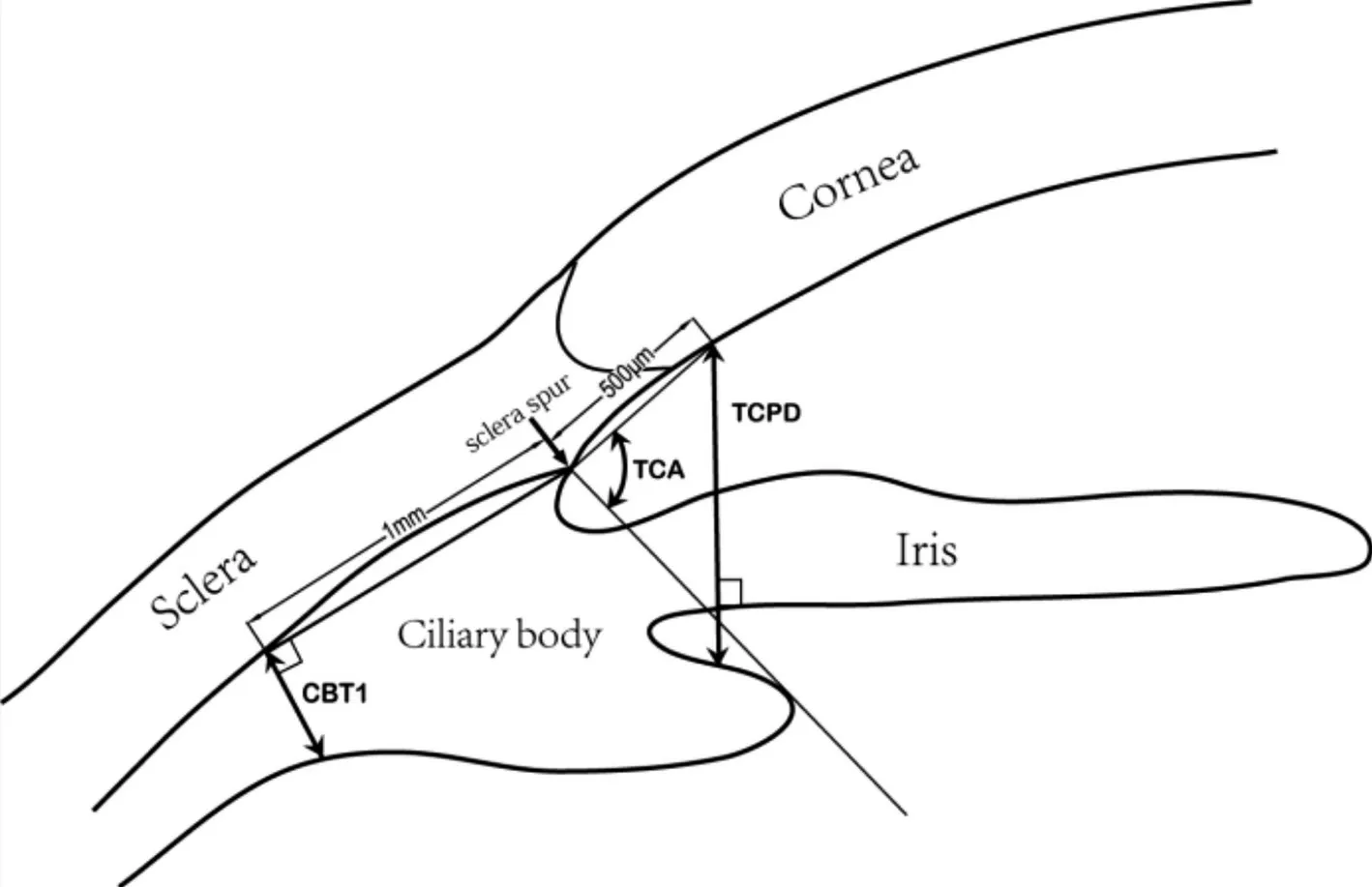
All images were measured by one masked observer (Chen SY), using an originally developed semiautomatic measuring software. This measuring software was improved on the basis of UBM PRO 2000 software (Paradigm Medical, Salt Lake City, UT, USA). The following parameters were measured as previously described
(Figure 1): 1) the ciliary body thickness at the position of 1 mm posterior to the scleral spur(CBT1); 2) the trabecular-ciliary process distance (TCPD)defined as the length of the line extending from the corneal endothelium 500 μm from the scleral spur perpendicularly through the posterior surface of the iris to the ciliary process;3) the trabecular-ciliary process angle (TCA) measured with the scleral spur as the apex, and the corneal endothelium along with the anterior surface of ciliary process as the two arms of the angle.
UBM examination was performed with a UBM (model P45, Paradigm Medical, Salt Lake City,UT, USA) equipped with a 50-MHz transducer probe allowing 5.0×5.0 mm
field of view and approximately 50 μm spatial resolution. All subjects were examined in the supine position in a dark room (illumination <1 lx, measured with an ST-92 luminance meter; Beijing Teachers University Photoelectricity Instrument Factory, Beijing, China). After topical anaesthesia,the globe was placed with an eyecup filled with hydroxyethyl cellulose as a coupling agent. Subjects were instructed to relax and focus on a fixation target about 1 m in front of the eyes to avoid the effect of accommodation. Each eye was examined in the way of radial scan through the centre of the pupil, and images of the 3, 6, 9, 12 o’clock were obtained to represent each quadrant respectively.
从理论上讲,汽油发动机尾气排气成分主要有碳氢化合物(HC)、一氧化碳(CO)、二氧化碳(CO2)和氮氧化合物(NOx)4种成分组成,其中有害物质主要有碳氢化合物(HC)、一氧化碳(CO)、氮氧化合物(NOx)。通过三元催化的氧化还原反应后,碳氢化合物变成水和二氧化碳,一氧化碳变成二氧化碳和氮。为了更好地达到氧化还原效果,需要汽油与空气完全燃烧,且没有残留氧气的理论空燃比为14.7∶1。利用空燃比反馈就是要将这一比例无限精确,才能保证三元催化转化器的转化效率最高。
Normal control subjects were consecutively recruited from general ophthalmologic clinic of Peking University Third Hospital between January 2009 and December 2009. The inclusion criteria for normal subjects included: 1) age between 40 and 80y; 2) willing and capable to attend this study. And one was excluded if presenting any intraocular diseases(including PAC/PACG, except for mild cataracts and refractive error with the spherical equivalent within -8 D and 4 D), or having previous intraocular surgery or laser treatment history,or any other condition conforming to the exclusion criteria for the PAC/PACG group.
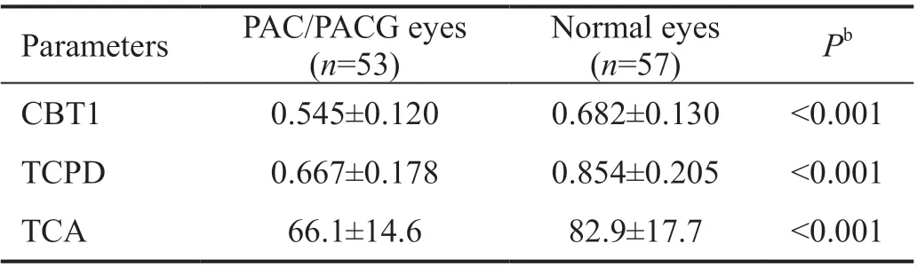
After excluding images of quadrants with PAS, 110 non-PAS images randomly selected from the matched two groups were measured, and the PAC/PACG group also showed the same features (Table 3). Moreover, the distributions of non-PAS images among the four quadrants were agreed in the two groups (Chi-square test,
=0.933).
This was a cross-sectional comparative study consisting of two groups: PAC/PACG patients and normal subjects. This study was performed at the Peking University Third Hospital, Peking University Eye Centre by 5 clinicians, including 2 full-time attending glaucoma specialists (Wu LL, Fan X) and 3 glaucoma fellows (Chen SY, He N, Yan YJ). Patients with PAC/PACG were consecutively enrolled in this study who underwent LPI at the glaucoma clinic of Peking University Third Hospital between January 2009 and December 2009. The inclusion criteria included: 1) age between 40 and 80y; 2) conformed to the diagnostic criteria of PAC/PACG. The diagnostic criteria of PAC were defined as follows
: an eye with an occludable drainage angle (an angle in which ≥270 degree of the posterior trabecular meshwork could not be seen with static gonioscopy) and features that indicated trabecular obstruction by the peripheral iris had occurred, such as peripheral anterior synechia (PAS), elevated intraocular pressure (IOP), or excessive pigment deposition on the trabecular surface, and without glaucomatous damage to the optic disc. While PACG was defined as presence of glaucomatous optic neuropathy(such as loss of neuroretinal rim with a vertical cup-to-disc ratio of >0.7 and/or notching with nerve fibre layer defect) with corresponding visual field loss on the basis of the diagnosis of PAC. Patients were excluded if any of the following conditions present: 1) secondary angle closure, such as neovascularization of the iris, uveitis, trauma, tumour, lens intumescence or subluxation; 2) any other ocular diseases (except for mild cataracts and refractive error with the spherical equivalent within -8 D and 4 D) or previous intraocular surgery; 3) unable to perform contact examination such as gonioscopy or UBM;4) sustained pilocarpine or prostaglandin administration which might affect ciliary body morphology
; 5) eyes with more than two quadrants of PAS and uncontrolled IOP with medications, which indicated for the filtration surgery; 6) acute attack history which might lead obvious uveal effusion
or pupil distortion and iris whirling.
The study followed the tenets of the Declaration of Helsinki and was approved by the Institutional Review Board of Peking University Third Hospital. Signed informed consent was obtained from each subject involved in this study.
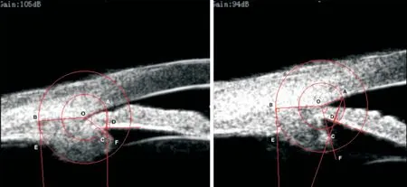
The measurement patterns by the software were presented in Figure 2. The semiautomatic measuring procedure was completed as follows: 1) randomly select one image for measurement; 2) mark the sclera spur by the observer and two circles centred on the sclera spur with radius of 500 μm and 1 mm were automatically drawn; 3) mark point A which is the intersection of the smaller circle and the inner surface of the cornea, then draw a line along the posterior surface of the iris and get a perpendicular line to iris through point A automatically, and mark the intersection of the perpendicular line and the anterior surface of the ciliary processes as point C; 4) mark point B as the intersection of the bigger circle and the outer surface of the ciliary body, and get a perpendicular line to line BO automatically, and then mark point E as the intersection of the perpendicular line and inner surface of ciliary body; 5) mark point F to make line OF the tangent lineof the anterior surface of the ciliary processes; 6) measure distances AC, BE, and angle AOF as TCPD, CBT1 and TCA,and then save the data automatically.
电力设计企业的核心竞争力是人才,支持PPP业务的发展需要PPP专业人才,电力设计企业应提前储备PPP专业人才,从而抢滩PPP市场。储备人才包括引进和培养两种模式,因为PPP是新领域,专业人才紧缺,引进人才不仅成本高昂,还可能水土不服,所以电力设计企业应重点从内部挖掘、培养人才,通过创新培养体系,注重理论与实践结合,邀请实战派专家进行实操培训、沙盘演练,结合实际问题进行讲授,重点培养PPP项目投融资、SPV公司组建及运营、财务税收筹划、经济模型、风险管理和代建管理等方面的能力,从而储备一支专业的PPP人才队伍,满足企业PPP业务发展需求,加快转型升级。
Statistical Analysis Description statistics and comparisons for general features of both groups were made by independent
-test (for quantitative data) and Chi-square test (for proportion data). If the age and gender were not matched between the PAC/PACG group and normal group, a propensity score matching (PSM, 1:1 matching with calliper set at 0.02, adjusting for the covariates of age and gender) should be performed to minimize the threat of selection bias. Comparisons of all the quantitative parameters between the matched two groups were performed using the independent
-test for the data was normally distributed. The comparisons included the general means of the four quadrants, means of each quadrant and the means of images without PAS. Meanwhile, the distributions of non-PAS images among the four quadrants between the matched two groups were compared by Chi-square test. The linear regression analysis was performed to investigate the association between CBT1 and the parameters for the ciliary body position (TCPD, TCA). At last, the intraclass correlation coefficient (ICC, with one-way random effects model) was applied to assess the intra-observer reproducibility.
The right eye was chosen to be the studied one unless only the left eye met the eligibility criteria. Images were removed if anterior uveal cysts presented, or the measurement area was beyond the image. Subjects without all four images (at 3, 6, 9, 12 o’clock) were excluded. In order to eliminate the potential influence of the PAS on measurement, we also made a random extraction of one image without PAS from each subject. Intra-observer reproducibility of UBM measurements for such parameters was pretty good as reported in previous studies
. A number of 100 randomly selected images were remeasured by the same masked observer 4wk later after the initial measurement to investigate the test-retest reliability.
RESULTS
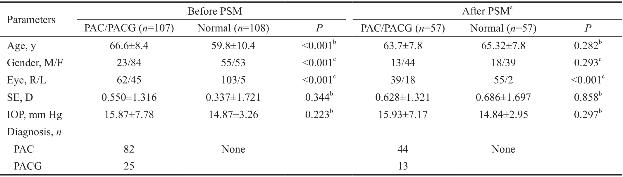
There were totally 217 PAC/PACG patients enrolled in this study according to the inclusion criteria, and 74 patients were excluded mainly due to acute attack history and other conditions such as combined ocular diseases, intolerable for UBM examination and so on. A number of 120 normal subjects were enrolled in the normal control group. During the UBM images measurement, 36 PAC/PACG patients and 12 normal subjects were excluded for presenting anterior uveal cysts or failed measurement beyond the image area in any quadrant. Then, a total of 215 eyes of 215 subjects (107 patients with PAC/PACG and 108 normal subjects) were eligible for analysis. General information of the subjects was summarized in Table 1. For the age and gender were not matched in the two groups, the PSM adjusting for age and gender was performed. After PSM,114 eyes of 114 subjects (57 per group) were analysed. The average spherical equivalent and IOP presented no significant differences between the two groups. In the PAC/PACG group,the PAC-to-PACG ratio was approximately 3:1.
Our previous work found that the Chinese ethnic had thinner ciliary body and more anteriorly positioned ciliary processes than the Caucasians
, which was consistent with the higher prevalence of angle closure in Chinese population. To further investigate the effect of ciliary body thickness on eyes with angle closure, this ultrasound biomicroscopy (UBM) study was carried out to quantitatively compare the ciliary body configuration between eyes with PAC/PACG and the normal eyes.
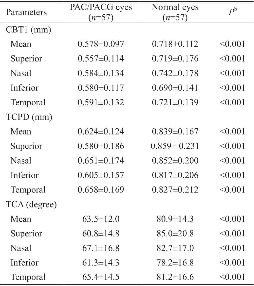
After PSM, there were totally 456 UBM images analysed from the 114 eyes. Comparisons of the parameters of ciliary body between the two groups were presented in Table 2. In general means comparison, eyes with PAC/PACG showed significantly thinner CBT1 (PAC/PACG, 0.578±0.097 mm; normal, 0.718±0.112 mm;
<0.001), shorter TCPD (0.624±0.124 mm;0.839±0.167 mm;
<0.001) and narrower TCA (63.5°±12.0°;80.9°±14.3°;
<0.001) than the normal eyes. And the results of the quadrant-based comparisons highly agreed with the general means analysis in each quadrant, which suggested that eyes with PAC/PACG had thinner ciliary body thickness and more anteriorly situated ciliary processes.
我心中充满了无限的宠溺之意,尽情地感受着昙花无限的魅力。我知道,到了早上,昙花的花冠就会慢慢闭合,它美丽的容颜将不复存在,仅剩一缕余香……
All subjects underwent comprehensive ocular examinations,including best-corrected visual acuity, IOP measurement by Goldmann applanation tonometry, slit-lamp biomicroscopy,direct ophthalmoscopy, static and dynamic gonioscopy, and UBM examination. For patients with PAC/PACG, all of the ocular examinations were performed one month after LPI. IOP record of the eyes studied was measured just before the UBM examination. Gonioscopy was performed in dark environment using a Goldmann single-mirror gonioscopy lens. Part of the eyes with PAC/PACG were under treatment with IOP-lowering medications (except prostaglandin), but administration of pilocarpine was discontinued for two weeks or more.
The simple linear regression analysis showed TCPD(
=0.537,
<0.001) and TCA (
=0.517,
<0.001) had significant correlation with CBT1, both in the whole and in each group (Table 4). The intra-observer reproducibility for the parameters was good in this study. The ICCs for the TCPD,TCA, CBT1 were 0.904, 0.925, 0.847, respectively.
DISCUSSION
In this study, ciliary body thickness was found to be significantly thinner in eyes with PAC/PACG than in normal eyes, after age and gender matched using PSM. Further quadrant-based comparisons and the comparison in images without PAS showed that eyes with PAC/PACG still presented the feature of thinner ciliary body after eliminating potential influence of quadrants and PAS. This study strongly demonstrated thinner ciliary body thickness in eyes with PAC/PACG, supporting the hypothesis that thin ciliary body thickness might be a potential factor for angle closure
.
This finding is also in agreement with several previous studies. Gohdo
found eyes with narrow angle (Shaffer classification, grade 0 to 2) had thinner ciliary body thickness than normal control eyes. However, Gohdo
’s
study only enrolled 36 eyes and was still limited in eyes with narrow but open angle. Li
showed eyes with APAC had thinner ciliary body compared with fellow eyes. Wang
also found eyes with APAC and malignant glaucoma presented thinner ciliary body thickness than the matched control eyes.Another study indicated the younger patients with PAC disease presented thinner and more anteriorly rotated ciliary body than the older ones
. These studies indicated that thinner ciliary body thickness might also be a predisposing factor for special kinds of angle closure glaucoma such as APAC and malignant glaucoma.
Contrary to the current study, some previous studies found eyes with angle closure had larger ciliary body
. And it was proved that Valsalva manoeuvre could cause thickening of the ciliary body and induce narrowing of the angle
. However,the parameters for ciliary body measured in above studies mainly represented the thickness of anterior part of ciliary body which included ciliary muscle and most stroma, and may be much more variable due to the effect of ciliary processes.While the parameter CBT1 applied in our study measured the relatively posterior ciliary body, mostly the longitudinal fibres.Therefore, we speculated that the reduction of the ciliary body thickness in eyes with angle closure mainly happened on the ciliary muscle. And thickening of the ciliary body during Valsalva manoeuvre was due to venous stasis and rise in episcleral venous pressure, which was not a common state and was avoided during the UBM examination in our study.The explanation for thinner ciliary body thickness in eyes with angle closure remains to be clarified. Previous studies showed ciliary body thickness was affected by various factors and might offer explanations. First, as we know old age is one of the risk factors of PACG
. Thus thinner ciliary body in eyes with PAC/PACG might be a manifestation of ciliary muscle atrophy related to aging, as showed in histological
andbiometric
studies. However, thinner ciliary body thickness has been found in younger patients with PAC disease
, which indicates age might not be major factor. Therefore, we applied PSM to diminish the effect of age in this study, and thinner ciliary body was still observed in eyes with PAC/PACG.Second, thinner ciliary body and anteriorly rotated ciliary process were also found in eyes with aniridia combined with ciliary body hypoplasia
. Therefore, we speculated that such thinner ciliary body in eyes with angle closure might also be related to inherent hypoplasia to some extent. Third, ciliary body thickness was proved to be positively correlated with axial length
, and thinner ciliary body might be explained by shorter axial length in eyes with angle closure. In this study, no significant difference in the spherical equivalent was found between the two groups, even though the data of axial length hadn’t been collected. Our previous work showed the difference of CBT1 between the Caucasians and Chinese was independent of axial length
. Therefore, shorter axial length might not be the only explanation for thinner ciliary body thickness in eyes with angle closure.

Besides thinner thickness, more anteriorly situated ciliary processes were also found in eyes with PAC/PACG than in normal eyes in the present study, which was widely agreed with previous studies
. And the reduction of the ciliary body thickness was significantly correlated with the anterior situation of the ciliary processes. This correlation has been previously found in normal eyes
, but not in eyes with angle closure before. As known, anterior situation of ciliary processes was proved to be associated with angle closure, while thinner ciliary body thickness might also be another predisposing factor for angle closure, and might induce angle closure through the same way as anterior situated ciliary processes do.One potential explanation for the effects of thinner ciliary body on angle closure was proposed as follows. Attaching firmly to the scleral spur, the ciliary muscle could prevent ciliary processes from anterior rotation. Thinner ciliary muscle may not be strong enough to attach to the scleral spur and may lead anterior situation of ciliary processes
, and then induce angle closure through the formation of plateau iris, or cause more anterior and thicker lens with greater lens vault and shallower anterior chamber depth by loosening the zonules
.
1.3.1 以Wbester评分标准为依据,对两组患者疗效进行判定。计算公式为:(治疗前积-治疗后积分)/治疗前积分×100%;计算结果为50%以上则表示痊愈、20%~49%表示显效、1%~19%表示有效、0%为无效。
There are several limitations in this study. First, UBM examinations for all PAC/PACG patients were performed after
LPI which differed from that in normal group. Some previous studies suggested widening of the anterior chamber angle(trabecular-iris angle, TIA, angle-opening distance, AOD),deepening of the anterior chamber depth after LPI in eyes with angle closure
, but the effect of LPI on the parameters of ciliary body has been poorly studied with controversial results.In one study TCPD was found to be increased
, while the other two studies indicated no significant changes of TCPD and CBT after LPI
. Even if TCPD is increased by LPI, the TCPD of the eyes with PAC/PACG is still shorter than that of normal eyes in this study. Second, the exclusion of the eyes with acute attack history or more than two quadrants of PAS may result in selection bias. Third, the data of axial length was not obtained for further analysis in this study, while the association between CBT1 and ethnicity difference adjusted for axial length was analysed in our previous work
.In conclusion, this UBM study demonstrated that eyes with PAC/PACG had thinner ciliary body thickness and more anteriorly situated ciliary processes than normal eyes. And thinner ciliary body thickness was associated with anterior situation of the ciliary processes. Thinning of the ciliary body might be one of the predisposing factors for angle closure.However further experimental study is required to comfirm this as a cause-and-effect relationship between ciliary body thinning and angle closure.
1.2.9 实时荧光定量RT-PCR检测肺组织TLR4 mRNA表达 Trizol法提取大鼠肺组织匀浆总RNA,取500 ng RNA进行逆转录,按说明书合成cDNA,引物序列见表1。按PCR反应试剂盒说明书进行反应,反应条件为:95℃ 30 s,95℃ 5 s,60℃ 10 s,共40个循环。每一例样本反应结束后由软件读出结果Ct值。
None;
None;
秦明月啊一声,心想罗伽在搞什么名堂?口中忙解释说:“没有啊,我真不知道这事,卢局你看,现在事情这么多,我哪有什么心情去搞什么聚会啊。”
None;
None;
None.
1 Zhang Y, Zhang Q, Thomas R, Li SZ, Wang NL. Development of angle closure and associated risk factors: the Handan eye study.
2022;100(1):e253-e261.
2 Mansoori T, Balakrishna N. Anterior segment morphology in primary angle closure glaucoma using ultrasound biomicroscopy.
2017;11(3):86-91.
3 Yan YJ, Wu LL, Wang X, Xiao GG. Appositional angle closure in Chinese with primary angle closure and primary angle closure glaucoma after laser peripheral iridotomy.
2014;55(12):8506-8512.
4 Li MW, Chen YH, Chen XX, Zhu WQ, Chen XL, Wang XL, Fang Y,Kong XM, Dai Y, Chen JY, Sun XH. Differences between fellow eyes of acute and chronic primary angle closure (glaucoma): an ultrasound biomicroscopy quantitative study.
2018;13(2):e0193006.
5 Kwon J, Sung KR, Han S, Moon YJ, Shin JW. Subclassification of primary angle closure using anterior segment optical coherence tomography and ultrasound biomicroscopic parameters.
2017;124(7):1039-1047.
6 Mansoori T, Sarvepally VK, Balakrishna N. Plateau iris in primary angle closure glaucoma: an ultrasound biomicroscopy study.
2016;25(2):e82-e86.
7 Gohdo T, Tsumura T, Iijima H, Kashiwagi K, Tsukahara S. Ultrasound biomicroscopic study of ciliary body thickness in eyes with narrow angles.
2000;129(3):342-346.
8 He N, Wu LL, Qi M, He MG, Lin S, Wang X, Yang F, Fan X.Comparison of ciliary body anatomy between American caucasians and ethnic Chinese using ultrasound biomicroscopy.
2016;41(4):485-491.
9 Wang ZH, Chung C, Lin JL, Xu JN, Huang JJ. Quantitative measurements of the ciliary body in eyes with acute primary-angle closure.
2016;57(7):3299-3305.
10 Li XY, Wang W, Huang WB, Chen SD, Wang JW, Wang ZH, Liu YM, He MG, Zhang XL. Difference of uveal parameters between the acute primary angle closure eyes and the fellow eyes.
2018;32(7):1174-1182.
11 Wang ZH, Huang JJ, Lin JL, Liang XW, Cai XY, Ge J. Quantitative measurements of the ciliary body in eyes with malignant glaucoma after trabeculectomy using ultrasound biomicroscopy.
2014;121(4):862-869.
12 Foster PJ, Buhrmann R, Quigley HA, Johnson GJ. The definition and classification of glaucoma in prevalence surveys.
2002;86(2):238-242.
13 Park S, Kang S, Lim J, Park E, Nam T, Jeong S, Seo K. Effects of prostaglandin-mediated and cholinergic-mediated miosis on morphology of the ciliary cleft region in dogs.
2018;79(9):980-985.
14 Yang JG, Li JJ, Tian H, Li YH, Gong YJ, Su AL, He N. Uveal effusion following acute primary angle-closure: a retrospective case series.
2017;10(3):406-412.
15 Lin SF, Zuo CG, Liu Y, Xiao H, Fang L, Su YH, Chen LM, Lin MK,Ling YL, Liu X. Ocular biometry of primary angle-closure disease in younger patients.
(
) 2021;8:772578.
16 Ku JY, Nongpiur ME, Park J, Narayanaswamy AK, Perera SA, Tun TA, Kumar RS, Baskaran M, Aung T. Qualitative evaluation of the iris and ciliary body by ultrasound biomicroscopy in subjects with angle closure.
2014;23(9):583-588.
17 You SQ, Liang ZQ, Yang KY, Zhang Y, Oatts J, Han Y, Wu HJ. Novel discoveries of anterior segment parameters in fellow eyes of acute primary angle closure and chronic primary angle closure glaucoma.
2021;62(14):6.
18 Li F, Gao K, Li XY, Chen SD, Huang WB, Zhang XL. Anterior but not posterior choroid changed before and during Valsalva manoeuvre in healthy Chinese: an UBM and SS-OCT study.
2017;101(12):1714-1719.
19 Jonas JB, Aung T, Bourne RR, Bron AM, Ritch R, Panda-Jonas S.Glaucoma.
2017;390(10108):2183-2193.
20 Tamm S, Tamm E, Rohen JW. Age-related changes of the human ciliary muscle. A quantitative morphometric study.
1992;62(2):209-221.
21 Sheppard AL, Davies LN. The effect of ageing on
human ciliary muscle morphology and contractility.
2011;52(3):1809-1816.
22 Li ZL, Meng ZQ, Qu WY, Li XY, Chang PJ, Wang DD, Zhao YE.The relationship between age and the morphology of the crystalline lens, ciliary muscle, trabecular meshwork, and schlemm’s canal: an
swept-source optical coherence tomography study.
2021;12:763736.
23 Okamoto F, Nakano S, Okamoto C, Hommura S, Oshika T.Ultrasound biomicroscopic findings in aniridia.
2004;137(5):858-862.
24 Gregory-Evans K, Cheong-Leen R, George SM, Xie J, Moosajee M, Colapinto P, Gregory-Evans CY. Non-invasive anterior segment and posterior segment optical coherence tomography and phenotypic characterization of aniridia.
2011;46(4):337-344.
25 Cevher S, Şahin T. Does anisometropia affect the ciliary muscle thickness? An ultrasound biomicroscopy study.
2020;40(12):3393-3402.
26 Okamoto Y, Okamoto F, Nakano S, Oshika T. Morphometric assessment of normal human ciliary body using ultrasound biomicroscopy.
2017;255(12):2437-2442.
27 He MG, Friedman DS, Ge J, Huang WY, Jin CJ, Cai XY, Khaw PT,Foster PJ. Laser peripheral iridotomy in eyes with narrow drainage angles: ultrasound biomicroscopy outcomes. The Liwan Eye Study.
2007;114(8):1513-1519.
28 Meduri E, Gillmann K, Bravetti GE, Niegowski LJ, Mermoud A,Weinreb RN, Mansouri K. Iridocorneal angle assessment after laser iridotomy with swept-source optical coherence tomography.
2020;29(11):1030-1035.
29 Gao XB, Zhou YY, Zuo CG, Chen LM, Ren JW, Lin HS, Liao YR,Gong HJ, Hu HL, Lin MK. Predictive equation for angle opening distance at 750 μm after laser peripheral iridotomy in primary angle closure suspects.
(
) 2021;8:715747.
30 Polikoff LA, Chanis RA, Toor A, Ramos-Esteban JC, Fahim MM,Gagliuso DJ, Serle JB. The effect of laser iridotomy on the anterior segment anatomy of patients with plateau iris configuration.
2005;14(2):109-113.
猜你喜欢
雷锋(2021年12期)2021-04-12
世界教育信息(2018年24期)2018-01-28
作文周刊·小学三年级版(2017年36期)2017-10-17
体育时空(2016年9期)2016-11-10
大学教育(2016年9期)2016-10-09
故事作文·低年级(2016年1期)2016-09-10
读者·校园版(2014年10期)2014-05-14
祝您健康(1989年1期)1989-12-30
 International Journal of Ophthalmology2022年9期
- International Journal of Ophthalmology的其它文章
- Clinical analysis of bilateral acute depigmentation of the iris: first reported case in China
- Interferon-gamma release assays in tuberculous uveitis:a comprehensive review
- Ocular stem cells: a narrative review of current clinical trials
- Different compression sutures combined with intracameral air injection for acute corneal hydrops
- Comparison of intravitreal aflibercept and dexamethasone implant in the treatment of macular edema associated with diabetic retinopathy or retinal vein occlusion: a Meta-analysis and systematic review
- Reproducibility of macular perfusion parameters in nonproliferative diabetic retinopathy patients by two different OCTA sweep modes