Involvement of glucose transporter overexpression in the protection or damage after ischemic stroke
Iván Alquisiras-Burgos,Penélope Aguilera
Cerebral ischemia and resultant energy collapse:The ischemic stroke is a complex neurological condition that can be devastating for patients and their families.This disease is the second leading cause of death worldwide and is characterized by a sudden decrease in cerebral blood flow due to major blood vessel blockage.Ischemic stroke generates two damaged zones with distinctive metabolic characteristics.The first is the ischemic core,the region directly irrigated by the occluded artery.A reduction to less than 20% of the baseline blood flow levels defines the area;therefore,depletion of glucose and adenosine triphosphate (ATP) cause an irreversible failure in the energy metabolism that leads to loss of ionic homeostasis,acidosis,and necrosis.The second is the region surrounding the ischemic core,called the penumbra,characterized by significantly depressed tissue perfusion that is barely sufficient to support basal oxygen,glucose,and ATP levels.The penumbra represents a region at risk that is functionally impaired but potentially recoverable and suitable for neuroregeneration.Therefore,physiological conditions must be reestablished rapidly after ischemia to avoid a permanent metabolic failure in the penumbra.
In a resting state,the adult human brain consumes 20–25% of the total body glucose;thus,glucose consumption must be maintained at high efficiency to allow the brain to perform adequately.Considering that acute arterial occlusion induces an immediate decrease in glucose concentration reaching the tissue,then,the brain’s pre-existing glucose level is an essential determinant of disease severity.Alterations in glucose metabolism have been identified as a pathological mechanism of the disease and have been visualized as a target for inducing neuroprotection.Consequently,it is interesting to understand how the brain manages glucose metabolism through the ischemic stroke.
Glucose transporters in the brain:During the ischemic stroke,various regulatory processes are activated to satisfy the energy demand of the brain.Brain cells up-regulate glucose metabolism to counteract the severe energy depletion.There is also a response that tends to reduce brain energy consumption rates.Interestingly,both reactions involve modifications in the facilitative diffusion glucose transporters (GLUTs) function.
GLUT1 and GLUT3 are the main GLUTs in the brain.Glucose from the blood transits through the GLUT1 (expressed in the endothelium of the blood-brain barrier) to enter the brain interstitium.Then,GLUT1,also broadly expressed in the dendritic end-feet of astrocytes that enwrap brain capillaries,allows glucose to enter into these cells where it is metabolized(e.g.,to lactate or glycogen) or moved to other regions of the brain parenchyma to be released into the interstitial space.Then,GLUT3 (a highaffinity glucose transporter) mediates the glucose uptake into neurons (Koepsell,2020).It is well document that cerebral ischemia induces changes in GLUT1 and GLUT3 expression (Monica et al.,2010,Gutiérrez Aguilar et al.,2020);h oweve r,the consequences of these changes are not entirely understood.
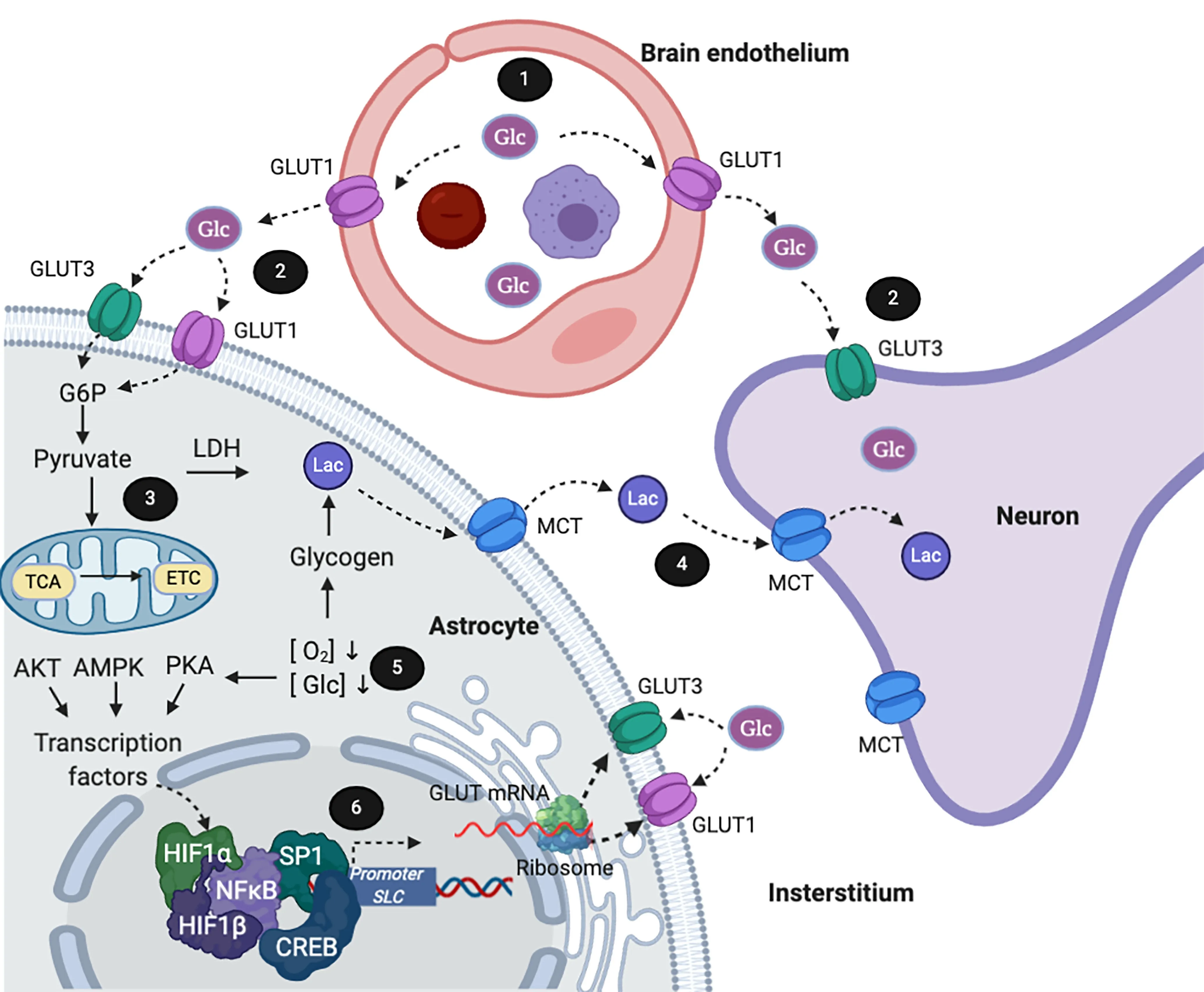
Beneficial effects of up-regulation of glucose transporters:Within the central nervous system,susceptibility to ischemia varies among different cell types,with astrocytes and vascular endothelial cells being more capable of withstanding ischemic conditions than neurons and oligodendrocytes.In the course of ischemia,the brain’s glucose uptake and glycolysis show an increasing trend,along with the conversion of higher levels of glucose-6-phosphate,fructose-6-phosphate,pyruvate,and lactate (Geng et al.,2019).This response is partially associated with a significant up-regulation of the glucose transporters expression trying to cover the neuronal energy demand.Models of oxygen and glucose deprivation show that signaling pathways that increase the expression of the GLUT1 are activated to optimize glucose uptake from the bloodstream in the cerebral vascular endothelial cells (Figure 1).Additionally,glycogen is immediately converted to lactate in astrocytes,from where it is released to the interstitium and taken up by neurons,ensuring,at least temporarily,their function.In addition to this response,there is an increase in GLUT1 expression that improves glucose uptake and restores the intracellular glycogen stores in astrocytes located at the penumbra.Interestingly,neurons and astrocytes also respond by up-regulating GLUT3 expression and promoting its translocation towards the cell membrane.Glutamate excitotoxicity induces this responsein vitro(primary cerebellar granular neurons) andin vivomiddle cerebral artery occlusion models of ischemia(Gutiérrez Aguilar et al.,2020,Koepsell,2020).Significantly,ischemic preconditioning(exposure to multiple mild ischemic stimuli previous to exposure to prolonged severe ischemia) induces the overexpression of both GLUT1 and GLUT3 in astrocytes and neurons eliciting a neuroprotective effect.Additionally,hyperglycemia up-regulates GLUT3 mRNA expression and GLUT3 protein synthesis and improves cerebral recovery during hypoxiaischemia in neonatal rats.Similarly,GLUT1 upregulation in cells from diabetic rats attenuates the exacerbation of cerebral ischemic injury induced by this disease (Iwata et al.,2014).
Regulation of cerebral glucose uptake plays a key role during cerebral ischemia to prevent neuronal damage;however,the endogenous response is not sufficient to prevent the injury because intermediates of the tricarboxylic acid cycle (i.e.,α-ketoglutarate,succinate,malate,and fumarate) decrease,revealing a compromised energy metabolism.Furthermore,β-hydroxybutyrate (a substitute energy source) raises,indicating an attempt by the brain to replace its energy supply.Most of the time,the brain bioenergetic crisis does not ameliorate since ATP stores are dramatically reduced,and the neuronal loss is evident 24 hours after onset of ischemia (Geng et al.,2019,Gutiérrez Aguilar et al.,2020).
Regulation of GLUT expression and induction of neuroprotection through signaling:The hypoxia-inducible factor 1 alpha (HIF-1α),the cAMP-response-element binding protein (CREB),and the specific protein 1 are transcriptional factors involved in GLUT3 gene expression(Figure 1).Interestingly,treatments that activate these factors induce transcription of several genes related to neural repair and recovery after stroke.Treatment with compounds such as S-aliylcysteine,curcumin,and progesterone enhances the expression of GLUT1 and GLUT3 and reduce neurological deficits,cerebral infarct volume,and brain edema following middle cerebral artery occlusion (Xia et al.,2018).N-acetyl cysteine increases the levels of HIF-1α and consequently increases GLUT3 expression.Suppression of HIF-1 activity with pharmacological inhibitors and specific knockout abolished N-acetyl cysteine’s neuroprotective effect in stroke (Zhang et al.,2014).Likewise,the phytoestrogen α-Zearalanol and salidroside analog induce neuroprotection by increasing the activity of CREB,up-regulating the expression of GLUT3 and improving the ATP and glutathione levels (Yu et al.,2014,Mohamed et al.,2019).At least three different pathways mediate GLUT3 expression calpain1/PKA/CREB,HIF-1α/AMPK/HSP70,and PI3K/Akt/mTOR/HIF-1α (Yu et al.,2014).Interestingly,mTOR-regulates HIF-1α accumulation in neurons independent of hypoxic conditions,revealing a post-ischemic regulation (Wu et al.,2012).Indeed,the signaling pathways involved in the endogenous neuroprotection mechanism are extraordinarily complex and have not been fully described;therefore,its effects are not well recognized.

Figure 1|Adaptive metabolic response in cerebral ischemic conditions.
Deleterious effects resulting from upregulation of glucose transporters:It has been suggested that increased glucose levels allow the metabolic shift towards increased glycolysis;thus,glucose is readily converted into lactate and exported to the extracellular media.There,lactate is captured by neurons to produce ATP without requiring oxygen.In this way,the uncoupling between glycolysis and mitochondrial full glucose oxidation leads to increased metabolic efficiency that protects cells,at least temporarily,from ischemicinduced damage.Nevertheless,other metabolic changes occur during acute brain ischemia that might influence the final damaging effects.For instance,stroke induces corticosterone release and activates glycogenolysis in the liver.Recently,Sá-Nakanishi et al.(2020) reported that hepatic anabolism and catabolism are altered during ischemia:gluconeogenesis,β-oxidation,and ketogenesis diminish after 24 hours of reperfusion and accelerate after 5 days when glycolysis has normalized.Although the entire response is orientated to increase the brain’s metabolism (a course needed to recover from the metabolic crisis),these changes also provoke alterations underlying the augmented tissue loss.Hypermetabolism has been detected in both experimental and clinical studies;and is linked to hyperglycolysis that promotes lactic acidosis,to compensate the impairment of the mitochondrial oxidative phosphorylation.Although reactive oxygen species (ROS) generation is observed in non-functional mitochondria,hyperglycolysis can also accelerate ROS production through lactic acidosis.These data agree with the response observed in the ischemic preconditioning,where the glycolytic pathway is downregulated,preferring the oxidative pentose phosphate pathway,where the reduced nicotinamide adenine dinucleotide phosphate is synthesized,favoring the antioxidant defense capacity of the cell.Interestingly,post-stroke physical rehabilitation has been recommended to reduce morbidity.Exercise induces hypermetabolism in the brain by increasing mRNA and protein levels of GLUT1 and GLUT3 and the activity of phosphofructokinase,lactate dehydrogenase,and phosphorylated adenosine monophosphate kinase.All these elements convey an enhanced brain’s capacity to utilize glucose.Regrettably,post-stroke exercise exacerbates brain injury when it is initiated shortly after the onset of ischemia (6 hours);conversely,late exercise (1–5 days) reduces brain infarct (Li et al.,2017).This situation reveals that neuronal damage is induced under circumstances in which the brain uses glycolysis excessively because ROS production is promoted.Therefore,metabolic depression is one of the main features of ischemic brain tolerance,while glucose incorporation at earlytimes after ischemia could favor hyperglycolysis.Interestingly,as mentioned above,some antioxidants promote up-regulation of GLUTs and induce neuroprotection.In contrast,resveratrol,which also has an antioxidant capacity and induces protection in cerebral ischemia,reduces the expression of GLUT3 in the brain at 24 hours of reperfusion (Gutiérrez Aguilar et al.,2020).Besides their antioxidant activity,these compounds have specific activities altering cell signals that might explain this controversy.Therefore,it is evident that a better description of these compounds’impact is necessary.For example,the complete temporal course of the mRNA and protein synthesis is needed.Additionally,localization and functionality of the transporter after the treatments should be characterized to understand their mechanism of action.
Similarly,the mechanisms regulating the response against damage-induced by ischemia involving glucose transport are not well established;other mechanisms may be associated with increases in intracellular glucose content.For instance,the sodium-glucose transporter,which is also over-expressed in cerebral ischemia,participates in the induction of the post hyperglycemia observed in stroke.In this case,the excessive influx of sodium ions into neuronal cells through the cerebral sodium-glucose transporter may exacerbate the development of cerebral ischemic neuronal damage (Koepsell,2020).
Summary:Convencig evidence demonstrates that glucose transporters are overexpressed in cerebral ischemia as part of an endogenous response against damage.This response tries to compensate for the energetic deficit.Inconveniently,the change is coupled to a state of hypermetabolism that provokes tissue injury.Therefore,it is essential to have a precise knowledge of brain metabolism in the short and long terms after the onset of ischemia to find therapeutic targets that promote neuroregeneration.
We are grateful to Dr.Arturo Hernández Cruz from Instituto de Fisiología Celular‚Departamento de Neurociencia Cognitiva‚Universidad Nacional Autónoma de México for valuable comments to the manuscript.
The present work was supported by the CONACYT (CB-2012-01-182266 to PA).Iván Alquisiras Burgos is a doctoral student from Programa de Doctorado en Ciencias Biomédicas‚Universidad Nacional Autónoma de México (UNAM) and beneficiary of scholarship No.275610 from CONACYT.
- 中国神经再生研究(英文版)的其它文章
- Towards a comprehensive understanding of p75 neurotrophin receptor functions and interactions in the brain
- Microglia regulation of synaptic plasticity and learning and memory
- Stroke recovery enhancing therapies:lessons from recent clinical trials
- Functional and immunological peculiarities of peripheral nerve allografts
- MicroRNA expression in animal models of amyotrophic lateral sclerosis and potential therapeutic approaches
- Significance of mitochondrial activity in neurogenesis and neurodegenerative diseases

