
Intervention effect and mechanism of Jisheng Shenqi Decoction plus Panax notoginseng and Bionjia on CCl4-induced hepatic fibrosis rat model
2022-07-28 01:47:48ZhenKangZhongXiaoLingZhouYueMingWangTengWuShiYinLuXinHuiShen
Journal of Hainan Medical College 2022年10期
Zhen-Kang Zhong, Xiao-Ling Zhou, Yue-Ming Wang, Teng Wu, Shi-Yin Lu, Xin-Hui Shen
1. Graduate School of Guangxi University of Traditional Chinese Medicine, Nanning 530001, China
2. Liuzhou Hospital of Traditional Chinese Medicine, Liuzhou 545001, China
Keywords:Jisheng Shenqi decoction with Panax notoginseng and Turtle shell Liver fibrosis Rats ACE-Ang Ⅱ-AT1R signaling pathway
ABSTRACT Objective: To investigate the intervention effect and specific mechanism of Jisheng Shenqi Decoction plus Panax notoginseng and Turmeric on carbon tetrachloride (CCl4)-induced liver fibrosis in rats. Methods: SPF SD rats were randomly divided into control group, model group,and Chinese medicine treatment (low, medium and high dose) groups. The model group and Chinese medicine treatment group were given intraperitoneal injection of 40% CCl4 olive oil solution twice a week. A rat model of liver fibrosis was replicated by the method for 8 weeks.After successful modeling, each drug-administered group was gavaged with corresponding drugs, and the control group and model group were gavaged with equal volume of distilled water, once a day, for 4 weeks. After the last intragastric administration, HE staining and Masson staining were used to observe the pathological morphology of liver tissue, and liver function (ALT, AST) was detected; ELISA method was used to determine transforming growth factor β (TGF-β), angiotensin II ( Ang-Ⅱ), chitinase 3-like protein 1 (CHI3L1), interleukin-1(IL-1), interleukin-6 (IL-6), tumor necrosis factor-α (TNF-α) content; The expression levels of type I, type III collagen (Col-Ⅰ, Col-Ⅲ) and TGF-β mRNA were determined by RT-PCR. Results: ①HE and Masson staining showed that a large number of liver cells in the model group were inflamed and necrotic, the collagen fibers in the liver tissue were extensively proliferated, the fibrous septa were interconnected, and the pseudolobules were formed. Compared with the model group, the levels of ALT, AST, TGF-β, Ang-Ⅱ, CHI3L1,IL-1, IL-6 and TNF-α in the traditional Chinese medicine treatment group were decreased,while the levels of Col-Ⅰ, Col-Ⅲ The expressions of Ⅲ and TGF-β mRNA were decreased,and the effect was most obvious in the middle and high dose groups of traditional Chinese medicine (P<0.01). Conclusion: Jisheng Shenqi Decoction combined with Panax notoginseng and Biejia can significantly improve liver fibrosis induced by CCl4 in rats, and its mechanism may be related to the down-regulation of angiotensin-converting enzyme (ACE)-angiotensin II(Ang-II)-angiotensin II receptor 1 (AT1R) signaling pathway related gene expression, reducing the level of related cytokines, promoting the degradation of extracellular matrix and so on.
1. Introduction
Hepatic fibrosis (HF) is a pathological change caused by excessive accumulation of extracellular matrix in liver tissue caused by various etiologies, resulting in abnormal liver tissue structure and function.The essential process of liver cirrhosis is a reversible excessive repair of liver tissue damage [1-5]. At present, Western medicine treatment of liver fibrosis mainly focuses on removing the cause and treating symptoms. However, due to its complex pathogenesis,involving multiple cells, cytokines, proteins and signaling pathways,single-target intervention is often ineffective. For example, the use
of nuclear Glycoside analog antiviral therapy, even if satisfactory virological effects have been obtained and liver inflammation has been effectively improved, some patients still cannot completely reverse liver fibrosis, and the disease can still develop progressively[6]. Definitive treatment of liver fibrosis is more difficult. If liver fibrosis progresses to decompensated cirrhosis or liver cancer, it will seriously threaten the life and health of patients [7]. Therefore, on the basis of etiological treatment, active and effective anti-hepatic fibrosis. Metabolism is an indispensable link in blocking disease progression [8].
Traditional Chinese medicine treatment has the advantages of multi-target, multi-level, overall regulation and relatively few adverse reactions, so it has become a hot spot in the research of anti-hepatic fibrosis drugs [9-11]. The author’s team’s previous experimental studies have shown that Jisheng Shenqi Decoction can promote the homing of stem cells to the liver after transplantation, thereby playing a role in the treatment of liver cirrhosis [12]. The patient's clinical symptom score can improve the patient's liver function and liver fibrosis indicators [13]. On the basis of previous research results, the author found that adopting economic living kidney tonga with promoting blood circulation to remove blood stasis syndrome elimination fights on the efficacy of notoginseng and turtle shell has a significant role in anti liver fibrosis, but the specific mechanism is not yet clear, so this study establishing the model of liver fibrosis in rats induced by CCI4 lavage treatment in Chinese medicine,detection related indicators,To observe the pathological changes of liver tissue in rats with liver fibrosis after treatment, and explore the specific intervention pathway, so as to provide more scientific theoretical basis for jisheng Shenqi Decoction plus Panax notoginseng and trionyx japonicus to resist liver fibrosis.
2. Materials and instruments
2.1 Animals
Fifty-four SPF grade SD rats, half male and half female, aged from 6 to 8 weeks and weighing (190±10) g, were purchased from the Experimental Animal Center of Guangxi Medical University. Experimental Animal Certificate No. : SCXK GUI 2009-0002, Use License No. : SYKG GUI 2009-2005. All rats were kept in the Animal Laboratory of Liuzhou Hospital of Traditional Chinese Medicine, with room temperature between 20℃ and 25℃, humidity between 50% and 65%, and natural light, and fed with ordinary feed and normal drinking water for 1 week. The feeding and experimental process of experimental rats followed the national regulations on experimental animal management.
2.2 Drugs
Jisheng Shenqi Decoction with Panax notoginseng and turtle shell(cooked Rehmannia glutinosa 40g, Cornus officinalis 20g, Chinese yam 20g, Alisma alisma 15g, Poria tucka15g, Cortex danpi 15g,cinnamon 5g, Aconite 5g, Plantain seed 10g, Niuzhu 10g, Panax notoginseng 3G, turtle shell 15g). The above drugs were agricultural granule preparations, produced by Peili (Nanning) Pharmaceutical Co., LTD. The above drug granule preparations were added into pure water to prepare 1.56g/ mL, 3.11g/ml and 6.22g/ml of low, medium and high concentration Traditional Chinese medicine suspensions,which were stored in a refrigerator at 4℃ for reserve.
2.3 Main reagents and instrument
Reagent: Gol extra virgin Olive oil (Spain, GB/T23347); CCl4(Tianjin Fuyu Fine Chemicals Co., LTD., GB/T688-2011); Hematoxylin-eosin (HE) staining kit and Masson tricolor staining kit were purchased from Beijing Solebo Technology Co., LTD., batch numbers: G1337, G1080, G1100). Alanine aminotransferase (ALT) and aspartate aminotransferase (AST)kits were purchased from Nanjing Jianceng Institute of Biological Engineering, the batch numbers were C009-2-1 and C010-2-1, respectively. Rat (TGF-β, Ang-Ⅱ, CHI3L1, IL-1, IL-6,TNF-α) ELISA Kit was purchased from Wuhan Beinley Biotechnology Co.,LTD., batch numbers: RA20599, RA20075,RA20662, RA20020, RA20607, RA20035; RNA Extraction Kit Trizol(Invitrogen, Lot no. 15596026), purchased from Thermo Fisher Technologies Co.,LTD. MonScript RTllI All-in-one One Mix with dsDNase, MonAmp SYBR Green qPCR Mix (MonA Biotechnology Co., LTD., Batch number:MR05101,MQ10101). Main instruments:High-speed refrigerated centrifuge (Changsha Xianggrui Centrifuge Co.,LTD.,TGD-20MC), ultra-speed cryogenic centrifuge (Sigma Company, USA), optical microscope Olympus BH2(Olympus Corporation, Japan), Automatic biochemical analyzer(Hitachi 7170S, Japan), full wavelength Enzyme reader (Thermo Fisher Technology Co., LTD., Model: 1510), Nucleic Acid Protein Quantitative Detector (Eppendorf, No. 6133), S1000TMTHermal Cycler Gene Amplifiers (BIO RAD, Inc.), 7500 Real PCR System(ABI, No. 4345241).
3. Method
3.1 Grouping and modeling
After one week of adaptive feeding, 10 rats were randomly selected as the control group, and the remaining 44 rats were intraperitoneally injected with 40%CCl4 olive oil solution (CCl4 and olive oil volume was allocated at 2:3), twice a week for 8 weeks to make the rat model of liver fibrosis. At the end of modeling, 4 rats were randomly selected for liver tissue biopsy. If liver cell necrosis, inflammatory cell infiltration, collagen fiber formation, and normal structure of liver tissue were destroyed, the liver fibrosis model was successfully constructed. After successful modeling, 40 rats were randomly divided into model group and Chinese medicine treatment (low,medium and high concentration) groups, with 10 rats in each group.
3.2 Drug treatment
The drug dose was calculated according to the body surface area conversion method between human and rat, and the drug solution volume was given at 1mL /100g. The groups treated with Traditional Chinese medicine (low, medium and high concentration) were given Jisheng Shenqi decoction plus notoginseng and turtle shell suspension 15.6g/kg, 31.1g/kg and 62.2g/kg, respectively.Control group and model group were given equal volume of distilled water intragastric administration. The drug was administered once a day for 4 weeks. Diet and water were not restricted during gavage.
3.3 Collection of specimens
After gavage, the patient was fasted overnight for 12h, and no water was allowed. 10% chloral hydrate was given for anesthesia by intraperitoneal injection at a dose of 0.5ml/100g. Blood was collected through the abdominal aorta and then placed for 1h,centrifuged at 3000r/min for 10min to obtain the upper serum, which was stored at -80℃ for future use. After blood collection, the liver was taken out quickly and tissue samples of 0.5cm×0.5cm×0.5cm were collected. A part of the liver was frozen with liquid nitrogen and stored at -90℃ for RT-PCR detection. One part was fixed with 10% formaldehyde, then dehydrated step by step by automatic dehydrator, immersed in wax and embedded, and sliced with 0.3μm thickness for the observation of pathological morphology of liver tissue after staining.
3.4 Histopathological observation of liver tissue
HE staining and Masson staining were performed on the liver tissue sections of rats in each group obtained in "2.3", and the inflammation and collagen fiber deposition of liver tissue were observed under light microscope.
3.5 Test of liver function
Serum collected in "2.3" was used to detect ALT and AST content of rats in each group by automatic biochemical analyzer.
3.6 Determination of TGF-β, Ang- Ⅱ, CHI3L1, IL-1,IL-6 and TNF-α contents
ELISA method was used to strictly operate the serum collected in "2.3" according to the instructions of the kit. The OD450 value was read at 450nm absorbance using a microplate reader and the contents of TGF-β, Ang- Ⅱ, CHI3L1, IL-1, IL-6 and TNF-α were calculated.
3.7 Determination of Col-ⅠmRNA, Col-Ⅲ mRNA and TGF-βmRNA expression levels
Rt-PCR was used to determine the total RNA. 30mg of liver tissue from "2.3" was extracted by Trizol one-step method. The total RNA concentration was determined by uv spectrophotometer, and the RNA was reversed into cDNA with reverse transcription kit,and then amplified with cDNA as template. QPCR system: SYBR Green qPCR Mix 10μ L, positive and negative primers 0.4μ L,cDNA 2μ L, nuclease-free Water 7.2μ L. Reaction conditions: using three-step method, pre-degeneration 95℃, 30s, degeneration 95℃,10s, annealing 55℃-65℃, 10s, extension 72℃, 30s, a total of 41 cycles. The expression levels of Col-ⅠmRNA, Col-Ⅲ mRNA and TGF-βmRNA were calculated by 2-△△Ctmethod with β-actin as internal reference gene. Primers for Col-ⅠmRNA, Col-ⅢmRNA and TGF-β mRNA were designed according to the known sequences of Genebank, and the primers were synthesized by Optimus Biotechnology Co., LTD. The serial numbers of primers are shown in Table 1.
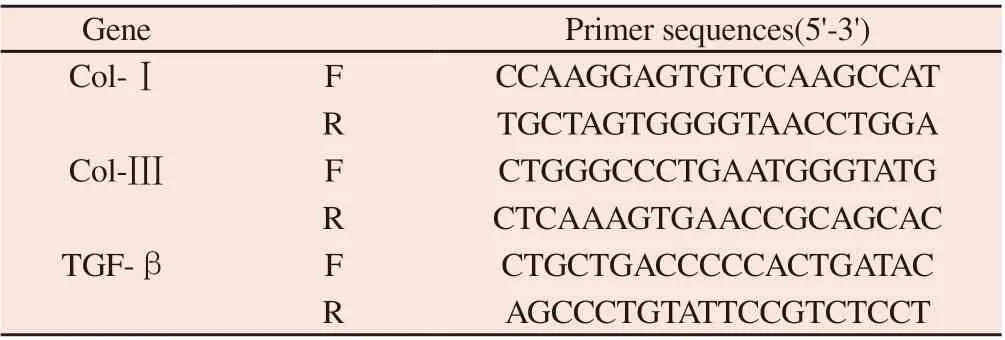
Table 1 Primer sequences of each gene
3.8 Statistical Methods
SPSS 24.0 software was used for statistical analysis of data,GraphPad Prism 5 was used for plotting, measurement data were expressed as (x±s), one-way anOVA was used for comparison between multiple samples, LSD-t test was used for homogeneity of variance, non-parametric test was used for heterogeneity, P < 0.05 was considered statistically significant.
4. Results
4.1 Effects of Jisheng Shenqi Decoction plus Panax notoginseng and trionyx sinensis on liver function of rats with CCL4-induced liver fibrosis
Compared with the control group, the levels of ALT and AST in model group were significantly increased (P < 0.01), indicating successful modeling; Compared with model group, ALT and AST levels in TCM groups were significantly decreased (P < 0.01), and the effect was most obvious in TCM high-dose group. The results are shown in Table 2 and Figure 1.
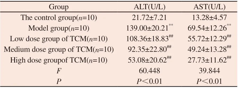
Table 2 Effects of Jisheng Shenqi Decoction plus Panax notoginseng and bionyx sinensis on liver function (ALT and AST) of rats with CCI4-induced liver fibrosis
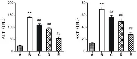
Figure1 Effect of Jisheng Shenqi Decoction plus Panax notoginseng and trionyx sinensis on liver function (ALT and AST) of rats with CCI4-induced liver fibrosis
4.2 Effects of Jisheng Shenqi Decoction plus Panax notoginseng and Trionyx sinensis on hepatic histopathology of rats with CCI4-induced hepatic fibrosis
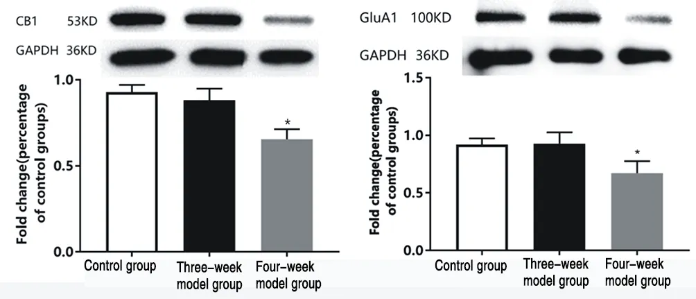
Figure 2 HE staining of liver tissue (×10),A:The control group,B:Model group,C:Low dose group of TCM,D:Medium dose group of TCM,E:High dose groupof TCM
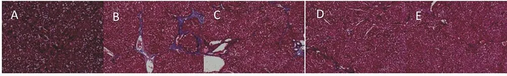
Figure 3 Masson staining of liver tissue (×10),A:The control group,B:Model group,C:Low dose group of TCM,D:Medium dose group of TCM,E:High dose groupof TCM
After intraperitoneal injection of 40%CCL4 8 weeks, liver histological sections HE and Masson staining results were shown in Figure 3 and Figure 4. Figure 2A and Figure 3A showed that the hepatic lobule structure of the control group was normal, liver cell cords were radially evenly and closely arranged by central vein,and no inflammatory or fibrosis changes were observed in liver tissue. Compared with the control group, Figure 2B and 3B showed that most liver cells in the model group had obvious degeneration and necrosis, and liver cells were disordered. The necrotic liver cells formed fibrous septum and divided liver parenchyma into liver cells of different sizes, forming false lobules, indicating that the modeling was successful. Compared with model group, Figure 2 (C, D, E) and Figure 3 (C, D, E) showed that inflammation and fibrosis degree of rats in low-dose, medium-dose and high-dose TCM groups were alleviated at different levels.
4.3 Effects of Jisheng Shenqi Decoction on serum IL-1, IL-6,TNF-α, TGF-β, Ang-Ⅱ and CHI3L1 in rats with Liver fibrosis induced by CCl4
Compared with model group, the levels of IL-1, IL-6, TNF-α,TGF-β and CHI3L1 in each dose group were significantly decreased (P < 0.05), and the intervention effect was most obvious in medium and high dose groups (P < 0.01). For Ang-Ⅱ, the level of Ang-Ⅱ in medium and high dose groups was significantly decreased(P < 0.01), while the effect of low dose group was not significant (P> 0.05). The results are shown in Table 3, Table 4 and Figures 4 and 5.
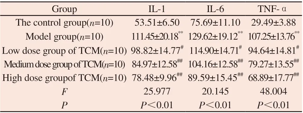
Table 3 Effects of Jisheng Shenqi Decoction plus Panax notoginseng and trionyx sinensis on serum IL-1, IL-6 and TNF-α in rats with CCI4-induced liver fibrosis
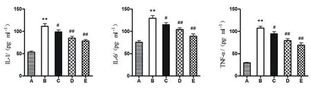
Figure 4 Effects of Jisheng Shenqi Decoction plus Panax notoginseng and trionyx sinensis on serum IL-1, IL-6 and TNF-α in rats with CCI4-induced liver fibrosis
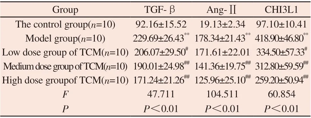
Table 4 Effects of Jisheng Shenqi Decoction on serum TGF-β, Ang-Ⅱ and CHI3L1 in rats with CCI4-induced liver fibrosis

Figure 5 Effects of Jisheng Shenqi Decoction plus Panax notoginseng and trichoderma on serum TGF-β, Ang-Ⅱ and CHI3L1 in rats with CCI4-induced liver fibrosis
4.4 Effects of Jisheng Shenqi Decoction with Panax notoginseng and trichoderma on the expression of Col-ⅠmRNA, Col-Ⅲ mRNA and TGF-β mRNA in CCI4-induced hepatic fibrosis rats
Compared with the control group, the mRNA expressions of Col-Ⅰ, Col-Ⅲ and TGF-β in liver tissue of model group were significantly increased (P < 0.01). Compared with model group, the mRNA expressions of Col-Ⅰ, Col-Ⅲ and TGF-β in medium and high dose groups were significantly decreased (P < 0.01); In addition to TGF-β mRNA, Col-ⅠmRNA and Col-Ⅲ mRNA expression were also decreased in low dose group (P < 0.05). The results are shown in Table 5 and Figure 6.
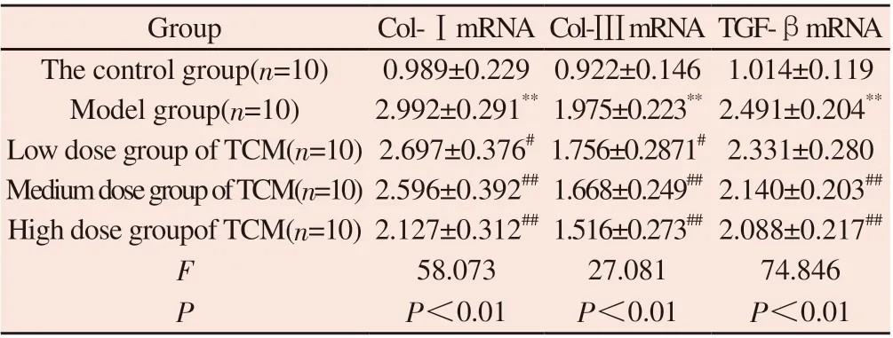
Table 5 Effects of Jisheng Shenqi Decoction plus Panax notoginseng and bionyx sinensis on the expression of Col-Ⅰ mRNA, Col-Ⅲ mRNA and TGF-β mRNA in liver tissue of rats with CCI4-induced liver fibrosis
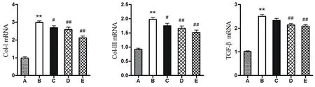
Figure 6 Effects of Jisheng Shenqi Decoction plus Panax notoginseng and turtle turtle on the expression of Col-Ⅰ mRNA, Col-Ⅲ mRNA and TGF-β mRNA in liver tissue of rats with cCI4-induced liver fibrosis
5. Discussion
According to its pathological characteristics and clinical manifestations, liver fibrosis belongs to the categories of "flank pain","accumulation" and "bloating" in traditional Chinese medicine."Medical School Must Read" says: "The accumulation of righteous qi is insufficient, and then evil qi ensnares it", "Neijing" says:"The warm qi does not work, the blood coagulation accumulates and does not disperse", liver fibrosis is a chronic disease, and the course of the disease is long. The basic pathogenesis of this disease should be the accumulation of deficiency and blood stasis [10],and the general treatment should be to invigorate the deficiency and strengthen the body, and promote blood circulation to remove blood stasis and eliminate accumulation. Zhongjing put forward the theory that "blood is not good for water". "Blood is not good"is mainly reflected in the state of blood stasis, and "water is bad" is mainly reflected in the stop of water accumulation. The connotation of this discussion is reflected in the physiological homology and mutualization of blood and water. Pathologically, they are mutually causal and closely related to each other. "Blood Syndrome": "Blood disease causes blood, and blood disease causes both water." "Blood stasis and water are also combined with water." From this, it can be seen that the treatment of blood stasis can also be started by diluting water and regulating qi. Jisheng Shenqi Decoction comes from the Song Dynasty physician Yan Yonghe's "Jisheng Fang".This recipe has the functions of warming yang and transforming qi, diuresis and swelling, and is mainly used to treat kidney deficiency, waist edema, and difficulty in urination. This recipe contains the method of three tonifying and three purging. The three tonifying is mainly to nourish the kidney yin, which means "tonifying the kidney and generating marrow to generate the liver". Among the large-dose Yinnourishing medicines, the product of Guifu is supplemented, which is intended to reduce fire and anger. When yang is supported by yin, the biochemical changes are infinite. It warms the kidneys and tonifies yang and transforms qi to help activate blood and diuretic water. The "three diarrheas" are Alisma, psyllium, and tuckahoe. It is to control water, control water is to control qi", "those who control blood must regulate qi", so the source of warming and nourishing yang qi and benefiting fire is given to this recipe, in order to apply qi and water, adjust the effect of triple burner qi machine, qi machine is smooth, The blood vessels are self-opening, and the addition of Panax notoginseng and turtle shells can not only eliminate symptoms and dissipate knots, but also remove blood stasis without harming the body.
ALT, AST, most abundant in the liver, the cytoplasm, mainly exist in the liver are sensitive indicators of reaction of liver cell damage, this study result shows that compared with the control group, model group rats liver cell damage necrosis occurs in the formation of liver fibrosis, ALT, AST in serum was significantly higher (P < 0.01), born in dhi kidney tonga notoginseng, turtle shell after the intervention, ALT and AST were significantly decreased(P < 0.01), indicating that Jisheng Shenqi Decoction plus Panax notoginseng and bionyx japonica could protect liver cells. Liver tissue pathology biopsy is the gold standard in the diagnosis of liver fibrosis [14], because of its belong to the invasive operation, potential infection, hemorrhage, such as poor repeatability, operating error characteristics not widely used in clinical, instantaneous elasticity imaging techniques can also be because of cholestasis liver fatty degeneration and liver inflammation, and eating and there is a certain error, Similarly, the results of the four traditional liver fiber tests are also prone to be affected by liver inflammation [10]. Chitinase 3-like protein 1 (CHI3L1), also known as chitinase 3-like protein 1, is a novel serological marker closely related to a variety of liver diseases and is most abundant in liver with high specificity [15-18]. CHI3L1 is positively correlated with the degree of liver fibrosis, and is involved in the formation of liver fibrosis mainly through inflammatory reaction, cell proliferation and differentiation, anti-apoptosis and promoting extracellular matrix remodeling and other pathological processes [19,20]. CHI3L1 is superior to hyaluronic acid (HA), typeⅢ procollagen (PⅢNP), laminin (LN) and type Ⅳ collagen (C-Ⅳ)in evaluating the degree of liver fibrosis, and has a higher diagnostic value in liver fibrosis [21-24]. Therefore, the measurement of CHI3L1 has certain guiding significance for evaluating the efficacy of liver fibrosis. The results of this study showed that the level of CHI3L1 was significantly reduced in the groups with different doses of Traditional Chinese medicine, indicating that Jisheng Shenqi Decoction plus Notoginseng and bionychia can effectively improve liver fibrosis. Liver regeneration and fibrous tissue hyperplasia secondary to liver cell degeneration and necrosis further lead to angiogenesis and thrombosis, aggravating liver cell injury and liver blood circulation disorders [25]. The manifestations of hepatic sinusoidal vascular lesions and excessive deposition of extracellular matrix in liver are consistent with the essential characteristics of"blood stasis" in traditional Chinese medicine [10]. The combination of panax notoginseng and Triticum triticum can promote blood circulation and dissipate blood stasis as well as strengthen and loosen the knot, and the two can play a significant anti-liver fibrosis effect by inhibiting the activation of HSC and promoting the degradation of extracellular matrix [26-29].
Studies have shown that the renin-angiotensin-aldosterone system(RASS) also exists in human liver, and RASS is involved in the occurrence and development of liver fibrosis [30]. The ACE-ANG-Ⅱ-AT1R axis is an important pathway promoting the formation of liver fibrosis. When the liver is damaged, Components such as Ang-Ⅱand AT1R in RASS are up-regulated or redistributed. For example,in liver fibrosis, the content of Ang-Ⅱ is significantly increased [31],which is positively correlated with the degree of liver fibrosis. ACE lyses Ang-Ⅰ into Ang-Ⅱ. As one of the most important active substances in RASS, Ang-Ⅱ has significant pro-inflammatory and pro-fibrotic effects. It not only participates in inflammatory reactions, but also activates HSC and promotes the release of cytokines such as TGF-β, TNF-α and IL [32,33]. The above cytokines in turn continuously stimulate HSC proliferation and activation and increase THE generation of ECM [34-40]. As a central link in the formation of liver fibrosis, HSC proliferates in large quantities, undergo phenotypic transformation and differentiation into myofibroblasts, which secrete ECM and produce too much ECM with relatively little degradation. Excessive deposition in liver tissue leads to the formation of liver fibrosis. In addition, Ang-Ⅱcan also activate HSC and up-regulate the mRNA expressions of typeⅠand Ⅲ collagen and TGF-β through AT1R [41,42]. Studies have shown that the use of AT1R antagonists or ACE inhibitors can inhibit liver fibrosis [30,43]. TGF-β is one of the most important cytokines regulating the formation and deposition of ECM, which can promote the activation of HSC and the expression of type typeⅠand Ⅲ collagen, and has a significant role in promoting liver fibrosis [44,45]. The results of this study showed that compared with the control group, the serum levels of Ang-Ⅱ, TGF-β, IL-1, IL-6 and TNF-α in the model group were significantly increased (P <0.01). The levels of Ang-Ⅱ, TGF-β, IL-1, IL-6 and TNF-α were all decreased, indicating that Jishengshenqi Decoction plus Panax notoginseng and triticum triticum can act on ace-Ang-Ⅱ-AT1R axis, reduce the content of Ang-Ⅱ and TGF-β, and then reduce the production of IL-1, IL-6, TNF-α and other related cytokines. As the main components of ECM, typeⅠand type Ⅲ collagen are involved in the formation of liver fibrous tissue structure. The results of this study showed that compared with the control group, the mRNA expressions of Col-Ⅰ, Col-Ⅲ and TGF-β in liver tissue of rats with hepatic fibrosis in model group were significantly increased (P< 0.01). The expression levels of the above genes all decreased to varying degrees, indicating that Jishengshenqi Decoction plus Panax notoginseng and trionyx japonicus can regulate the balance of ECM generation and degradation, inhibit ECM deposition, promote ECM degradation and thus alleviate liver fibrosis.
In conclusion, Jishengshenqi Decoction plus Panax notoginseng and bionjia can improve liver fibrosis. The specific mechanism may be related to improving liver inflammation and necrosis, regulating ACE-ANG-Ⅱ-AT1R signal transduction pathway, reducing the level of related cytokines, HSC activation and proliferation, and downregulating the expression of Col-Ⅰ, Col-Ⅲ and TGF-β genes. The reduction of ECM overproduction and accumulation in liver tissue is related.
Author’s contribution Zhong Zhenkang was responsible for the design and co-completion of experiments, index detection, paper writing and data statistical analysis; Wang Yueming, Wu Teng and Shen Xinhui assisted in completing the experiment; Zhou Xiaoling and Lu Shiyin were responsible for experimental guidance and paper revision.
 Journal of Hainan Medical College2022年10期
- Journal of Hainan Medical College的其它文章
- Study on the medication rules of traditional Chinese medicine in the treatment of sleep disorder after stroke based on data mining
- Effects of acupuncture combined with Kaijingtongmai Decoction on ATP sensitive potassium channel related proteins Kir6.1 and Kir6.2 in myocardial infarction rats
- Exploring the molecular biological mechanism of Shugan Jianpi Decoction in the treatment of depression-related breast cancer based on network pharmacology
- Clinical effect of enriching qi, activating blood circulation, clearing away dampness and heat combined with western medicine in the treatment of idiopathic membranous nephropathy: A meta-analysis
- Effects of LPS-induced cholangitis on the cytoskeleton morphology of bile duct epithelium and the intervention mechanism of Dahuang Lingxian formula for these changes
- Effects of stress of different duration on depression-like behavior and expression of CB1 and GluA1 in medial prefrontal cortex of rats