
miRNA-451 regulates rhesus choroid-retinal endothelial cell function and proteome profile
2022-06-22 03:12:50HongLianWuYanShaoZhenNaChenHuiZhangXiaoMinZhangXiaoRongLi
INTRODUCTION
Each experiment at least comprised three independent repeats. All the quantitative value was presented as mean±standard deviation (SD) and Student’s
-test was performed to analyze the differences among groups. Prism software v. 8.4.0 (GraphPad Prism, San Diego, CA, USA) was used for the statistical analyses.
-value <0.05 was considered statistically significant.
miRNA-451 (miR-451), a small non-coding RNA molecule, is involved in the regulation of cell proliferation
. Based on our previous data, the expression of miR-451a was down-regulated both in the retina of
mice and 4-hydroxynonenal(4-HNE)-treated RPE cells
. We further verified that miR-451a inhibited the proliferation and migration of retinal pigment epithelium (RPE)
and showed a protective effect on mitochondria function of RPE in diabetic conditions
. Studies on patients’ blood have demonstrated that miR-451 could become a prognostic biomarker in DR since its expression level gradually decreased with the progression of the disease
. In fact, miR-451 is expected to be a therapeutic target for different cancers since it could suppress tumor proliferation, invasion, and metastasis
. For example, Nan
revealed that miR-451 could suppress glioma cell growth and invasion through mTOR/HIF-1α/VEGF pathway,and repressed the epithelial-mesenchymal transition (EMT)and metastasis of cancer
PI3K/Akt/Snail pathway
. Liu
also exemplified that the proliferation and invasion of hepatocellular carcinoma, a highly vascularized tumor,could be inhibited by miR-451
IL-6R-STAT3 pathway or caspase-3- and MMP9-dependent pathway
. Thus, we expected that miR-451 might also play a therapeutic role in fundus neovascular disease by regulating the function of retinal endothelial cells.
In fact, the biological function and regulation of miR-451 on different cells is extremely intricate. Despite increasing studies illustrated that miR-451 primarily repressed cell growth in various cancers, there were still a small portion of research that reported miR-451 could promote the proliferation and migration of tumor cells
. Proteomics offers an effective method for the large-scale and in-depth analysis of the intracellular protein composition; hopefully, the new protein targets which are critical for cellular interaction can be discovered and quantified. Liquid chromatography and tandem mass spectrometry (LC-MS/MS) is a powerful proteomic technique that combines the separation capabilities of liquid chromatography with the high sensitivity and selective quality analysis capabilities of triple quadrupole mass spectrometry.isobaric tags for relative and absolute quantification (iTRAQ)is a newly established method to analyze proteins using mass spectrometry. By specially attach 4 or 8 isotope tags to the amino group of polypeptides, iTRAQ can simultaneously compare the relative or absolute content of proteins in 4 or 8 different samples. In this study, we intend to explore the potential effect of miR-451 on the function and proteomic profile of rhesus macaque choroid-retinal endothelial(RF/6A) cell, a choroid retinal endothelial cell line. iTRAQ coupled with LC-MS/MS was used to identify and quantify the differentially expressed proteins (DEPs) in RF/6A cells after miR-451 overexpression or inhibition. To elucidate the altered cellular function and proteins’ interactions, Gene Ontology (GO), Kyoto Encyclopedia of Genes and Genomes(KEGG), and protein-protein interaction (PPI) web analysis were performed.
MATERIALS AND METHODS
RF/6A cell line was purchased from Chinese Academy of Science (Shanghai, China) and has been characterized previously
. Cells were maintained in MEM(Gibco, Carlsbad, CA, USA) supplemented with 10% fetal bovine serum (Gibco), 100 U/mL penicillin, and 100 μg/mL streptomycin (Gibco) at 37℃ in the cell incubator with 5%CO
and 95% air.
笔者在本文主要以《我是什么》这一课文为例,创建项目学习活动。首先,结合课文内容而提出真实的问题或者创设真实的情境。将语文课堂置于真实情境之中,可以让学生产生真实的体验,让学生完成真实的任务,利于学生感受到项目学习的现实意义。根据部编版新教材小学二年级《我是什么》这篇课文,教师可提出真实问题:“水汽为何会变成云?有哪些决定条件?”“为什么雪是在冬日降临,而并非在夏日出现?”“云变成雨或者雹子需具备什么条件?”通过这些真实的问题,可以引导学生关注自然环境和生活,利于丰富课堂学习资源,又可激发学生自主探究,甚至是实地考察的欲望,从中获得相应的知识及技能。
Of all the DEPs in RF/6A transfected with miR-451 inhibitor compared with inhibitor control, 13 proteins (PCYT1A,MGAT1, TUBB, MCU, SIL1,
.) are up-regulated, and 17 proteins (BID, GNL3, MSH6, PNPLA6, PTPN1
.) are down-regulated. PCYT1A is a rate-limiting enzyme that participated in the phosphatidylcholine biosynthesis. Xiong
revealed that expression silencing of PCYT1A led to the remarkable proliferation suppression of B-lymphoma cells. However, another research found that PCYT1A knockdown promoted the proliferation and migration of lung adenocarcinoma cells
. These seemingly controversial results might be due to the heterogeneous response of different cells to PCYT1A. MGAT1 promoted glioma cell proliferation and migration
, and inhibition of MGAT1 tended to suppress breast cancer metastasis
. Higher expression level of TUBB,the major constituent of microtubules, was correlated with the worse prognosis of lung adenocarcinoma
. In addition,MCU, locating on the mitochondrion inner membrane and mediating calcium uptake into mitochondria, enhanced cell growth remarkably and played a critical role in the progression of various cancers such as colorectal cancer
, esophageal cancer
and gastric cancer
. MCU also contributed to the angiogenesis of breast cancer
negatively regulating the secretion of miR-4488 into extracellular vesicles (EVs); that is,increased MCU led to the lower level of miR-4488 in the EVs,thus resulted in the angiogenesis in the metastatic niche
.Furthermore, SIL1, a regulator of endoplasmic reticulum function, promoted the progression of breast cancer
regulating cell proliferation, migration and invasion
. These up-regulated DEPs explained the positive effect of miR-451 mimic on RF/6A cells to some extent.
miR-451 mimic: 5’-AAACCGUUACCAUUACUGAGU U-3’; miR-451 mimic control: 5’- UUCUCCGAACGUGU CACGUTT-3’; miR-451 inhibitor: 5’- AACUCAGUAA UGGUAACGGUUU-3’; miR-451 inhibitor control: 5’-UUGUACUACAAAAGUACUG-3’.
Cells were inoculated on a 24-well plate at an appropriate density (1×10
) and were transfected at about 70% confluence.Lipofectamine 2000 (Life Technologies, USA) was used during cell transfection according to manufacturer’s protocol.RF/6A cells were transfected by miR-451 mimic, miR-451 mimic control, miR-451 inhibitor, and miR-451 inhibitor control separately.
The standard growth curve of RF/6A cells was prepared according to the manufacturer’s protocol (CCK-8; Beyotime, Shanghai, China). RF/6A cells in the logarithmic growth phase were inoculated into 96-well plates with 5×10
cells/well in 100 μL complete medium then incubated with 5% CO
at 37℃ in humidified 95% room air overnight. About 70% confluent RF/6A cells were transfected with miR-451 mimic, miR-451 mimic control, miR-451 inhibitor, and miR-451 inhibitor control for 24 and 48h respectively. Then, after aspirating out the previous medium,90 μL fresh serum-free medium with 10 μL CCK-8 solution was added into each well. Cells were incubated at 37℃ for 4h, and the absorbance of each well was measured at 450 nm using a microplate reader.
Totally 12 samples consisting of three biological replicates of 4 groups including miR-451 mimic, miR-451 mimic control, miR-451 inhibitor, miR-451 inhibitor control transfected RF/6A cells were prepared. Proteins were extracted with lysis buffer containing 8 mol/L urea, 50 mmol/L NH
HCO
and 0.2% SDS and followed by 5min of ultrasonication on ice. The lysate was centrifuged at 15 000× g for 15min at 4℃,and the supernatant was moved to a clean tube. Samples were further reduced with 10 mmol/L DTT for 1h, then alkylated with 40 mmol/L IAM for 1h at room temperature in the dark.Samples mixed with pre-cold acetone were incubated at -20℃for 2h. After that, according to the manufacture protocol,proteins were dissolved, denatured, and blocked on cysteines.The protein solution (100 μg) was then diluted with 100 mmol/L TEAB, digested with trypsin Gold, and desalted with a Strata X C18 column. Peptide labeling was performed with iTRAQ Reagent 8-plex kit following the manufacturer’s instruction.
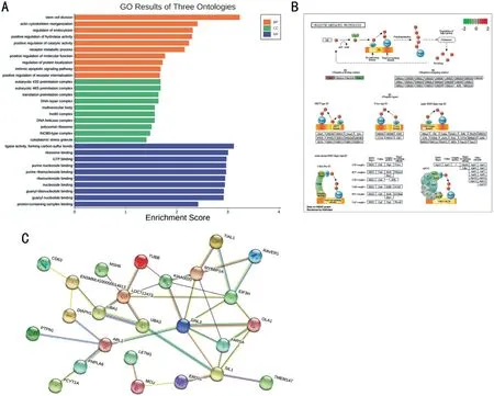
DEPs were analyzed in GO, KEGG, and STRING database for functional annotation (Figure 5A-5C). The BP participated by DEPs included stem cell division, actin cytoskeleton reorganization,regulation of endocytosis, regulation of hydrolase and catalytic activity, receptor metabolic process, and regulation of protein localization,
. The MF included ligase activity (forming carbon-sulfur bonds), ribosome binding, GTP binding,nucleoside binding, and protein-containing complex binding,
. The CC included eukaryotic 43S and 48S preinitiation complex, DNA repair complex, multivesicular body, lno80 complex, DNA helicase complex, polysomal ribosome,cytoplasmic stress granule. The KEGG pathway enrichment analysis revealed that the DEPs mainly took part in the regulation of ubiquitin mediated proteolysis. STRING database revealed that there was one major protein interaction network composing of the most DEPs, of which GNL3 acts as a hub protein in the network.
The combined peptide mixture was fractionated with a C18 trap column using an Eksigent nanoLC system (SCIEX, Framingham, MA, USA) at a flow rate of 10 μL/min, and was eluted from the trap column by the gradient solvent B. The peptides were subjected to MS/MS analysis with a Triple TOF 6600 System (SCIEX,Framingham, MA, USA). Data-dependent acquisition mode as below was applied for data collection.
综上所述,凝固酶阴性葡萄球菌在前列腺炎中的致病作用应引起重视,而且应对患者前列腺液中的病原菌进行检测,以便临床医师对患者进行更好的诊治,促进患者痊愈。
Raw data from iTRAQ LC-MS/MS was analyzed by Proteome Discoverer Software 1.4 (Thermo Fisher Scientific, San Jose, CA, USA),and was searched against the UniProt Macaca mulatta database(https://www.uniprot.org/proteomes/UP000006718). The confidently identified protein requires at least one unique peptide with FDR <1%. IQuant software was used for quantitative analysis
. We defined proteins with fold change>1.2 or <0.83 and
<0.05 as DEPs.
DEPs were submitted to the Gene Ontology database (http://www.geneontology.org/) to interpret their annotations in biological process (BP), cellular component (CC), and molecular function (MF). The potential pathway analysis of DEPs was performed by the KEGG (http://www.genome.jp/kegg/mapper.html). And the PPI network was carried out by STRING database (https://string-db.org/).Cytoscape software 3.8.2 was used for the visualization of GO enrichment results.
Abnormal neovascularization is the common pathological process of neovascular diseases of fundus, such as diabetic retinopathy (DR) and age-related macular degeneration (AMD). DR is a common microvascular lesion of diabetes mellitus (DM), accounting for the majority of acquired blindness among the working-age population
.Chronic hyperglycemia could damage the basement membrane of endothelial cells, promote the pathological proliferation of endothelial cells, and eventually cause the formation of retinal neovascularization
. The emergence of retinal neovascularization is the hallmark of proliferative diabetic retinopathy (PDR), and it’s also an important indication of the rapid progress of the patient’s fundus pathology and the sharp decline of visual acuity. Similarly, in the evolutionary stage of wet AMD (nAMD), neovascularization is the major feature that could result in vascular leakage or retinal hemorrhagic detachment, and eventually cause the sharp decrease of vision
. Since anti-angiogenesis therapy is a critical aspect in the treatment of these diseases, research targeting the intervention of abnormal proliferation of retinal endothelial cells is undoubtedly valuable.
RESULTS
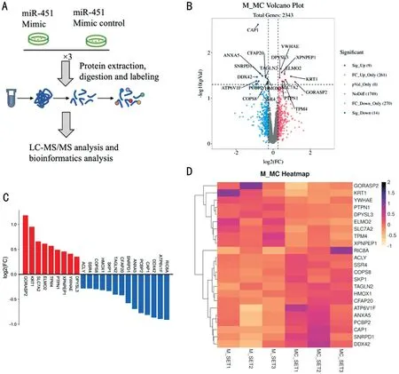
To investigate the biological characteristic changes of endothelial cells in the presence of miR-451, RF/6A cells were transfected with miR-451 mimic and inhibitor respectively (Figure 1A). CCK-8 test was performed at different co-culture times. Compared with miR-451 mimic control, OD450 value of RF/6A cells transfected miR-451 mimic was lower both at 24h and 48h, indicating the decreased proliferation ability induced by miR-451 on RF/6A cells (Figure 1B). On the contrary,in comparison to the inhibitor control, miR-451 inhibitor promoted RF/6A cell proliferation after 48h of transfection despite it showed no effect at 24h (Figure 1B).
iTRAQ LC-MS/MS Analysis of RF/6A Cells Transfected with miR-451 Mimic Versus Mimic Control
To explore the DEPs following miR-451 overexpression, total cell lysates of RF/6A transfected with miR-451 mimic and mimic control were collected and analyzed by iTRAQ and LC-MS/MS (Figure 2A). A total of 2343 proteins were detected and quantified in both mimic-transfected and mimic control-transfected RF/6A cells. We considered proteins with fold change over 1.2 or less than 0.83 and
-value <0.05 as DEPs. Therefore, in the comparison of miR-451 mimic and mimic control, 23 DEPs were identified, with 9 up-regulated proteins and 14 downregulated proteins as volcano plot (Figure 2B), fold change of DEPs (Figure 2C) and heatmap (Figure 2D) exhibited. Among these DEPs, up-regulated proteins were golgi reassembly stacking protein 2 (GORASP2 ), cytokeratin-1 (KRT1), [solute carrier family 7 (cationic amino acid transporter, y+ system),member 2 (SLC7A2)], engulfment and cell motility protein 2(ELMO2), tropomyosin 4 (TPM4), and the five most downregulated proteins were RIC8 guanine nucleotide exchange factor A (RIC8A), ATPase H+ transporting V1 subunit F(ATP6V1F), ATP-dependent RNA helicase DDX42 (DDX42),Adenylyl cyclase-associated protein (CAP1), [poly(rC) binding protein 2 (PCBP2)].
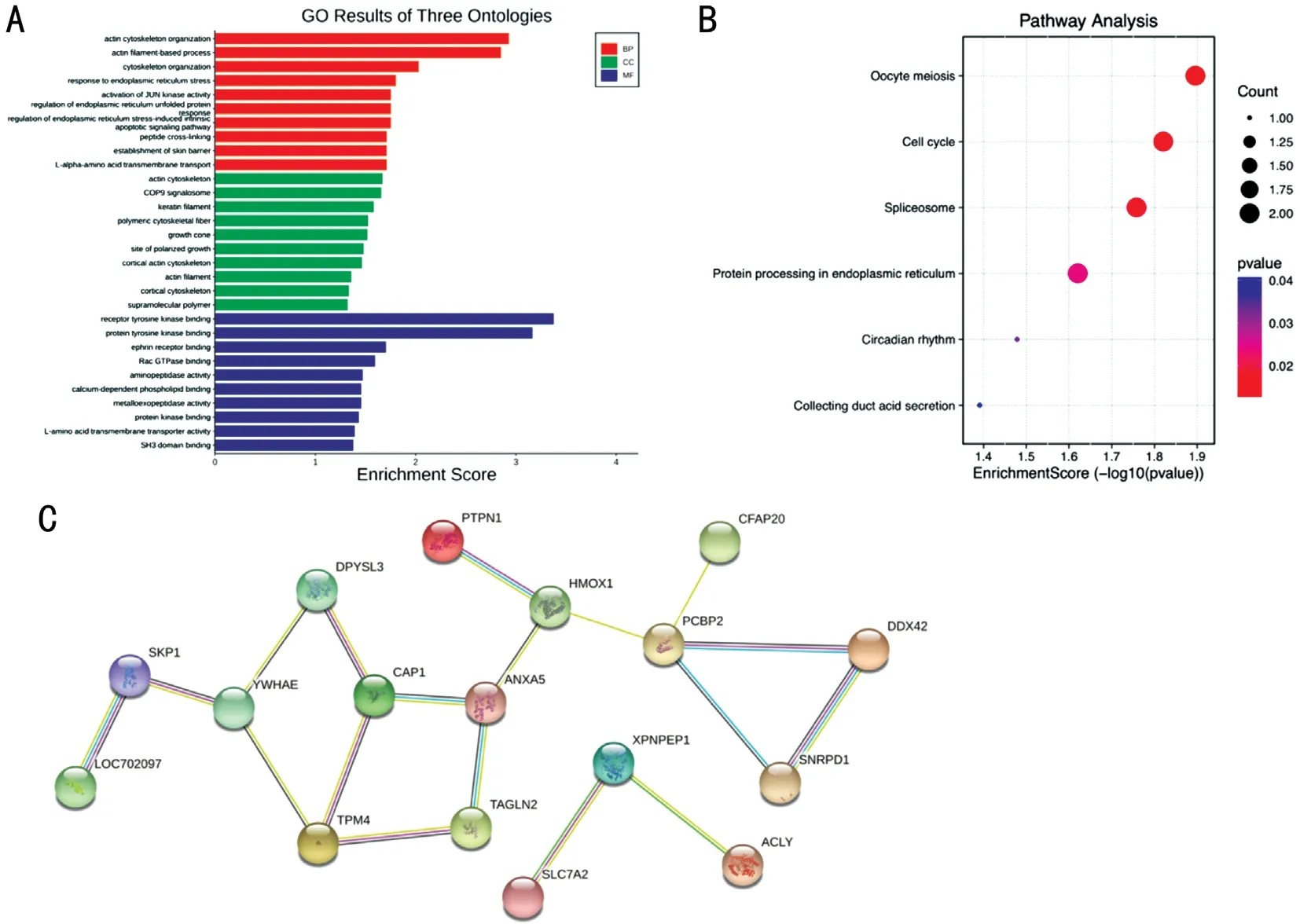
To illuminate the biological characterization of the DEPs, both up-regulated and down-regulated proteins were subjected to GO (Figure 3A) and KEGG (Figure 3B) enrichment analysis. The BP enrichment analysis revealed that the DEPs mainly participated in the cytoskeleton organization, endoplasmic reticulum stress,intrinsic apoptotic signaling pathway, JUN kinase stress,peptide cross-linking, and L-alpha-amino acid transmembrane transport. The MF enrichment analysis exhibited that the DEPs were mainly associated with receptor tyrosine kinase binding,protein tyrosine kinase binding, ephrin receptor binding,Rac GTPase binding, aminopeptidase activity, L-amino acid transmembrane transport activity,
. The CC enrichment analysis showed that the DEPs were mainly involved in the actin cytoskeleton, COP9 signalosome, keratin filament,polymetric cytoskeletal fiber, growth cone, site of polarized growth,
. The KEGG pathways involved by the DEPs included oocyte meiosis, cell cycle, spliceosome, protein processing in endoplasmic reticulum, circadian rhythm, and collecting duct acid secretion. STRING database was applied to investigate the interaction between these DEPs (Figure 3C). There were two groups of strongly interacted proteins including YWHAE-DPYSL3-CAP1-TPM4-TAGLN2-ANXA5-HMOX1-PCBP2-DDX42-SNRPD1, and SLC7A2-XPNPEP1-ACLY. PCBP2 and XPNPEP1 act as hub proteins in their respective groups.
但是,他对这些大道理想得并不长久。他蹲在苔藓地上,嘴里衔着一根骨头,吮吸着仍然使骨头微微泛红的残余生命。甜蜜蜜的肉味,跟回忆一样隐隐约约,不可捉摸,却引得他要发疯。他咬紧骨头,使劲地嚼。有时他咬碎了一点骨头,有时却咬碎了自己的牙,于是他就用岩石来砸骨头,把它捣成了酱,然后吞到肚里。匆忙之中,有时也砸到自己的指头,使他一时感到惊奇的是,石头砸了他的指头他并不觉得很痛。

iTRAQ LC-MS/MS Analysis of RF/6A Cells Transfected with miR-451 Inhibitor Versus Inhibitor Control
iTRAQ LC-MS/MS was also applied to analyze the proteome profile of RF/6A cells after miR-451 knockdown. According to the criteria of fold change >1.2 or <0.83 and
-value <0.05, we identified 30 DEPs, including 13 up-regulated and 17 downregulated proteins (Figure 4A-4D). The top five up-regulated proteins were phosphate cytidylyltransferase 1, choline,alpha (PCYT1A), alpha-1,3-mannosyl-glycoprotein 2-beta-N-acetylglucosaminyltransferase (MGAT1), tubulin beta chain (TUBB), mitochondrial calcium uniporter (MCU),and Nucleotide exchange factor SIL1 (SIL1), and the top five down-regulated proteins were (BH3 interacting domain death agonist (BID), [guanine nucleotide binding protein-like 3 (nucleolar) (GNL3)], mutS homolog 6 (MSH6), patatinlike phospholipase domain containing 6 (PNPLA6), protein tyrosine phosphatase non-receptor type 1 (PTPN1).
As one of the small non-coding RNA molecules, miR-451 has participated in the various pathophysiological process
and is expected to be a promising therapeutic target in different diseases
. In fundus oculi disease, endothelial cell dysfunction induced by hypoxia, ischemia, or hyperglycemia is responsible for retinal neovascularization
. Previous studies revealed that miR-451 has a therapeutic effect in suppressing angiogenesis in hepatocellular carcinoma
and other vascular diseases
. The latest research by Trotta
found that circulating miR-451a was gradually reduced as DR progressed and could be a promising biomarker for DR. And our research of miR-451a on RPE found that miR-451a inhibited the proliferation and migration of RPE cells, exhibiting the potential effect to inhibit the formation of the epiretinal membrane which was a critical feature of the severe stage of PDR
. However, whether miR-451 could regulate the biological function of retinal endothelial cells and play a protective role in neovascular disease of the fundus is still unknown. Therefore, we intend to reveal the potential role of miR-451 in regulating the function and proteome profile of RF/6A, to explore whether miR-451 could be a therapeutic target for neovascular diseases of the fundus.

装药准备工作做好后,质检员检查药卷的线装药密度是否符合设计要求,药卷绑扎是否牢固,导爆索与药卷衔接是否紧密,并复核药卷编号与预裂孔编号是否相符。药卷验收合格后,进行预裂孔装药,药卷与孔位一一对应,使竹片靠结构面侧放置。联网施工时,炮工严格按爆破设计网络图连线,导爆索连接采用扭结法,搭接长度要求>30cm,且要求主干索、支干索和引爆索顺传爆方向的夹角<90º,以避免出现拒爆现象。
In this experiment, we found that up-regulation of miR-451 could inhibit RF/6A cell proliferation, and the inhibitory effect could last from 24h to 48h after transfection, suggesting that miR-451 itself played an inhibitory effect on cell proliferation.In addition, the downregulation of miR-451 level did not affect RF/6A cell proliferation at 24h but promoted cell proliferation at 48h. We believe that this phenomenon was probably related to the specific mechanism of miR-451 mimic and inhibitor function. miR-451 mimic can act directly on the target mRNA such as inhibiting mRNA translation after entering RF/6A cells. However, miR-451 inhibitors may need to combine with miR-451 pieces before playing the inhibiting role, leading to the occurrence of functional delay. On the other hand, downregulation of miR-451 may be insufficient for the inhibition of RF/6A cell proliferation before causing cell biology behavior change.
DISCUSSION
利用Design-Expert8.0.6软件对表3进行多元回归拟合,得到山羊发酵乳水解度与菌种添加量/%(X 1)、后熟时间(X 2)、发酵时间(X 3)的二次方程模型为:Y=8.29+0.82X 1-0.069X 2+0.80X 3-0.33X 1X 2+0.57X 1X 3+0.092X 2X 3-1.10X 12-0.81X 22-0.84X 32回归模型的方差分析结果见表4。
miR-451 mimic and inhibitor were supposed to play an opposite role in regulating cell function. So, protein, whose fold change presented an opposite expression level between mimic
mimic control and inhibitor
inhibitor control,might exert an essential effect in cell regulation. By comparing the significantly increased and reduced proteins among the four groups, PTPN1 was the only protein exhibiting an opposite expression tend (up-regulated when miR-451 mimic versus mimic control and down-regulated when miR-451 inhibitor versus inhibitor control; Figure 6A-6B). According to the STRING database, the most intensively correlated proteins with PTPN1 in human were SRC, BCAR1, GRB2, IRS1,EGFR, JAK2, IGF1R, INSR, CDH2, and RMDN3 (Figure 6C). These proteins were involved in the insulin receptor signaling pathway, platelet-derived growth factor receptor signaling pathway, peptidyl-tyrosine autophosphorylation,ERBB2 signaling pathway, and positive regulation of glucose metabolic process (Figure 6D).
DEPs detected by iTRAQ LC-MS/MS in RF/6A after miR-451 overexpression included 9 up-regulated proteins(GORASP2, KRT1, SLC7A2, ELMO2, TPM4,
.), and 14 down-regulated proteins (RIC8A, ATP6V1F, DDX42,CAP1, PCBP2,
.). GORASP2 also known as GRASP55,is a Golgi stacking protein involved in autophagy, protein and lipid trafficking, sorting, processing, and modification
.Ahat
found that depletion of GRASP increased the growth of HeLa cells though exact mechanism has not been revealed. In our research, the upregulation of GORASP2 might be partly accountable for the inhibitory effect of miR-451 on RF/6A cell proliferation. KRT1, a member of the keratin gene family, constituted the skin epidermis structure.Blanckaert
revealed that increased KRT1 was related to the inhibition of docosahexaenoic acid (DHA) on the invasion of MDA-MB-231 breast cancer cells. SLC7A2, a member of the solute carrier family, was related to the amino acid transmembrane transporter activity. A latest research by Xia
, demonstrated that overexpression of SLC7A2 suppressed the invasion and metastasis of hepatocellular carcinoma by regulating myeloid-derived suppressors cell recruitment. However, increased ELMO2
and TPM4
were related to the proliferation and metastasis of different cancers respectively, suggesting that they might contributed little to the regulation of miR-451 on RF/6A cell proliferation.

RIC8A, was involved in the regulation of cell adhesion and migration. Researchers found that knockdown of RIC8A gene inhibited the tumorigenesis in a mouse model of melanoma
.Therefore, we supposed that miR-451 probably inhibited the proliferation of RF/6A cells through down-regulating RIC8A expression. ATP6V1F, participating in the transport of hydrogen ions, was confirmed to be associated with the prognosis of rectal cancer
and renal clear cell carcinoma
.However, there was no study that verified its role in the regulation of cell proliferation. DDX42, participated in the protein localization and regulation of apoptotic process,
.Sohn and Chay
, found that DDX42 inhibited the apoptosis of Ba/F3 cells (a mouse pro-B cell line). Besides, CAP1, involved in the regulation of the actin cytoskeleton, was extensively studied in various cancers. The upregulation of CAP1 was responsible for the proliferation, migration and invasion of lung cancer
, gastric cancer
, and breast cancer
.Moreover, increase of PCBP2, one of the major cellular poly(rC)-binding proteins, significantly promoted cell viability,metastasis and invasion in hepatocellular carcinoma
. Thus,these down-regulated DEPs were very likely to take part in the regulation of miR-451 on RF/6A cell function.
miR-451 mimic, mimic control, inhibitor, and inhibitor control were purchased from Genepharma (Suzhou,China). The coding sequences were as below.
3)确定指标的隶属度函数类型。参数间的函数关系可采用二次函数、指数函数以及对数函数等形式拟合分析。其中,三次函数的拟合程度最高,二次与三次函数的拟合程度相近。考虑到ELV充电站选址的评分关系的复杂性,采用二次函数y=a1x2+a2x+a3作为备选方案与评价指标的评分函数。其中,y为评价得分,x为评价指标值,a2、a1分别为一、二次项系数,a3为随机误差项。
For the down-regulated DEPs, different proteins also exemplified various effect on cell viability. BID is related to the cellular apoptotic process. Bi and Wang
recently exemplified that LINC00472, a tumor inhibitor, could increase apoptosis and repress the proliferation of pancreatic cancer cells by promoting BID expression. Since BID ranked first among the down-regulated proteins, it might exert critical effect in the regulation RF/6A cell viability. However, GNL3,participated in GTP binding and mRNA 5’-UTR binding, was up-regulated in tumor progression. Li
confirmed that knockdown of GNL3 inhibited the growth, migration and invasion of osteosarcoma cell lines. MSH6 (DNA mismatch repair protein) is one of the components of the post-replicate DNA mismatch repair system (MMR). Palassin
found that NRIP1 (nuclear receptor interacting protein 1) mutant participated in the progression of colorectal cancer
downregulating MSH6 expression. Therefore, the decrease of MSH6 expression might partly be responsible for the proliferation of RF/6A. Besides, PNPLA6, was closely related to the normal development of nervous system. Although the mutant of PNPLA6 was verified in different neurological syndrome
,there was no research on its potential role in cell proliferation and migration.
当主程序检测到铆接力和位移大于设定的初始阈值时,则铆接位置识别程序启动。首先采集一幅图像并保存,然后将图像送入在线识别子程序中,输出铆接位置。
Among all the DEPs, PTPN1 was the only protein that showed an opposite trend between miR-451 mimic and inhibitor group.As a member of the protein tyrosine phosphatase (PTP) family,PTPN1 specifically catalyzes the dephosphorylation of tyrosine residues, involved in the cell growth, differentiation, mitotic cycle, and oncogenic transformation
. The role of PTPN1 in the regulation of cell growth was inconsistent referring to different cancer cells. For colon cancer, Radhakrishnan
reported that curcumin increased the activity of PTPN1,resulting in the reduced migration of HCT116 and SW480 cells
dephosphorylating pTyr(421)-cortactin. Nunes-Xavier
also found that in neuroblastoma, PTPN1 deficiency led to an increase of SH-SY5Y cell proliferation. In lung adenocarcinoma, PTPN1 inhibited cell growth and metastasis through the dephosphorylation of c-Met and PIK3R2
. The latest research on bladder cancer by Monoe
revealed that PTPN1 knockdown resulted in the increase of cancer cell proliferation and migration, and miR-130-targeted LNA(locked nucleic acid) increased PTPN1 and exhibited as a promising therapeutic agent. However, PTPN1 was also found to be positively correlated with the progression of melanoma
and glioma
. These seemingly controversial results might partly be due to the heterogeneity of different cells. Therefore,we suppose that miR-451 mimic probably inhibited RF/6A cell growth through up-regulation of PTPN1, while miR-451 inhibitor promoted cell proliferation
down-regulating PTPN1.
在临床思维能力、操作能力、团队协作能力方面,SBMEPBL组的赞成比例明显高于PBL组(P<0.05);在提高自学能力、学习兴趣方面,SBME-PBL组和PBL组两组的赞成比例差异无统计学意义(P>0.05)。详见表3。
In conclusion, our study revealed that when miR-451 was overexpressed, the proliferation ability of RF/6A cells was inhibited; on the contrary, miR-451 inhibitor enhanced cell growth of RF/6A. Data obtained from iTRAQ LC-MS/MS showed that a variety of proteins were involved in RF/6A cell growth and angiogenesis. GO and KEGG pathway analysis of DEPs depicted the cellular function influenced by miR-451.PTPN1, as the unique protein showing opposite expression trend between miR-451 mimic and inhibitor, was most likely to be the target protein of miR-451 regulating the proliferation of RF/6A. We suppose that miR-451 inhibited RF/6A cell growth
up-regulating the expression level of PTPN1, and miR-451 might be a promising therapeutic target for retinal neovascularization. However, further research is required to exemplify the exact protective role of miR-451 in neovascular disease of fundus based on the animal model.
Supported by grants from National Natural Science Foundation of China (No.81900891); Global Ophthalmology Awards Program 2020 (No.482667).
None;
None;
None;
None;
None;
None.
1 Cheung N, Mitchell P, Wong TY. Diabetic retinopathy.
2010;376(9735):124-136.
2 Antonetti DA, Klein R, Gardner TW. Diabetic retinopathy.
2012;366(13):1227-1239.
3 Bhutto I, Lutty G. Understanding age-related macular degeneration(AMD): relationships between the photoreceptor/retinal pigment epithelium/Bruch’s membrane/choriocapillaris complex.
2012;33(4):295-317.
4 Bai H, Wu SH. miR-451: a novel biomarker and potential therapeutic target for cancer.
2019;12:11069-11082.
5 Shao Y, Dong LJ, Takahashi Y, Chen J, Liu X, Chen Q, Ma JX, Li XR.miRNA-451a regulates RPE function through promoting mitochondrial function in proliferative diabetic retinopathy.
2019;316(3):E443-E452.
6 Nan Y, Guo HB, Guo LY, Wang L, Ren BC, Yu K, Huang Q, Zhong Y.MiRNA-451 inhibits glioma cell proliferation and invasion through the mTOR/HIF-1α/VEGF signaling pathway by targeting CAB39.
2018;29(3):156-166.
7 Nan Y, Guo LY, Lu YL, Guo GC, Hong RJ, Zhao LW, Wang L, Ren BC,Yu K, Zhong Y, Huang Q. miR-451 suppresses EMT and metastasis in glioma cells.
2021;20(13):1270-1278.
8 Liu XM, Zhang AP, Xiang JX, Lv Y, Zhang XF. miR-451 acts as a suppressor of angiogenesis in hepatocellular carcinoma by targeting the IL-6R-STAT3 pathway.
2016;36(3):1385-1392.
9 Liu XM, Zhang XF, Xiang JX, Lv Y, Shi JH. miR-451: potential role as tumor suppressor of human hepatoma cell growth and invasion.
2014;45(2):739-745.
10 Chen MB, Wei MX, Han JY, Wu XY, Li C, Wang J, Shen W, Lu PH.MicroRNA-451 regulates AMPK/mTORC1 signaling and fascin1 expression in HT-29 colorectal cancer.
2014;26(1):102-109.
11 Zhang LJ, Dong LJ, Liu X, Jiang YF, Zhang LJ, Zhang XM, Li XR,Zhang Y. A-Melanocyte-stimulating hormone protects retinal vascular endothelial cells from oxidative stress and apoptosis in a rat model of diabetes.
2014;9(4):e93433.
12 Wen B, Zhou R, Feng Q, Wang Q, Wang J, Liu S. IQuant: an automated pipeline for quantitative proteomics based upon isobaric tags.
2014;14(20):2280-2285.
13 Liu Y, Li H, Li LH, Tang JB, Sheng YL. Mir-451 inhibits proliferation and migration of non-small cell lung cancer cells
targeting LKB1/AMPK.
2019;23(3 Suppl):274-280.
14 Park MC, Kwon OC, Lee SW, Song JJ, Park YB. MiR-451 suppresses inflammatory responses in ankylosing spondylitis by targeting macrophage migration inhibitory factor.
2020;38(2):275-281.
15 Khordadmehr M, Jigari-Asl F, Ezzati H, Shahbazi R, Sadreddini S, Safaei S, Baradaran B. A comprehensive review on miR-451: a promising cancer biomarker with therapeutic potential.
2019;234(12):21716-21731.
16 Liang C, Gao L, Liu Y, Liu YZ, Yao R, Li YP, Xiao LL, Wu LM, Du BB, Huang Z, Zhang YZ. miR-451 antagonist protects against cardiac fibrosis in streptozotocin-induced diabetic mouse heart.
2019;224:12-22.
17 Bharadwaj AS, Appukuttan B, Wilmarth PA, Pan Y, Stempel AJ,Chipps TJ, Benedetti EE, Zamora DO, Choi D, David LL, Smith JR.Role of the retinal vascular endothelial cell in ocular disease.
2013;32:102-180.
18 Zhang WG, Liu DM, Han XW, Ren JZ, Zhou PL, Ding PX.microRNA-451 inhibits vascular smooth muscle cell migration and intimal hyperplasia after vascular injury
Ywhaz/p38 MAPK pathway.
2019;379(2):214-224.
19 Trotta MC, Gesualdo C, Platania CBM, de Robertis D, Giordano M,Simonelli F, D’Amico M, Drago F, Bucolo C, Rossi S. Circulating miRNAs in diabetic retinopathy patients: prognostic markers or pharmacological targets?
2021;186:114473.
20 Tang DM, Wang YZ. Cell cycle regulation of Golgi membrane dynamics.
2013;23(6):296-304.
21 Ahat E, Xiang Y, Zhang XY, Bekier ME 2nd, Wang YZ. GRASP depletion-mediated Golgi destruction decreases cell adhesion and migration
the reduction of α5β1 integrin.
2019;30(6):766-777.
22 Blanckaert V, Kerviel V, Lépinay A, Joubert-Durigneux V,Hondermarck H, Chénais B. Docosahexaenoic acid inhibits the invasion of MDA-MB-231 breast cancer cells through upregulation of cytokeratin-1.
2015;46(6):2649-2655.
23 Xia SH, Wu JW, Zhou WD, Zhang MY, Zhao K, Liu JM, Tian DA,Liao JZ. SLC7A2 deficiency promotes hepatocellular carcinoma progression by enhancing recruitment of myeloid-derived suppressors cells.
2021;12(6):570.
24 Wang YC, Li HY, Li F. ELMO2 association with G α i2 regulates pancreatic cancer cell chemotaxis and metastasis.
2020;8:e8910.
25 Luo YS, Yu PJ, Zhao JH, Guo QJ, Fan BH, Diao YZ, Jin YL, Wu J,Zhang CW. Inhibitory effect of crocin against gastric carcinoma via regulating TPM4 gene.
2021;14:111-122.
26 Patel BR, Tall GG. Ric-8A gene deletion or phorbol ester suppresses tumorigenesis in a mouse model of
(Q209L)-driven melanoma.
2016;5(6):e236.
27 Qi X, Yuxin L, Liu XY, Chen JJ, Shen BR. Biomarker discovery for the carcinogenic heterogeneity between colon and rectal cancers based on lncRNA-associated ceRNA network analysis.
2020;10:535985.
28 Li X, Li H, Yang C, Liu L, Deng S, Li M. Comprehensive analysis of ATP6V1s family members in renal clear cell carcinoma with prognostic values.
2020;10:567970.
29 Sohn SO, Chay KO. The ATP-dependent RNA helicase, DDX42 interacts with paxillin and regulates apoptosis and polarization of Ba/F3 cells.
(
) 2019;23(1):1-9.
30 Zeng J, Li X, Liang L, Duan HX, Xie SS, Wang CH. Phosphorylation of CAP1 regulates lung cancer proliferation, migration, and invasion.
2022;148(1):137-153.
31 Wang WH, Chen SK, Huang HC, Juan HF. Proteomic analysis reveals that metformin suppresses PSMD2, STIP1, and CAP1 for preventing gastric cancer AGS cell proliferation and migration.
2021;6(22):14208-14219.
32 Hasan R, Zhou GL. The cytoskeletal protein cyclase-associated protein 1 (CAP1) in breast cancer: context-dependent roles in both the invasiveness and proliferation of cancer cells and underlying cell signals.
2019;20(11):2653.
33 Ma ZL, Li S, Wang YQ, Zhang JX, Zeng XF. Upregulation of a novel LncRNA AC104958.2 stabilized by PCBP2 promotes proliferation and microvascular invasion in hepatocellular carcinoma.
2021;407(1):112791.
34 Xiong J, Wang L, Fei XC, Jiang XF, Zheng Z, Zhao Y, Wang CF, Li B,Chen SJ, Janin A, Gale RP, Zhao WL. MYC is a positive regulator of choline metabolism and impedes mitophagy-dependent necroptosis in diffuse large B-cell lymphoma.
2017;7(7):e0.
35 Yu J, Wu C, Wu Q, Huang J, Fu W, Xie X, Li W, Tang W, Xu C, Jin G. PCYT1A suppresses proliferation and migration via inhibiting mTORC1 pathway in lung adenocarcinoma.
2020;529(2):353-361.
36 Li YN, Liu YC, Zhu HD, Chen XN, Tian M, Wei YY, Gong Y,Jiang JH. N-acetylglucosaminyltransferase I promotes glioma cell proliferation and migration through increasing the stability of the glucose transporter GLUT1.
2020;594(2):358-366.
37 Yu R, Longo J, van Leeuwen JE, Zhang C, Branchard E, Elbaz M,Cescon DW, Drake RR, Dennis JW, Penn LZ. Mevalonate pathway inhibition slows breast cancer metastasis via reduced N-glycosylation abundance and branching.
2021;81(10): 2625-2635.
38 Yu XJ, Zhang YQ, Wu BG, Kurie JM, Pertsemlidis A. The miR-195 axis regulates chemoresistance through TUBB and lung cancer progression through BIRC5.
2019;14:288-298.
39 Zhao YL, Wang YY, Zhao J, Zhang ZH, Jin MP, Zhou F, Jin C, Zhang J, Xing JL, Wang N, He XL, Ren TT. PDE2 inhibits PKA-mediated phosphorylation of TFAM to promote mitochondrial Ca
+-induced colorectal cancer growth.
2021;11:663778.
40 Miao Y, Wang XF, Lai YF, Lin W, Huang Y, Yin H, Hou RR, Zhang FX. Mitochondrial calcium uniporter promotes cell proliferation and migration in esophageal cancer.
2021;22(3):686.
41 Wang XF, Song XD, Cheng G, Zhang JW, Dong LR, Bai J, Luo D,Xiong YJ, Li S, Liu F, Sun YY, Wang X, Li YY, Huang YN. The regulatory mechanism and biological significance of mitochondrial calcium uniporter in the migration, invasion, angiogenesis and growth of gastric cancer.
2020;13:11781-11794.
42 Zheng XC, Lu ST, He ZX, Huang HH, Yao ZC, Miao YT, Cai CQ,Zou F. MCU-dependent negative sorting of miR-4488 to extracellular vesicles enhances angiogenesis and promotes breast cancer metastatic colonization.
2020;39(46):6975-6989.
43 Li ZF, Xu WW, Li JD, Tao FL, Chen JX, Xu JH. Nucleotide exchange factor SIL1 promotes the progress of breast cancer cells via regulating the cell cycle and apoptosis.
2020;103(1):36850419891046.
44 Bi C, Wang G. LINC00472 suppressed by ZEB1 regulates the miR-23a-3p/FOXO3/BID axis to inhibit the progression of pancreatic cancer.
2021;25(17):8312-8328.
45 Li TY, Li L, Wu XY, Tian KX, Wang YZ. The oncogenic role of GNL3 in the progression and metastasis of osteosarcoma.
2019;11:2179-2188.
46 Palassin P, Lapierre M, Pyrdziak S,
A truncated NRIP1 mutant amplifies microsatellite instability of colorectal cancer by regulating MSH2/MSH6 expression, and is a prognostic marker of stage III tumors.
2021;13(17):4449.
47 Liu F, Ji YM, Li GM, Xu C, Sun Y. Identification of Oliver-McFarlane syndrome caused by novel compound heterozygous variants of PNPLA6.
2020;761:145027.
48 Sen K, Finau M, Ghosh P. Bi-allelic variants in PNPLA6 possibly associated with Parkinsonian features in addition to spastic paraplegia phenotype.
2020;267(9):2749-2753.
49 Bourdeau A, Dubé N, Tremblay ML. Cytoplasmic protein tyrosine phosphatases, regulation and function: the roles of PTP1B and TCPTP.
2005;17(2):203-209.
50 Radhakrishnan VM, Kojs P, Young G, Ramalingam R, Jagadish B,Mash EA, Martinez JD, Ghishan FK, Kiela PR. pTyr421 cortactin is overexpressed in colon cancer and is dephosphorylated by curcumin:involvement of non-receptor type 1 protein tyrosine phosphatase(PTPN1).
2014;9(1):e85796.
51 Nunes-Xavier CE, Aurtenetxe O, Zaldumbide L,
. Protein tyrosine phosphatase PTPN1 modulates cell growth and associates with poor outcome in human neuroblastoma.
2019;14(1):134.
52 Chen YC, Tang JQ, Lu T, Liu F. CAPN
promotes malignant behavior and erlotinib resistance mediated by phosphorylation of c-Met and PIK3R2 via degrading PTPN1 in lung adenocarcinoma.
2020;11(7):1848-1860.
53 Monoe Y, Jingushi K, Kawase A, Hirono T, Hirose R, Nakatsuji Y,Kitae K, Ueda Y, Hase H, Abe Y, Adachi J, Tomonaga T, Tsujikawa K. Pharmacological inhibition of miR-130 family suppresses bladder tumor growth by targeting various oncogenic pathways via PTPN1.
2021;22(9):4751.
54 Liu JQ, Luan WJ, Zhang Y, Gu JY, Shi YD, Yang YW, Feng ZH,Qi FZ. HDAC6 interacts with PTPN1 to enhance melanoma cells progression.
2018;495(4):2630-2636.
55 Jin T, Li DB, Yang T, Liu F, Kong J, Zhou YF. PTPN1 promotes the progression of glioma by activating the MAPK/ERK and PI3K/AKT pathways and is associated with poor patient survival.
2019;42(2):717-725.
猜你喜欢
现代制造技术与装备(2023年11期)2024-01-17 07:41:18
现代制造技术与装备(2023年9期)2023-11-14 12:04:44
广州化工(2020年18期)2020-09-28 06:46:14
保健医苑(2019年5期)2019-05-15 01:07:34
同煤科技(2018年3期)2018-07-24 02:03:32
中华胃食管反流病电子杂志(2017年2期)2017-10-27 01:47:16
中华胃食管反流病电子杂志(2016年1期)2016-10-19 08:25:12
当代化工研究(2016年9期)2016-03-20 16:22:08
火工品(2014年2期)2014-10-11 07:46:12
济宁医学院学报(2014年4期)2014-08-16 13:44:19
 International Journal of Ophthalmology2022年6期
- International Journal of Ophthalmology的其它文章
- Intraocular lens removal or not during vitrectomy for acute infectious endophthalmitis after cataract surgery
- Vitreous function and intervention of it with vitrectomy and other modalities
- Short-term outcomes of mitomycin C-augmented excisional bleb revision with capsulectomy for failed Ahmed glaucoma valve
- Evaluation of nintedanib as a new postoperative antiscarring agent in experimental extraocular muscle surgery
- Multimodal imaging of experimental choroidal neovascularization
- A novel Nance-Horan syndrome mutation identified by next-generation sequencing in a Chinese family