Optimization of adipose tissue-derived mesenchymal stromal cells transplantation for bone marrow repopulation following irradiation
Min-Jung Kim,Won Moon,Jeonghoon Heo,Sangwook Lim,Seung-Hyun Lee,Jee-Yeong Jeong,Sang Joon Lee
Min-Jung Kim,Jee-Yeong Jeong,Department of Biochemistry,Cancer Research Institute Kosin University College of Medicine,Seo-gu 49267,Busan,South Korea
Won Moon,Department of Internal Medicine,Kosin University College of Medicine,Seo-gu 49267,Busan,South Korea
Jeonghoon Heo,Department of Molecular Biology and Immunology,Kosin University College of Medicine,Seo-gu 49267,Busan,South Korea
Sangwook Lim,Department of Radiation Oncology,Kosin University College of Medicine,Seo-gu 49267,Busan,South Korea
Seung-Hyun Lee,Department of General Surgery,Kosin University College of Medicine,Seogu 49267,Busan,South Korea
Sang Joon Lee,Department of Ophthalmology,Gospel Hospital,Kosin University College of Medicine,Seo-gu 49267,Busan,South Korea
Abstract BACKGROUND Bone marrow(BM)suppression is one of the most common side effects of radiotherapy and the primary cause of death following exposure to irradiation.Despite concerted efforts,there is no definitive treatment method available.Recent studies have reported using mesenchymal stromal cells(MSCs),but their therapeutic effects are contested.AIM We administered and examined the effects of various amounts of adipose-derived MSCs(ADSCs)in mice with radiation-induced BM suppression.METHODS Mice were divided into three groups:Normal control group,irradiated(RT)group,and stem cell-treated group following whole-body irradiation(WBI).Mouse ADSCs(mADSCs)were transplanted into the peritoneal cavity either once or three times at 5 × 10⁵ cells/200 μL.The white blood cell count and the levels of,plasma cytokines,BM mRNA,and BM surface markers were compared between the three groups.Human BM-derived CD34+hematopoietic progenitor cells were co-cultured with human ADSCs(hADSCs)or incubated in the presence of hADSCs conditioned media to investigate the effect on human cells in vitro.RESULTS The survival rate of mice that received one transplant of mADSCs was higher than that of mice that received three transplants.Multiple transplantations of ADSCs delayed the repopulation of BM hematopoietic stem cells.Anti-inflammatory effects and M2 polarization by intraperitoneal ADSCs might suppress erythropoiesis and induce myelopoiesis in sub-lethally RT mice.CONCLUSION The results suggested that an optimal amount of MSCs could improve survival rates post-WBI.
Key Words:Adipose tissue-derived stem cells;Bone marrow suppression;Mesenchymal stromal cells;Radiation;Cell therapy
INTRODUCTION
Acute exposure to high doses of ionizing radiation(IR)can lead to acute radiation syndrome(ARS),which affects the hematopoietic,gastrointestinal,and neurovascular systems[1,2].Hematopoietic symptoms,particularly those induced by ARS,develop into severe neutropenia and thrombocytopenia,which eventually lead to bleeding,infection,and death[3].The same clinical results are obtained when myeloablative therapy is performed in patients with conditions such as leukemia,lymphoma,and myeloma[4].Although significant advances have been made in the development of safe,non-toxic,and effective radiation over the past six decades,no ARS drug has yet been approved by the United States Food and Drug Administration[5].
Bone marrow(BM)suppression is one of the most common side effects of radiotherapy and the primary cause of death post-exposure to a moderate or high dose of whole-body irradiation(WBI)[6].The main therapeutic treatment is the administration of cytokines,such as granulocyte-colony stimulating factor(G-CSF),granulocyte-macrophage CSF(GM-CSF),and transplantation of hematopoietic stem cells(HSCs).Among these,G-CSF is a well-established and effective drug for the treatment of IR-induced acute BM suppression[7].However,recent data have demonstrated that G-CSF could exhaust BM capacity during radiotherapy[8-10].Currently,the modulation of the BM microenvironment post-irradiation has emerged as a novel target for hematopoiesis reconstruction in patients with ARS[11,12].
Novel cytokine-based treatments have been proposed for accidentally irradiated(RT)victims,particularly to prevent the apoptosis of hematopoietic stem and progenitor cells(HSPCs)[13-16].The concept of “emergency anti-apoptotic cytokine therapy” is based on the administration of a four-cytokine cocktail:Stem cell factor(SCF),FLT3-ligand,thrombopoietin(TPO),and interleukin(IL)-3,to mitigate the apoptosis of HSPCs and promote balanced differentiation[14].MSCs can secrete a number of cytokines required for hematopoietic re-proliferation post-irradiation,including IL-6,IL-11,leukemia inhibitor factor,SCF,FLT3 ligand,and stromal cell-derived factor(SDF),and can potentially be used as therapeutic agents[17].
BM-derived mesenchymal stem cells(BM-MSCs)are the major components of the hematopoietic microenvironment and play key roles in hematopoiesis.In pre-clinical studies,BM-MSCs have been shown to protect against radiation-induced liver injury[18],promote healing in RT murine skin wounds[19],improve survival in RT mice[20],mitigate gastrointestinal syndrome in mice[21],and restore the intestinal mucosal barrier in RT mice[22].Superoxide dismutase gene-transfected MSCs improved survival in RT mice[23];however,several other reports using similar models showed that MSCs alone did not affect survival[19,20,24].Thus,the effectiveness of MSCs in promoting the proliferation of the RT BM remains controversial and requires investigation of the exact mechanisms to achieve consistent results in the MSC-based treatment of radiation-induced damage.
Adipose-derived mesenchymal stromal cells(ADSCs)were first identified in 2001 by Zuket al[25],as cells with multilineage differentiation characteristics.ADSCs are characterized as a type of adult stem cells,i.e.,pluripotent cells with limited differentiation capacity.The International Fat Applied Technology Society defines any fat-derived pluripotent cell population capable of adherent,proliferative,and differentiating capacity as an ADSC[26].These ADSCs are able to differentiate into adipogenic,osteogenic,chondrogenic,myogenic,cardiac,and neuronal cellsin vitro[27-29].The plasticity and pluripotency of these ADSCs,as well as their clinically abundant and non-invasive harvestability,have enabled researchers to regenerate dead or damaged cells or organs.
ADSCs have been used in several studies to alleviate the side effects of radiation,particularly cicatricial changes in the salivary glands,liver,and skin[30-33].A few studies have demonstrated that ADSCs can support hematopoiesis bothin vitroandin vivo[34-36].ADSCs injected into the BM cavity of RT mice promoted BM reconstitution by supporting the homing,proliferation,and differentiation of BM progenitors,especially those for megakaryocytes[35],granulocytes,and monocytes[34,36].Unfortunately,none of these studies examined the effect of ADSC transplantation on erythropoiesis,which is one of the most important aspects of hematopoiesis.
In the present study,we demonstrated that during BM repopulation post-WBI,mouse ADSC(mADSC)transplantation into the abdominal cavity delayed differentiation into red blood cells(RBCs)and accelerated the granulocyte/macrophage(GM)lineage differentiation and M2 macrophage polarization.Compared to low-dose,high-dose mADSC transplantation reduced survival rates.Therefore,the number of ADSCs for transplantation needs to be optimized to improve post-irradiation survival.
MATERIALS AND METHODS
Animal model
Male C57BL/6 mice(7-8-wk-old,weighing 21-23 g)were purchased from Central Laboratory Animals(Seoul,South Korea).The mice were kept under controlled conditions at a constant temperature(23.6°C)and a 12 h light/dark cycle.The mice were provided with regular chow and water ad libitum.All animal experiments were approved by the Institutional Animal Care and Use Committee of Kosin University College of Medicine,and the animals were maintained and treated according to the regulations of the Association for Research in Vision and Ophthalmology.
The mice were randomly divided into three groups(n= 10/group):Normal control(NC),RT,and stem cell-treated(ST).The mice were RT with a single dose of 900 cGy using therapeutic 6 MV photon beams from a linear accelerator(Clinac iX,Varian Medical Systems,Palo Alto,CA,United States).The irradiation dose was calculated using a treatment planning system(Eclipse v13.0,Varian Medical Systems).During irradiation,the mice were anesthetized for immobilization to ensure homogeneous delivery of the photon beams.After WBI,mice in the ST group were intraperitoneally injected with mADSCs[5 × 10⁵cells/200 μL phosphate buffer solution(PBS)]once(#1)on day 1 or three times(#3)on days 1,5,and 12.The mice in the RT group were intraperitoneally injected with 200 μL PBS;those in the NC group were not RT.The animals were placed in sterile cages immediately after the experiments.All experiments were repeated three times.To assess survival,animals were monitored for four weeks postirradiation and subsequently euthanized using CO2.
Preparation of ADSCs
ADSCs were isolated as previously described,with some modifications[37].Briefly,mouse adipose tissues were obtained from epididymal and dorsal subcutaneous fat layers;human adipose tissues were obtained from fat discarded during elective breast surgeries.All procedures using human adipose tissue were conducted with informed consent under the Kosin University Gospel Hospital IRB approval protocol(protocol number 09-36).
Raw adipose tissue was processed to obtain the stromal vascular fraction(SVF).To isolate SVF,adipose tissues were washed extensively with equal volumes of PBS(Lonza,Basel,Switzerland).The extracellular matrix was digested at 37 °C for 30 min with 0.075% type I collagenase(Worthington Biochemical Corporation,Lakewood,NJ,United States).Enzyme activity was neutralized with Dulbecco’s modified Eagle’s medium/nutrient mixture F-12(DMEM/F-12;GibcoBRL,Grand Island,NY,United States)containing 10% fetal bovine serum(FBS;GE Healthcare,Little Chalfont,United Kingdom)and centrifuged at 1200 × g for 10 min to obtain a high-density SVF pellet.The cell pellet was resuspended in DMEM/F-12 supplemented with 10% FBS;filtered through a 100 mm mesh to remove cellular debris;seeded in culture plates;and incubated overnight at 37 °C with 5% CO2in DMEM/F-12 medium containing 10% FBS,1% penicillin/streptomycin solution(Gibco,MD,United States),10 ng/mL epidermal growth factor(Sigma-Aldrich,St.Louis,MO,United States),and 2 ng/mL basic fibroblast growth factor(bFGF)(Sigma-Aldrich).Following incubation,the plates were washed extensively with PBS to remove residual non-adherent RBCs.To prevent spontaneous differentiation,ADSCs were maintained at sub-confluent levels.Cells were subcultured with 0.05% trypsin and 1 mmol/L ethylenediaminetetraacetic acid(EDTA);and passaged at a ratio of 1:4.
Multilineage differentiation assay of ADSCs
In vitrodifferentiation of ADSCs was performed as previously described[37].Briefly,P3 ADSCs were trypsinized and seeded in 6-well plates at a density of 5 × 10⁴cells/well.The cells were divided into four groups,i.e.,control,adipogenic,chondrogenic,and osteogenic,and were incubated for three weeks in control,adipogenic(A10070-01,Gibco),chondrogenic(A10071-01,Gibco),and osteogenic(A10072-01,Gibco)differentiation media,respectively.The hanging drop method was used for chondrogenic differentiation[38].In the adipogenic,chondrogenic,and osteogenic groups,the culture medium was replaced every 2 d and the cells were stained with Oil Red O(Sigma-Aldrich),Alcian Blue(Sigma-Aldrich),and Alizarin Red S(Sigma-Aldrich),respectively,to confirm differentiation.
Peripheral blood cell evaluation
Peripheral blood was drawn from the tail vein,streaked as thin smears across a sterile slide,and airdried.Peripheral blood was stained with Wright stain(Muto,Tokyo,Japan)for 2 min,rinsed with PBS,and dried before examination.The number of WBCs was counted in three random fields at 200 ×magnification.Wright staining was performed once on the day before and twice per week post-WBI,until euthanasia.
For complete blood cell counting,blood was drawn from cardiac puncture using a 26-gauge needle syringe and collected in an EDTA-coated bottle(BD Biosciences,Franklin Lakes,NJ,United States).Total differential blood cell counts(WBCs,RBCs,and platelets)were obtained using a veterinary hematology analyzer(Hemavet 950;CDC Technologies,Oxford,CT,United States)at three weeks postirradiation.
Histology
Femurs were fixed in 10% formalin and decalcified in 5% nitric acid(Ducksan,Gwangju,Korea)for 2 h.The femurs were then embedded in paraffin wax and sectioned.The blocks were cut into 5-μm sections using a rotary microtome(Leica RM2235,Leica Biosystems,Germany).The slides were either stained with hematoxylin and eosin or processed for immunohistochemistry(IHC).
For IHC staining,paraffin-embedded sections were deparaffinized in xylene(Duksan,Ansan,Korea)and rehydrated using ethanol(Duksan)gradients and PBS.Paraffin slides were immersed in 1 × citrate buffer(pH 6.0;Sigma-Aldrich),heated for 2 min in a microwave oven,and rinsed three times with PBS.Next,the sections were blocked for 1 h with blocking buffer containing 0.5% goat serum(Sigma-Aldrich)and 0.5% Tween-20 in PBS,and incubated overnight with primary antibodies(mouse anti-PCNA[BD Biosciences,San Jose,CA,United States],rabbit anti-Iba-1(Wako,Tokyo,Japan),and rabbit anti-CD34(GeneTex,Irvine,CA,United States).After incubation,the sections were washed three times with PBS and incubated for 1 h at room temperature with secondary antibodies[goat anti-mouse Alexa Fluor™ 568 and 488(Invitrogen,Carlsbad,CA,United States),goat anti-rabbit Alexa Fluor™ 568 and 488(Invitrogen)].Next,the sections were stained with 4’,6-diamidino-2-phenylindole(DAPI,Sigma-Aldrich),washed three times with PBS,mounted using 2-3 drops of a water-soluble mounting medium(Biomeda Crystal mount,Foster City,CA,United States),and then stored at 4 °C for immediate use or at-70 °C.Images were acquired using a fluorescence microscope(Nikon,Tokyo,Japan).Slides processed without the primary antibody were used as negative controls.
For cryosections,samples were fixed in 4% paraformaldehyde in 0.1 M phosphate buffer for 20 min,followed by three washes with 0.1 M phosphate buffer.The tissues were sequentially cryoprotected with 5%,10%,and 15% sucrose for 1 h and 20% sucrose overnight.Next,the tissues were embedded using polyvinyl alcohol and a 2:1 ratio of polyethylene glycol-based optimal cutting temperature reagent(OCT;Tissue-Tek,Tokyo,Japan)and 20% sucrose.Serial sections of 10 μm were made using a cryostat and mounted on slides.Frozen sections were prepared by dissolving the OCT compound in a slide warmer(Chang Shin Science,Seoul,Korea).Immunostaining was performed after incubating the slides with cold 100% acetone for 10 min.
The numbers of CD34-,PCNA-,and Iba-1-positive cells were calculated as follows:For each group,three or more tissues stained with primary/secondary antibodies and DAPI were randomly selected,and their images captured at 400 × magnification.The number of positive cells in the merged images was counted using a preset area(190 μm × 170 μm).Immunostaining was considered positive if > 50%of the cells fluoresced,regardless of cell size.The percentage of positive cells was calculated as a proportion of the total DAPI-positive cells,multiplied by 100.
Polymerase chain reaction
The mRNA expression levels in mouse BM or human BM CD34+cells were evaluated using reverse transcription polymerase chain reaction(PCR).Total RNA was extracted with a TRIzol™ reagent(Invitrogen)or an RNAqueous™-4PCR kit(Ambion,Austin,TX,United States).Total RNA(1-3 μg)in a 20 μL reaction volume was reverse-transcribed into cDNA using a TOPscript™ cDNA synthesis kit(Enzynomics,Daejeon,Korea).Reactions were performed in a 20 μL volume using SYBR®Premix Ex Taq™ II(Takara,Dalian,China)with 2 μL cDNA.The sequences of human PCR primers for IL-4,IL-10,IL-1β,tumor necrosis factor-α(TNF-α),CD68,CD80,CD206,and β-actin as well as mouse PCR primers for IL-1β,TNF-α,CD14,CD68,CD163,CD206,IL-10,M-CSF,GM-CSF,SDF-1,TPO,IL-7,and glyceraldehyde 3-phosphate dehydrogenase(GAPDH),are listed in Supplementary Table 1.Quantitative realtime PCR was performed using an ABI PRISM 7300 sequence detection system(Applied Biosystems,Foster City,CA,United States)with thermal cycling consisting of denaturation for 10 s at 95 °C,followed by 40 cycles of 5 s at 95 °C,and 31 s at 60 °C.All values were normalized to GAPDH or β-actin and are presented as the average of three independent experiments.
For mouse BM samples,standard reverse transcription PCR was used for IL-1β,TNF-α,CD14,CD68,CD163,and CD206 primers.PCR was performed with TB Green™ Premix Ex Taq™ II(Takara)in a 20 μL volume containing 2 μL of cDNA,0.25 μM of primer,and 10 μL of 2 × PCR Master Mix,using the following conditions:35 cycles of 15 s at 95 °C for denaturation and 30 s at 60 °C for primer annealing and extension.The PCR products were analyzed using agarose gel electrophoresis.
Flow cytometry
To characterize ADSCs,trypsinized cells were incubated with monoclonal antibodies conjugated to either fluorescein isothiocyanate(FITC)or phycoerythrin(PE)for 30 min at room temperature.The following antibodies were used for mouse cells:CD31(Abcam,Cambridge,United Kingdom),CD34,CD105,CD73(Bioss Antibodies,Woburn,MA,United States),CD45,and CD90(BD Biosciences).The antibodies used for human cells were CD34,CD45,CD90,CD105(Beckman Coulter,Brea,CA,United States),and CD73(BD Pharmingen,San Diego,CA,United States).The cells were assessed using flow cytometry(BD Accuri C6 Plus Flow Cytometer)and the data were analyzed using the BD Accuri C6 software(BD Biosciences).
The RT mice were euthanized with CO2.The BM was removed from the femurs and tibiae by flushing with 1 mL DMEM/F-12 media using a 27-gauge needle and resuspended in PBS containing 1% bovine serum albumin(Sigma-Aldrich).The BM cells were filtered using a 30 μm polyamide filter(Myltenyi Biotec,Auburn,CA,United States),and labeled with Sca-1-PE(BD Biosciences),c-Kit-FITC(CD117;MACS,Cambridge,United Kingdom),CD34-FITC(Bioss Antibodies),IL-3R-PE(Miltenyi Biotec,Bergisch Gladbach,Germany),IL-7R-PE(Miltenyi Biotec),CD45RA-PE(Miltenyi Biotec),CD80-PE(eBioscience,San Diego,CA,United States),and CD206-FITC(R&D Systems,Minneapolis,MN,United States)antibodies.
Human BM CD34+stem cells were washed with PBS and labeled with CD34-PE,CD45-APC,CD15-PE,CD71-FITC,and CD235a(Gly-A)-PE(Miltenyi Biotec)antibodies at 4 °C for 30 min after 7 d of culturing.
Cytokine microarray
To determine the plasma cytokine profiles of the mice in each treatment group,blood samples were collected in heparin tubes and centrifuged at 2000 × g for 20 min at 4 °C.The plasma was separated,kept on ice,and stored at -80 °C.Next,the plasma samples were incubated on a pre-coated Proteome Profiler array membrane(ARY006,Mouse Cytokine Antibody Array kit;R&D Systems)and processed according to the manufacturer’s instructions.Briefly,the array membranes were treated with blocking buffer for 1 h at room temperature on a rocking platform shaker.The blocking buffer was removed,and a mixture of plasma and antibody cocktail was added and incubated overnight at 2-8 °C.The incubated membranes were washed and incubated with streptavidin-horseradish peroxidase for 30 min at room temperature.After washing,the membranes were incubated with Chemi Reagent Mix and exposed to an X-ray film for 4-7 min.Densitometry analysis of dot blots was performed using the FluorChemHD2 software(ProteinSimple,Santa Clara,CA,United States).The density of each sample was normalized to that of a reference spot.The cytokine levels in each sample were determined relative to the cytokine levels expressed in the plasma samples of the NC group.A hierarchical clustering algorithm was applied to measure the Euclidean distance for the similarity metric and average linkage clustering method using the Cluster 3.0 open-source software.
Co-culture of human CD34+ HSCs with human ADSCs
Frozen human BM CD34+HSPCs were purchased from Lonza(Walkersville,MD,United States).According to flow cytometry analysis with anti-CD34-PE conjugates(Miltenyi Biotec),the purity of the CD34+cells was consistently > 95%.To produce conditioned media(CM),human ADSCs(hADSCs)at P4 were cultured in DMEM/F-12(Corning,Corning,NY,United States)containing 10% FBS(Hyclone,Logan,UT,United States)and arrested with 20 μg/mL mitomycin C(Sigma-Aldrich)for 2.5 h at 37 °C in 5% CO2atmosphere.After cell growth inhibition,the cells were washed twice with PBS and incubated for 72 h in serum-free StemSpan™ medium(STEMCELL Technologies,Vancouver,Canada)supplemented with recombinant human SCF(50 ng/mL,Peprotech,Rocky Hill,NJ,United States),IL-3(10 ng/mL,Peprotech),and low-density lipoprotein(LDL,25 μg/mL,STEMCELL Technologies).The CM was collected,centrifuged at 500 × g for 20 min to remove cellular debris,and then concentrated 10-fold by means of centrifugation using a Centriprep®Centrifugal Filter Unit(Millipore,Bedford,MA,United States)with a 10 kDa cutoff membrane.
For co-culturing with CD34+cells,the hADSCs(1 × 10⁵cells/well)were seeded in 12-well plates(Corning)and incubated until about 80% confluence.hADSC growth was arrested by treating the cells with 20 μg/mL mitomycin C for 2.5 h,followed by two washes with PBS and incubation for an additional 24 h prior to co-culturing with CD34+cells.BM CD34+cells(1 × 10⁴/well)were cultured in StemSpan™ medium supplemented with SCF(50 ng/mL),IL-3(10 ng/mL),LDL(25 μg/mL),and TPO(100 ng/mL,Peprotech)in three different conditions:(1)In a control medium(control group);(2)In 1 ×CM(CM group);or(3)In direct contact by co-culturing on a layer of hADSC(contact group).The fresh medium and cytokines were replaced on days 3 and 5 of the culture.On day 7,the cells were counted to determine the total cell number,and the proportion of CD34+cells was analyzed using flow cytometry and colony-forming cell(CFC)assays.
hADSC layers for co-culture were prepared as described above.BM CD34+cells(1 × 10⁴cells/well)were cultured in StemSpan™ medium supplemented with SCF(50 ng/mL),IL-3(10 ng/mL),LDL(25 mg/mL),GM-CSF(10 ng/mL),and erythropoietin(EPO,1 U/mL)in three different culture conditions:(1)In a control medium(control group);(2)In 1 × CM(CM group);or(3)In direct contact by coculturing on a layer of hADSC(contact group).The fresh medium and cytokines were added on days 3 and 5 of the culture.On day 7,the cells were counted to determine the total cell number,and the expression of surface markers was analyzed using flow cytometry,as described above.
CFC assay
CFC assays were performed in triplicate for the control,CM,and contact groups.CD34+cells(1 × 10³cells/dish)were plated in methylcellulose medium(MethoCult™ H4100,STEMCELL Technologies)supplemented with SCF(50 ng/mL),IL-3(10 ng/mL),GM-CSF(10 ng/mL,Peprotech),and EPO(1 U/mL)in 35 mm culture dishes and incubated at 37 °C in 5% CO2atmosphere for 14 d.Colonies were counted and analyzed using a scoring grid and an inverted microscope(Olympus,Tokyo,Japan).Colonies were classified based on their morphology as colony-forming unit erythroid(CFU-E),burstforming unit erythroid(BFU-E),CFU-GM,and granulocyte/erythroid/macrophage/megakaryocytes.
Statistics
Data are expressed as mean ± SD.Statistical significance was set atP< 0.05.Five sections were evaluated per sample,and cell counts were used for immunohistochemical analysis.Each analysis was repeated three times.The Kaplan-Meier method was used to estimate the distribution of survival rates over time.A repeated measures two-way repeated measures ANOVA with Sidak’s or Tukey’s multiple comparison test was used to compare groups over time.A Kruskal-Wallis test and Dunn’s multiple comparison test were used to compare the three mouse and human experimental groups,respectively.The GraphPad Prism 7 software for windows(San Diego,CA,United States)was used for statistical analysis.
RESULTS
Characterization of mouse and hADSCs
Mouse ADSCs(mADSCs)and hADSCs appeared as flat cubic cells in the initial culture and changed to spindle-shaped cells similar to fibroblasts with successive subcultures(Supplementary Figures1A and 1F).ADSCs were differentiated into adipose,cartilage,and bone,as evidenced by staining with Oil Red O,toluidine blue,and von Kossa,respectively(Supplementary Figures 1B-D and 2G-I).Surface markers of cultured hADSCs,passaged 3-5 times,were > 95% positive for CD73 and CD105,but negative for CD45 and CD34(Supplementary Figure 1E).The surface markers of mADSCs were positive for CD29,CD73,and CD90,and negative for CD34,CD45,and CD31(Supplementary Figure 1J).
Survival rates
Kaplan-Meier plots showed that WBI induced a mortality rate of 49.2% in the RT group by day 30(Figure 1).The survival rates of the ST group transplanted with mADSCs once(ST#1)and three times(ST#3)were 92.3% and 55.6%,respectively.The survival rate of the ST#1 group was significantly higher than that of the ST#3(P= 0.043)and RT groups(P= 0.021).The reduced survival rates of the ST#3 group are similar to the results reported by Huet al[20],which showed that the mortality rates of RT BALB/c mice increased post-transplantation with BM-MSCs.Due to the relatively high survival rates,the ST#1 group was used in subsequent analyses.
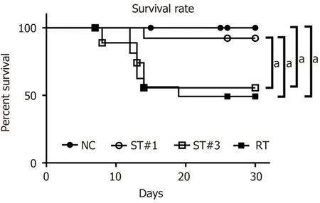
Figure 1 Survival curves of normal control,stem cell-treated,and irradiated mice.Mice were irradiated with a single dose of 900 cGy.After irradiation,mouse adipose tissue-derived stem cells(mADSCs)were transplanted intraperitoneally once(#1)or three times(#3)on days 1,5,and 12.The irradiated(RT)group was injected intraperitoneally with the same volume of saline.The survival rates are presented as Kaplan-Meier plots.The stem cell-treated(ST)group that received one transplant(ST#1)showed higher survival than the RT group.There was no difference in survival between the ST#3 and RT groups.aP < 0.05.NC:Normal control;ST:Stem cell-treated;RT:Irradiated.
Peripheral blood analysis
WBI significantly reduced the number of WBCs,as determined using Wright staining(Figure 2).Compared to the NC group on day 2 post-WBI,the number of WBCs was reduced to 25.7% and 23.8%in the ST#1 and RT groups,respectively.Moreover,the number of WBCs in the ST#1 group was significantly higher(P< 0.01)than in the RT group on days 14-21 post-WBI(Figure 2).However,by day 28,the number of WBCs in the ST and RT groups was partially restored to 70.8% and 57.8%,respectively,compared to that in the NC group.These results indicated that 9 Gy WBI induced a significant reduction in peripheral leukocyte counts,whereas peritoneal transplantation of mADSCs partially restored the number of peripheral leukocytes.
Hematocrit(Hct)and hemoglobin(Hb)levels and RBC and platelet counts were significantly lower in the ST#1 group than in the other groups on day 21 post-WBI(Figure 3A,3B and 3C).However,there were no differences in the mean corpuscular volume,mean corpuscular Hb,and mean corpuscular Hb concentration between the groups(Figure 3D,3E and 3F).These results suggested that mADSCs injected into the abdominal cavity secreted factors that affected the repopulation of BM post-WBI,thus changing the peripheral blood parameters.
Delayed repopulation of CD34+ HSCs in the BM post-WBI
CD34-expressing BM cells were assessed in the ST and RT groups on day 21 post-WBI.Our results indicated that administration of stem cells delayed the emergence of CD34+HSCs(Figure 3G-M).Next,we compared the percentage of proliferating cell nuclear antigen(PCNA)+cells in the CD34+HSCs of all three groups.At week 3 post-WBI,the percentage of PCNA+cells in CD34+HSCs was significantly higher(P≤ 0.0001)in the ST#1 group than in the RT group(Figure 4A,4B and 4C).These findings suggest that the secretome from mADSCs delays the repopulation of CD34+BM progenitor cells and promotes the proliferation of CD34+progenitor cells in the third week post-WBI.Furthermore,the number of ionized calcium-binding adaptor molecule-1(Iba-1)+cells was similar between the RT and ST#1 groups;however,the number of cells expressing both PCNA and Iba-1 was significantly higher in the ST group(P≤ 0.01,Figure 4D,4E and 4F).It has previously been reported that Iba-1 is expressed in the BM of rat monoblastic cells[39].Upon mADSC administration,CD34+HSCs were still observed a week later than in the RT group and the number of proliferating Iba-1+monoblastic cells was significantly higher in the ST#1 group than in the RT group.To identify the cell lineages that were proliferating during repopulation post-WBI,we performed a fluorescence-activated cell sorting analysis.
Characterization of repopulated BM cells post-mADSC transplantation
To compare the differentiation of HSCs during repopulation,Sca-1,c-Kit,and CD34 for HSCs;IL-3R for common myeloid progenitor cells;IL-7R for common lymphoid progenitor cells;CD45RA for common granulocyte-macrophage progenitor cells;and CD80 and CD206 for macrophage polarization were evaluated in BM cells harvested 3 wk post-WBI(Figure 5A-I).Sca-1 levels were significantly reduced in the RT and ST#1 groups compared to those in the NC group.Interestingly,CD34+HSCs were more abundant in the ST#1 group than in the RT group.These findings were consistent with our IHC results where the analyzed samples were also harvested 3 wk post-WBI.There was no difference in the number of BM cells expressing IL-3R between the RT and ST#1 groups.At the same time,IL-7R levels were significantly lower and CD45RA levels were significantly higher in the ST#1 group than those in the RT group.These results suggested that intraperitoneal mADSCs induced an increase in the number of CD34+HSCs and differentiation toward the common granulocyte-macrophage lineage during repopulation post-WBI.The polarization of the granulocyte-macrophage lineage toward the M2 phenotype was also observed using flow cytometry.In particular,the CD80:CD206 ratio was reversed in the ST#1 group compared to that in the RT group,indicating M2 polarization during repopulation post-WBI in mice transplanted with mADSCs(Figure 5H and 5I)[40].
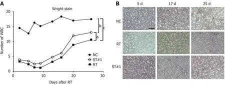
Figure 2 Evaluation of white blood cell count in peripheral blood using Wright staining.A:The number of white blood cells were compared between the normal control NC,stem cell-treated,and irradiated groups;B:Representative images of the cells harvested on days 5,17,and 25 after 9 Gy whole-body irradiation.Magnification 200 ×;scale bar:20 μm;aP < 0.01,bP < 0.001,and cP < 0.0001 vs controls,as indicated.NC:Normal control;ST:Stem cell-treated;RT:Irradiated;WBC:White blood cell.
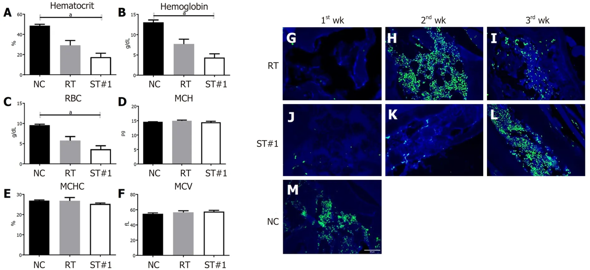
Figure 3 Characterization of red blood cell parameters in peripheral blood and CD34+ hematopoietic stem cells in the bone marrow.A-C:Hematocrit level,hemoglobin level,and red blood cells count in the control,irradiated(RT),and stem cell-treated(ST)#1 groups at three weeks post-whole-body irradiation(WBI);D-F:Mean corpuscular hemoglobin,mean corpuscular hemoglobin concentration,and mean corpuscular volume for the control,RT,and ST#1 groups at three weeks post-WBI;G-M:Immunohistochemistry for CD34+ hematopoietic cells(green)in bone marrow;nuclei were counterstained with 4’,6-diamidino-2-phenylindole(blue).Magnification 400 ×;scale bar 50 μm;aP < 0.01 vs controls.RBC:Red blood cell;MCH:Mean corpuscular hemoglobin;MCHC:Mean corpuscular hemoglobin concentration;MCV:Mean corpuscular volume;NC:Normal control;ST:Stem cell-treated;RT:Irradiated.
The mRNA expression levels of CD163,CD206,and IL-10 were higher in the BM of the ST#1 group than in the other groups.At the same time,IL-1β and TNF-α mRNA levels were lower in the ST#1 group than in the other groups during the first and second week post-WBI(Figure 5J and Supplementary Figure 2).These results indicated that intraperitoneal ADSCs induced M2 polarization during repopulation post-WBI.The mRNA levels of GM-CSF,SCF,SDF-1,and IL-7 in the ST#1 group increased mainly in the first and second weeks post-WBI compared to those in the NC and RT groups(Supplementary Figure 2).
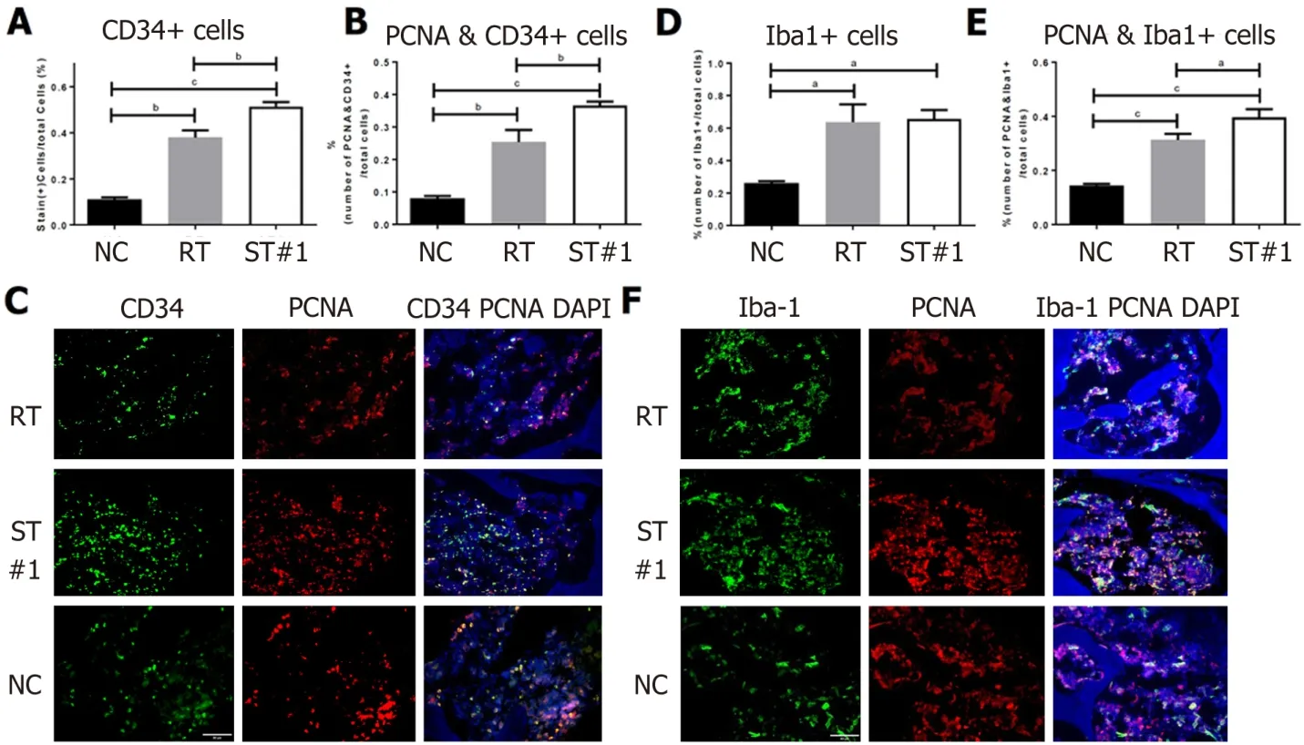
Figure 4 Immunohistochemical assessment of CD34,proliferating cell nuclear antigen,and ionized calcium-binding adaptor molecule-1 expression in bone marrow.Three weeks following whole-body irradiation,mouse femurs were fixed,decalcified,and embedded in paraffin wax.The sections were deparaffinized,stained with antibodies,and imaged under 400 × magnification.A-C:The number of CD34+ cells in the stem cell-treated(ST)#1 group was higher than that in the irradiated(RT)group.The number of proliferating cell nuclear antigen(PCNA)+ cells in CD34+ hematopoietic stem cells was significantly higher in the ST#1 group than in the RT group;D-F:The number of ionized calcium-binding adaptor molecule-1(Iba-1+)cells,a marker of monoblastic cells,was similar between the ST and RT groups;however,the number of cells expressing both PCNA and Iba-1 was significantly higher in the ST group.Magnification 400 ×;scale bar:50 μm;aP < 0.001,bP < 0.001,and cP < 0.0001 vs controls.NC:Normal control;ST:Stem cell-treated;RT:Irradiated;CD:Cluster of differentiation;PCNA:Proliferating cell nuclear antigen;Iba-1:Ionized calcium-binding adaptor molecule-1;DAPI:4′,6-diamidino-2-phenylindole.
Assessment of plasma cytokine levels
To evaluate the effect of stem cell transplantation on plasma cytokine levels in RT mice,cytokine array analysis was performed using a mouse cytokine antibody kit and plasma collected from mice in the NC group,RT group at week 2,and ST group at week 2(ST2w)and 3(ST3w)post-WBI.The protein levels in the plasma of the RT,ST2w,and ST3w groups were normalized to those of the NC group.According to the clustering analysis of all 39 proteins detected in the RT,ST2,and ST3 groups,expression patterns in the RT group were more similar to that of the ST3w group than those in the ST2w group(Figure 6A).The expression patterns of significant proteins(those showing more than 1.5-fold difference in expression level)in the ST3w group varied from those in the RT and ST2w groups(Figure 6B).Interestingly,G-CSF,C-X-C Motif Chemokine Ligand 13,and IL-2 levels increased in the ST2w group at week 2 post-WBI,which,with the exception of IL-2,was not observed in the ST3w group.
In vitro co-culture of human CD34+ HSCs with hADSCs
BM CD34+cells were cultured in StemSpan™ medium supplemented with SCF,IL-3,LDL,and TPO in the absence(control group)or presence of the hADSC CM group or directly on a hADSC layer(contact group).After 7 d of incubation,the total cell numbers in the control,CM,and contact groups were(32.6± 3.75)× 104,(62.3 ± 2.95)× 104,and(81.7 ± 1.65)× 104,respectively(Figure 7A),indicating that cell proliferation was the highest in the contact group.The proportions of CD34+cells are illustrated in Figure 7B.The number of CD34+cells was(7.9 ± 1.38)× 104in the control group,(13.4 ± 0.89)× 104in the CM group,and(24.9 ± 2.21)× 104in the contact group(Figure 7C).Therefore,the number of CD34+cells was highest in the contact group.
Next,we evaluated colony-forming ability by incubating CD34+cells in three different culture conditions for 7 d.As shown in Figure 7D,the differentiation of CD34+cells cultured in the presence of hADSC CM(CM group)or on an hADSC layer(contact group)into CFU-E and BFU-E was lower than that in the control group;however,differentiation into CFU-GM was higher in the CM and contact groups than in the control group.These results suggest that secretory factors from hADSCs suppress erythroid differentiation and promote GM differentiation.
We also investigated the effects of culture conditions(control,CM,and contact groups)on the differentiation of fresh BM CD34+cells in a suspension culture.After 7 d of differentiation,the total cell numbers in the control,CM,and contact groups were(26.4 ± 1.32)× 105,(32.5 ± 3.15)× 105,and(25.6 ±0.90)× 105,respectively.Each surface marker was evaluated as a percentage of positive cells and total cell number.Both the percentage and number of CD15+and CD45+cells(granulocyte markers)were significantly higher in the CM and contact groups than in the control group(Figure 7E-J).While the percentage of CD71+and Gly A+cells(erythroid markers)were lower in the CM and contact groups than in the control group,the number of CD71+and Gly A+cells were lower only in the contact group compared to the control group(Figure 7K-P).These results suggest that the secretory factors of hADSCs may be sufficient to promote GM differentiation,but not enough to suppress erythroid differentiation under liquid differentiation conditions.
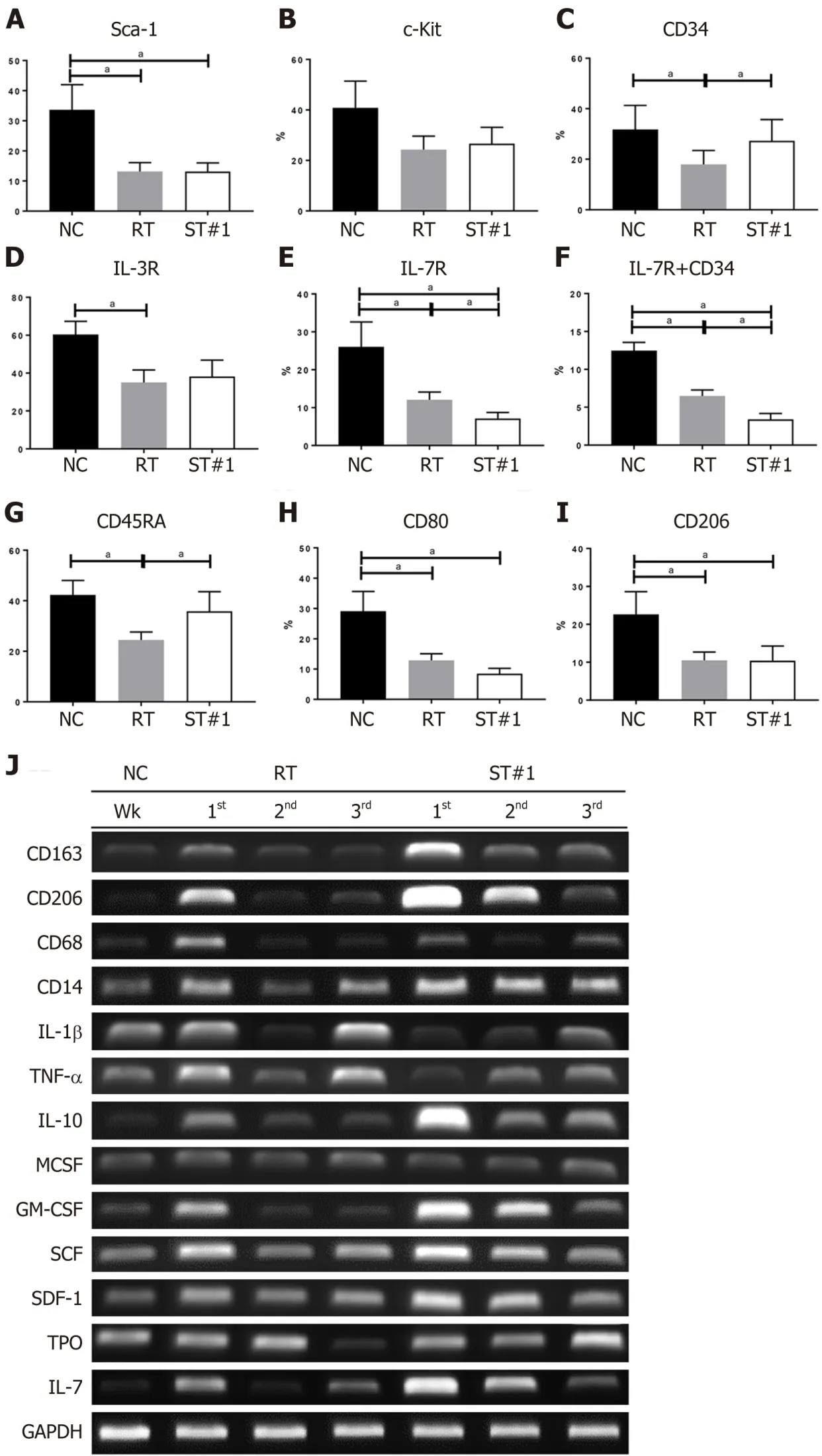
Figure 5 Characterization of repopulated bone marrow post-whole-body irradiation in the normal control,irradiated,and stem cell-treated groups.A-I:The expression levels of Sca-1,c-Kit,CD34,interleukin(IL)-3R,IL-7R,CD45RA,CD80,and CD206 in the bone marrow(BM)at 3 wk after post-wholebody irradiation(WBI)was examined using flow cytometry;J:mRNA expression in the BM at weeks 1,2,and 3 post-WBI was evaluated using reverse transcription PCR.aP < 0.05 vs controls,as indicated.NC:Normal control;ST:Stem cell-treated;RT:Irradiated;Sca-1:Stem cells antigen-1;c-KIT:Tyrosine-protein kinase KIT;CD:Cluster of differentiation;IL:Interleukin;TNF-α:Tumor necrotic factor-a;M-CSF:Macrophage colony-stimulating factor;GM-CSF:Granulocyte-macrophage colony-stimulating factor;SCF:Stem cell factor;SDF-1:Stromal cell-derived factor 1;TPO:Thrombopoietic growth factor;GAPDH:Glyceraldehyde 3-phosphate dehydrogenase.
Macrophage polarity of BM CD34+ cells differentiated in liquid culture
We examined the gene expression of several cytokines and surface markers in BM CD34+cells differentiated in liquid culture for 7 d.As shown in Figure 8,the gene expression levels of IL-4 and CD68 were not affected by the CM or contact conditions;however,the gene expression levels of IL-10,IL-1β,TNF-α,CD80,and CD206 were higher in the contact group than in the control group.Interestingly,CM alone was sufficient to increase the gene expression of IL-10 and CD206,markers that are highly expressed in M2 polarized macrophages,suggesting that secretory factors from hADSCs may promote macrophage polarization into the M2 phenotype.
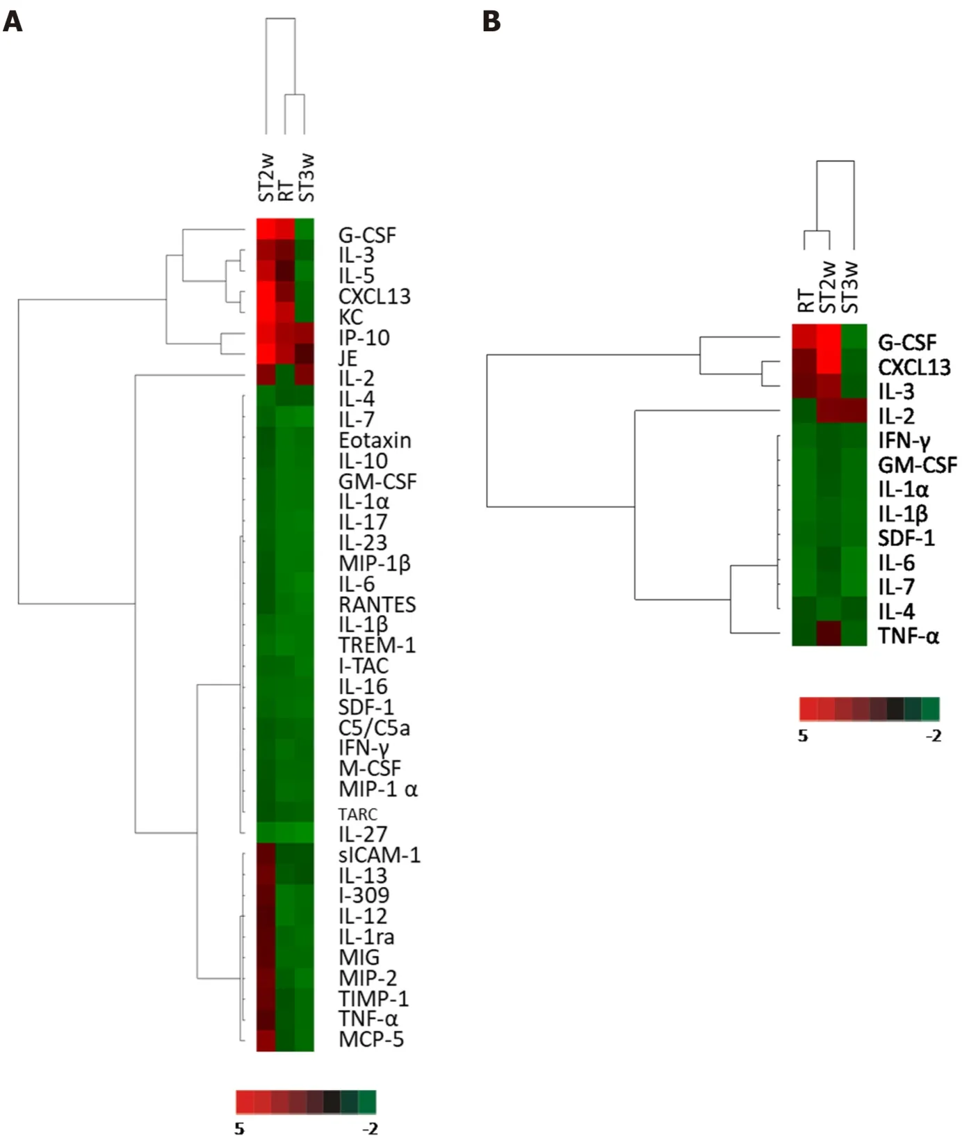
Figure 6 Clustering analysis of cytokine array from plasma samples.A:Thirty-nine proteins in the plasma were investigated in normal control(NC)and irradiated(RT)groups at week 2 and the stem cell-treated(ST)group at weeks 2 and 3(ST2w and ST3w,respectively).Proteins in the plasma of RT and ST groups were normalized to the NC group before comparison;B:The proteins with 1.5-fold higher expression in the three groups than in the NC group were selected and compared.NC:Normal control;ST:Stem cell-treated;RT:Irradiated;IL:Interleukin;TNF-α:Tumor necrotic factor-α;GM-CSF:Granulocyte-macrophage colonystimulating factor;SDF-1:Stromal cell-derived factor 1;IFN:Interferon.
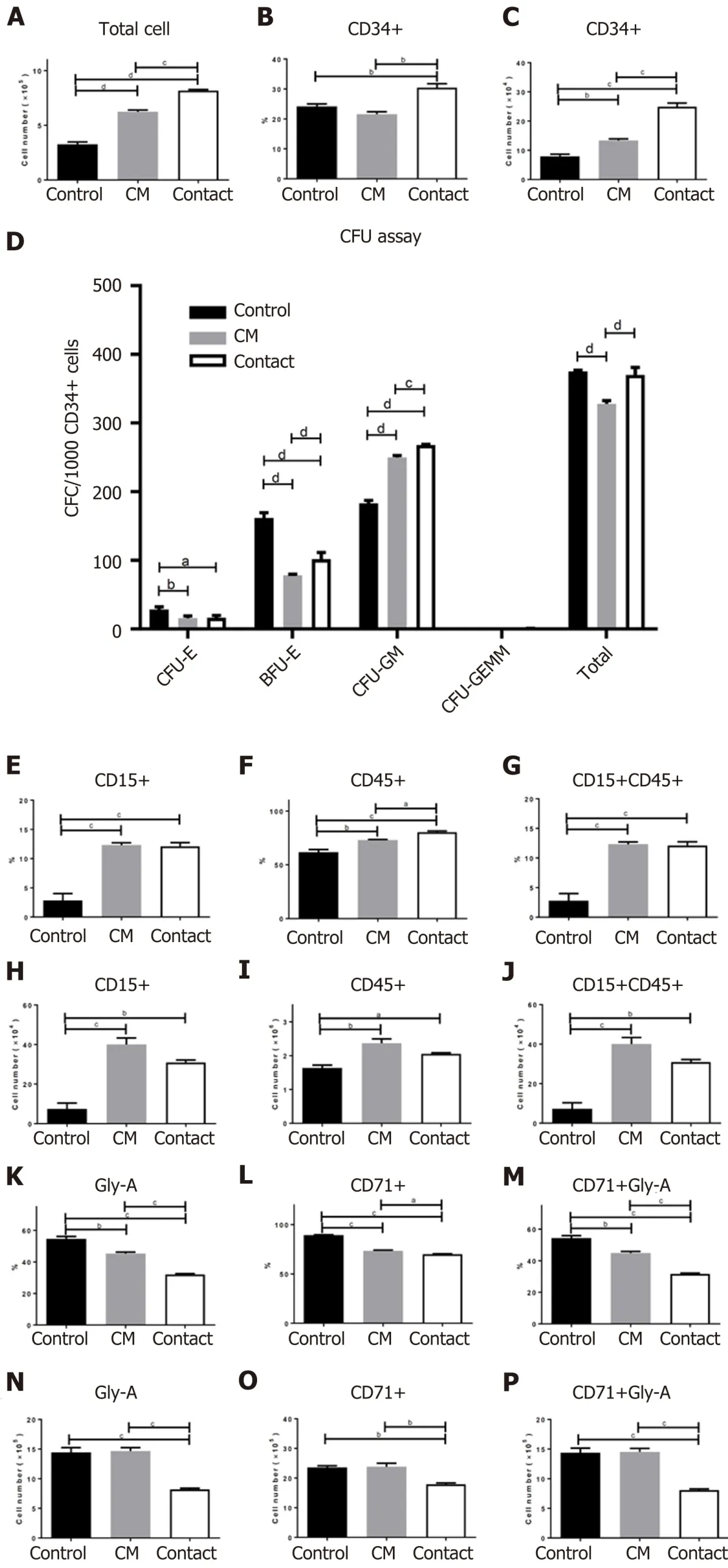
Figure 7 Effect of co-culture on CD34+ cell fate.CD34+ cells were incubated in the absence(control group)or presence of human adipose tissue-derived stem cells(hADSCs)conditioned medium(CM group)or directly on a hADSC layer(contact group).A:Total cell number;B:Percentage of CD34+ cells;C:The number of CD34+ cells were determined after 7 d of incubation;D:Colony-forming unit assays were performed using MethoCult™ by plating an equal number of CD34+ cells.The number of colonies was counted after 14 d of incubation;E-P:Differentiation assays of bone marrow CD34+ cells in liquid cultures,in the absence(control group)or presence of hADSC CM group or directly on a hADSC layer(contact group).The percentage of each surface marker was analyzed using flow cytometry(upper panel)and the marker-positive cell numbers were calculated(lower panel).aP < 0.05,bP < 0.01,cP < 0.001,and dP < 0.0001 vs controls.CFU:Colony-forming unit;CM:Conditioned media;CFU-E:Colony-forming unit erythroid;BFU-E:Burst-forming unit erythroid.
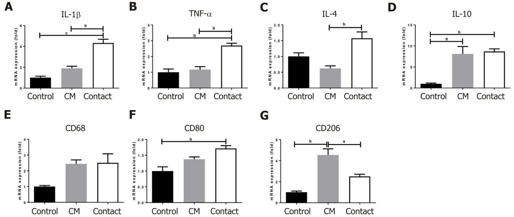
Figure 8 Gene expression analysis of bone marrow CD34+ cells cultured in the absence(control)or presence of human adipose tissuederived stem cell conditioned medium or directly on a human adipose tissue-derived stem cell layer(contact).Relative expression levels of the indicated genes were determined using qRT-PCR.aP < 0.05,bP < 0.01,and cP < 0.001 vs controls.CM:Conditioned media;IL:Interleukin;TNF-α:Tumor necrotic factor-α.
DISCUSSION
In the present study,we present evidence that transplantation of ADSCs into the abdominal cavity of RT mice delayed the repopulation of CD34+HSCs and simultaneously promoted the proliferation of the granulocyte/monocyte lineage,particularly toward M2 polarization.Similar results were observed in thein vitroco-culture of hADSCs and human CD34+HPSCs.However,RT mice with multiple transplantations of mADSCs showed a lower survival rate than those with a single transplantation.Therefore,the dosage of MSCs could be an important determinant of survival rates.
Different survival rates have been reported for post-irradiation MSC treatment[20,23,35,41].Shimet al[41]suggested that treatment with human umbilical cord blood-derived MSCs(2 × 10⁶)was more effective than that with G-CSF for hematopoietic reconstitution,following sublethal dose(7 Gy)radiation exposure.Abdel-Mageedet al[23]proposed that extracellular superoxide dismutasetransduced mouse BM-MSCs could improve survival rate post-WBI.However,the control BM-MSC group did not display an enhanced survival rate compared to the WBI group[23].Compared to RT and untreated animals,Kovalenkoet al[24]found considerably increased survival rates in animals that received 2 × 10⁸human umbilical cord blood mononucleated cells with antibiotics.However,Huet al[20]showed that transplantation of more than 1.5 × 10⁸cells/kg BM-MSCs increased mortality rates compared to those at lower BM-MSC concentrations,though the exact mechanism underlying these contradictory results is unclear.Similarly,our results indicated that survival rates after three MSC transplantations were lower than that after a single transplantation.After WBI,mice with multiple transplantations of mADSCs showed delayed CD34+hematopoietic repopulation and recovery of Hct and Hb levels in peripheral blood,which could be associated with increased mortality.We hypothesize that the anti-inflammatory effects of MSCs on BM could mediate this delay.
Previous studies have shown that MSCs exert their immunosuppressive effects through the release of IL-6,IL-10,transforming growth factor β(TGF-β),tumor necrosis factor-inducible gene-6(TSG-6),and indoleamine 2,3-dioxygenase[42,43].MSC spheroids displayed anti-inflammatory activity mediated by prostaglandin E2(PGE2),thus altering the phenotype of lipopolysaccharide - or zymosan-stimulated macrophages[44].MSCs can also alleviate inflammation in macrophages by secreting TSG-6 and stanniocalcin-1[45].Furthermore,MSCs decrease TNF-α secretion by mature DC type 1(DC1),increase IL-10 secretion by mature DC2 cells,reduce interferon-γ(IFN-γ)production by Th1 cells,increase the secretion of IL-4 by Th2 cells,expand the proportion of regulatory T cells,and decrease IFN-γ secretion by natural killer cells[46].The anti-inflammatory effects of MSCs have also demonstrated a beneficial effect on organ damage caused by irradiation.Donget al[47]confirmed that the systemic infusion of human ADSCs ameliorated lung fibrosis in rats that received semi-thoracic irradiation(15 Gy)by upregulating hepatocyte growth factor(HGF)and PGE2,and downregulating TNF-α and TGF-β1.Sahaet al[21]found that,in male C57BL/6 mice that received 10 Gy WBI or 16–20 Gy of abdominal irradiation,a bone mesenchymal stem cell(BMSC)transplant increased the levels of R-spondin1,keratinocyte growth factor,platelet-derived growth factor,bFGF,and anti-inflammatory cytokines in the serum,with reduced levels of pro-inflammatory cytokines.The anti-inflammatory effects of MSCs were also examined in our experiments.IL-1β and TNF-α mRNA levels were lower in the ST#1 group than in the RT group.At the same time,the mRNA level of the anti-inflammatory cytokine IL-10 was higher in the ST#1 group than in the RT group.
Although the reduction of pro-inflammatory cytokines can relieve sequelae caused by radiation irradiation,these inflammatory factors are essential for the proliferation of HSCs during repopulation.Interferons act as major regulators of HSC[48,49].TNF-α and its receptors play a critical role in facilitating HSC engraftment and function[50,51].TNF-α signaling is also necessary for the maintenance of HSCs,suggesting that baseline inflammatory signaling is crucial for the maintenance of proper HSC division and,subsequently,function[52].In our study,mRNA levels of TNF-α and IL-1β were suppressed shortly after WBI in the ST#1 group compared to those in the other groups.Therefore,the anti-inflammatory effects derived from the secretome of mADSCs might delay reconstitution.It is also plausible that multiple doses(or an overdose)of MSCs lead to a severe delay in BM repopulation and,eventually,poor survival rates.
In the present study,the number of WBCs in the peripheral blood was higher in the ST#1 group than in the RT group at three weeks post-WBI;the number of proliferating myeloblasts(Iba1+and PCNA+)in the BM was higher in the ST#1 group than in the RT and NC groups;and the number of CD45RA+cells(GM progenitors)was higher in the ST#1 group than in the RT group.mADSCs indirectly affected myeloid differentiation in the BM.In previous studies,intraosseous or retrobulbar transplants of ADSCs with BMSCs promoted the differentiation of the myeloid lineage in the BM[34,36,53,54].In the present study,the mRNA levels of GM-CSF and SDF-1 in the BM and G-CSF in the plasma were higher in the ST#1 group than in the RT group.G-CSF and GM-CSF are well-known hematopoietic factors that induce HSC differentiation into the myeloid lineage[55].SDF-1 also promotes myeloid differentiation[56],and is expressed by stromal cells in several tissues and organs,including the skin,thymus,lymph nodes,lung,liver,and BM.Nakaoet al[34]reported that SDF-1 secreted from ADSCs can induce the differentiation of CD34+HSCs into myeloid cells.De Toniet al[36]showed that the co-culture of ADSCs and CD34+HSCsin vitrowithout any cytokine supplement promoted CD34+HSC differentiation into granulocyte/monocyte lineages.In the present study,in vitroexperiments demonstrated that hADSC CM or direct contact with the hADSC layer increased differentiation in CD34+HSCs toward the granulocyte-macrophage lineage.
ADSCs have been reported to have an effect on M2 polarization[43,57,58].M1 macrophages are known to stimulate umbilical cord-MSCs and increase the expression of IL-6:A cytokine that upregulates IL-4R,promotes phosphorylation of STAT6 in macrophages,and eventually polarizes macrophages toward the M2 phenotype[43].In mice with mutations in the gene encoding for IL-6Ra,IL-6 was found to be an important determinant of M2 macrophage activation[57].These results suggest that the secretion of IL-6 by MSCs plays a key role in mediating macrophage polarization[43,57].MSCs can also promote M2 macrophage polarizationviaIL-10[58].In our study,M2 macrophage markers,such as CD206,CD164,and IL-10,were upregulated in the BM of the ST#1 group.IL-6,M-CSF,and IL-10,which can be secreted by mADSCs,can also induce M2 polarization in the BM.Therefore,M2 polarization could act as an anti-inflammatory suppressor in BM during the first week following radiation.
MSCs are multipotent cells that were originally isolated from BM and subsequently from other tissues,including fat[59],cardiac tissue[60],umbilical cord[61],and oral tissue[62].BM-MSCs,which have been widely used for treating various diseases,traditionally required invasive harvesting procedures with low yields.On the other hand,ADSCs are abundant in the human body and have multiple differentiation potentials,making them a promising source for wound healing and tissue engineering,with low-risk cell harvesting and easy processing[63].MSCs derived from the BM or adipose tissue share many similar biological characteristics,such as immunophenotype,multipotent differentiation,profiles of cytokine secretion,and immunomodulatory effects[64,65].However,depending on tissue sources,donors,and isolation and culture protocols,the properties of MSCs may vary slightly[65-67].Given the convenience of harvesting and multitude of sources,ADSCs appear to have numerous clinical advantages over cells derived from BM or other sources.
Umbilical cord derived MSCs(UC-MSCs)from fetal tissue have better proliferation capacity than ADSCs,which are MSCs from adult tissue[68].However,compared to ADSC,UC-MSC is difficult to collect and secure,and the culture success rate is low[68,69].It is not yet known whether MSCs derived from various tissues will have the same effect on BM repopulation.However,according to previous studies,UC-MSC,BM-MSC,and ADSC showed similar immunomodulatory effects[43,46,70].In this regard,future research should evaluate variation in treatment efficacy according to the origin,age,and passage of MSCs.In treating BM repopulation with MSCs in humans,future clinical trials should specifically consider the dose to be administered.In the present study,mADSCs and hADSCs were used in thein vivoandin vitroexperiments,respectively.In mice,P3-5 of mADSC were used,and P5-7 of hADSC were used;we recognize this to be a limitation in the interpretation of our results.
CONCLUSION
We present evidence that multiple intraperitoneal transplantations of mADSCs increased mortality post-WBI.The anti-inflammatory effects and M2 polarization promoted by the intraperitoneal administration of mADSCs might suppress erythropoiesis and induce myelopoiesis in sub-lethally RT mice.To improve survival rates post-WBI,the amount of MSCs should be optimized for administration.
ARTICLE HIGHLIGHTS
Research background
Bone marrow(BM)suppression is one of the most common side effects of radiotherapy and the primary cause of death following exposure to irradiation.Despite concerted efforts,no definitive treatment method is available.Transplantation of adipose tissue-derived stromal cells has been proposed as a promising therapy for BM suppression.
Research motivation
Although adipose tissue-derived stromal cell transplantation has shown reasonable efficacy in studies of BM suppression,the therapeutic effects are controversial.
Research objectives
We administered and examined the effects of various amounts of adipose-derived mesenchymal stromal cells(ADSCs)in mice with radiation-induced BM suppression.
Research methods
Mice were divided into three groups:Normal control group,irradiated(RT)group,and stem celltreated group after whole-body irradiation(WBI).Mouse ADSCs were intraperitoneally transplanted either once or three times at 5 × 10⁵cells/200 μL.The white blood cell count and levels of plasma cytokines,BM mRNA,and BM surface markers were compared between the three groups.Human BMderived CD34+hematopoietic progenitor cells were co-cultured with human ADSCs(hADSCs)or incubated in the presence of hADSC conditioned media(CM)to investigate the effect on human cellsin vitro.
Research results
The survival rate of mice that received one ADSC transplant was higher than that in the three-transplant group.Multiple transplants of ADSCs delayed the repopulation of BM hematopoietic stem cells.Antiinflammatory effects and M2 polarization by intraperitoneal ADSCs might suppress erythropoiesis and induce myelopoiesis in sub-lethally RT mice.
Research conclusions
To improve survival rates post-whole-body(WBI)irradiation,the amount of mesenchymal stromal cells should be optimized for transplantation.
Research perspectives
We demonstrated the effects of ADSC doses on BM suppression and suggest that the mechanisms involved can determine the success of future experiments and clinical applications.
ACKNOWLEDGEMENTS
The authors would like to thank Myung-Joo Lee and Eun-Sook Kim for their assistance with immunostaining,genetic analysis,and animal breeding.
FOOTNOTES
Author contributions:Lee SJ and Jeong JY contributed to the conception and design of the study,data interpretation,and funding acquisition;Lee SH,Lim S,Moon W,and Kim MJ contributed to the methodology,data acquisition,and analysis;Lee SJ wrote the original draft of the article;Lee SJ,Jeong JY,and Heo J drafted,reviewed and edited the manuscript,and contributed to project administration and supervision;all authors read and approved the final version of the manuscript.
Supported byThe Basic Science Research Program Through The National Research Foundation of Korea(NRF)Grant Funded By The Korean Government To Lee S.J.,No.2021R1F1A1052084.
Institutional review board statement:The animal study was reviewed and approved by the Institutional Animal Care and Use Committee of Kosin University College of Medicine(KMAP-16-18)and all procedures for human adipose tissue were conducted with informed consent under the Kosin University Gospel Hospital IRB approval protocol(protocol number 09–36).
Institutional animal care and use committee statement:All animal experiments were approved by the Institutional Animal Care and Use Committee of Kosin University College of Medicine,and the animals were maintained and treated according to the regulations of the Association for Research in Vision and Ophthalmology.
Conflict-of-interest statement:The authors declare no conflicts of interest.
Data sharing statement:No additional data are available.
ARRIVE guidelines statement:The authors have read the ARRIVE guidelines,and the manuscript was prepared and revised according to the ARRIVE guidelines.
Open-Access:This article is an open-access article that was selected by an in-house editor and fully peer-reviewed by external reviewers.It is distributed in accordance with the Creative Commons Attribution NonCommercial(CC BYNC 4.0)license,which permits others to distribute,remix,adapt,build upon this work non-commercially,and license their derivative works on different terms,provided the original work is properly cited and the use is noncommercial.See:https://creativecommons.org/Licenses/by-nc/4.0/
Country/Territory of origin:South Korea
ORCID number:Min-Jung Kim 0000-0002-3283-5737;Won Moon 0000-0002-3963-8680;Jeonghoon Heo 0000-0002-7176-2411;Sangwook Lim 0000-0003-3788-5108;Seung-Hyun Lee 0000-0002-5610-5237;Jee-Yeong Jeong 000-0001-9482-4526;Sang Joon Lee 0000-0001-6673-569X.
S-Editor:Wang JJ
L-Editor:A
P-Editor:Zhang YL
 World Journal of Stem Cells2022年3期
- World Journal of Stem Cells的其它文章
- In vitro induced pluripotency from urine-derived cells in porcine
