Effect of Majie Pingchuan cataplasm on epithelial-derived cytokines in asthmatic mice based on the "lung-skin-intestine" axis
Han-Fen Shi,Tian-Yi Feng,Shuang Zhang,Wen-Ting Ji,Qing-Guo Wang,Xue-Qian Wang✉
1.Beijing University of Chinese Medicine,Beijing 100105,China
2.Chengdu University of Traditional Chinese Medicine,Chengdu 610075,China
Keywords:Allergic asthma mice MJPC cataplasm Lung-skin-intestine axis TSLP IL-33
ABSTRACT Objective:To observe the effect of MJPC cataplasm on the content of epithelial-derived cytokines in lung,skin and intestine of asthmatic mice.Methods:C57/BL6 female mice were randomly divided into four groups:control group,asthma model group,dexamethasone group and MJPC cataplasm group.Ovalbumin sensitized and challenged asthmatic mouse models were established.The spleen index was calculated,and HE staining was used to observe the pathological change in lung tissues.The ova-specific IgE in the mouse serum and the content of TSLP in lung,skin and intestine were detected by enzyme-linked immunosorbent assay(ELISA).The expressions of TSLP mRNA and IL-33 mRNA in skin and intestinal tissue were detected by qRT-PCR.Results:Compared with the control group,the spleen index of mice in asthma model group was increased.Vascular congestion and edema,inflammatory cell infiltration and bronchial wall thickening were observed.The expressions of IgE in the mouse serum were significantly increased,and the content of TSLP in lung and skin tissue increased,but that in intestine tissue did not change significantly.The expression of TSLP mRNA was up-regulated in skin and intestinal tissues.The expression of IL-33 mRNA was up-regulated in skin tissue,but not in intestine,and the differences were statistically significant(P<0.05);Compared with the model group,MJPC cataplasm could decrease the spleen index and the expression of IgE in the mouse serum,improve the pathological damage of lung tissue in asthmatic mice,reduce the content of TSLP in lung,skin and intestinal tissue,increased the expression of TSLP mRNA in skin tissue,and down-regulate the expression of Il-33 mRNA in skin tissue and the expression of TSLP mRNA and IL-33 mRNA expression in intestinal tissue(P<0.05).Pearson correlation coefficient (r) of the content of TSLP between lung and skin was 0.689,that between lung and intestinal was -0.163,and that between skin and intestinal was -0.163,and the differences were not statistically significant(P>0.05).Conclusion:MJPC cataplasm improve airway inflammation by inhibiting the content of epithelial-derived cytokines on the "lung-skin-intestine" axis of asthmatic mice,and achieve the effect of treating asthm
1.Introduction
Epithelial cells in the skin,intestine and lungs have long been considered as protective barriers against infection and physical or chemical damage.Epithelium also plays a key role in the immune system.Studies on intestinal and respiratory mucosa have shown that epithelial cells can secrete specific cytokines,thus pushing inflammation to type 2 immune response.Studies have shown that epithelial derived cytokines thymic stromal lymphopoietin(TSLP) and interleukin-33 (IL-33) play a role in driving type 2 response[1].In allergic diseases,type 2 inflammation can lead to skin allergic dermatitis (AD),gastrointestinal dysfunction and food allergy and eosinophilic esophagitis (EOE),or asthma,allergic rhinitis and chronic sinusitis in the respiratory system.Pruritus,mucus production and bronchoconstriction may also be part of type 2 allergic reaction.For external application,the effective components of Majie cataplasm are absorbed from the skin into the body,so as to play the role of anti-inflammatory and relieving airway hyperresponsiveness.Previous studies have shown that Majie cataplasm has a significant inhibitory effect on Th2 inflammatory response in the lung,and under the condition of the same dosage,the effective components in blood after application and administration of Majie cataplasm is only 1/ 4 of that of the oral group,but the effect of improving lung function is better than that of the oral group[2].Therefore,its pharmacodynamic components pass through the skin into the blood,acting on the lungs is only one of the mechanisms to exert its efficacy.Based on the characteristics of "percutaneous treatment of the lung" of Majie cataplasm,the immune function of epithelial cells has come into our sight.Epithelial cells are the common material basis of lung,skin and intestine.The epithelial derived cytokines released by epithelial cells are considered to be the common factors related to the immune response of the above three parts.The purpose of this experiment is to explore the possible mechanism of Majie cataplasm on asthma based on the "lung-skinintestine" axis through the effect of Majie cataplasm on epithelial cytokines in lung,skin and intestine of asthmatic mice sensitized by ovalbumin (OVA).
2.Materials and Methods
2.1 Materials
SPF grade 8 to 10 weeks old female C57/ BL6 mice weighing(20-22) g were purchased from Sibeifu Biotechnology Co.,Ltd.(Beijing,China,No.:SCXK 2019-0010).This study was approved by the Animal Experiment Ethics Committee of Beijing University of Traditional Chinese Medicine (No.:bucm-4-2020102202-4022).
2.2 Reagents
Majie cataplasm containing ephedrae herba,sinapis semen,corydalis rhizoma,armeniacae semen amarum and zingiberis rhizoma recens (1:1:1:1),is 25g/ tablet and 63 cm2 (length 9 cm x width 7 cm),which is converted into the application area of mice is about 0.2 cm2.Phosphate buffered saline (PBS) was purchased from Thermo Fisher Scientific of the United States,and ovalbumin (OVA)was purchased from sigma Aldrich of the United States,imject™ Alum adjavant was purchased from Thermo Fisher Scientific,dexamethasone sodium phosphate injection was purchased from Chenxin Pharmaceutical Co.,Ltd.(GYZZ H37021967),and 4%paraformaldehyde was purchased from Beijing Oubei Biotechnology Co.,Ltd.The ELISA kit for detecting immunoglobulin E (IGE)and thymic stromal lymphopoietin (TSLP) was purchased from Jiangsu Meibiao Biotechnology Co.,Ltd.Hipure unviersal RNA kit was purchased from Guangzhou Magen Biotechnology Co.,Ltd.,cDNA reverse transcription kit and SYBR Green reagents were purchased from Thermo Fisher Scientific,USA,and DEPC water was purchased from Beyotime,China.
2.3 Equipment
Centrifuge (Brand:Eppendorf,model:5810,Germany),multifunctional enzyme labeling instrument (Brand:Bio TEK,model:cpoch,USA),PCR instrument (Brand:Bio rad,model:cfx96 touch,USA),PTC-150 PCR amplification instrument (Mjr company of the United States),NanoDrop2000 ultra micro spectrophotometer(Brand:Hangzhou Aosheng Instrument Co.,Ltd.,model:Nana 3000,China) .
2.4 Methods
2.4.1 Ovalbumin (OVA)-induced asthma model and drug intervention
Mice (n=32) were randomly divided into 4 groups,including the control group (n=8),the asthma model group (n=8),the dexamethasone group (n=8) and the Majie cataplasm group (n=8) .According to the previous research method,we constructed asthma mouse model[3].Except for the mice of the control group,others received an intraperitoneal injection (i.p.) of a solution containing 0.05 mg OVA and 1 mg Alum on Days 0,7 and 14.And on days 15-25,they were challenged with intranasal injection of OVA,2.5 mg/ml diluted in phosphate-buffered saline (PBS),40 μL/mouse.In contrast,mice in the control group were induced the same way just with PBS.
On the fifteenth day,mice in the control group and the asthma model group were taken no intervention,mice in the dexamethasone group were injected intraperitoneally with the dexamethasone sodium phosphate injection 0.2mL/mouse (2mg/kg),and mice in the Majie cataplasm group were patched with cataplasm on the mice’s backs,each cataplasm staying for 24 hours.After 10 days,all the mice were anesthetized and sacrificed for follow-up examination.
2.4.2 Spleen index measurement
After weighing,the spleen was removed,the surface fluid was sucked dry,and the spleen was weighed.Spleen index=spleen weight (mg)/body weight (g) of mice [4,5].
2.4.3 Hematoxylin eosin staining (H&E) detecting lung histopathology
The left lung tissues of mice were taken after perfusion with normal saline and 4% paraformaldehyde within 24h after the last asthma attack.The left lung tissues were dehydrated and fixed with 4% paraformaldehyde,embedded in paraffin,and sectioned 6 μm for H&E staining.The pathological changes of lung tissues were observed under light microscope.
2.4.4 Enzyme Linked immunosorbent assay (ELISA) detecting the content of IgE in serum and TSLP in lung,skin and intestinal tissues
The IgE content in serum and TSLP content in lung,skin and intestinal tissues of mice were detected according to the instructions of ELISA kit.
2.4.5 Quantitative rea-time PCR (QRT-PCR) detecting the expression of TSLP mRNA and IL-33 mRNA in skin and intestinal tissues
According to instruction,total RNA was collected from lung tissue using HiPure Unviersal RNA Mini Kit,and cDNA was prepared by reverse transcription after dilution in water without RNA enzyme using the cDNA synthesis kit.qPCR was performed three times on each sample with SYBR Green reagents.The relative expression of the target genes was calculated by the 2 ct method and normalized using GAPDH.Primers used in this study were as follows (Table 1).

Table 1 Primer sequences used in animal experiments
2.5 Statistical analysis
Data are expressed as mean ± standard deviation (SD).GraphPad Prism 8 software was used for one-way ANOVA,and multiple comparison (LSD) test was used for pairwise comparison between groups.P<0.05 was considered statistically significant.Statistical correlation analysis method (Pearson correlation coefficient) was used to analyze the correlation of TSLP content in lung,skin and intestine.
3.Results
3.1 Effect of Majie cataplasm on spleen index of allergic asthma model mice
Compared with control group,spleen index of mice in asthmatic model group increased (P<0.0001);Compared with asthmatic model group,the spleen index of Majie cataplasm group decreased(P<0.01),dexamethasone had no significant effect,as shown in Table 2.
Table 2 Spleen index in asthmatic mice(mg/g,n=8,)

Table 2 Spleen index in asthmatic mice(mg/g,n=8,)
Note:Compared with control group,# P<0.0 5,##P<0.01,###P<0.001,####P<0.0001;Compared with asthma model group,*P<0.05,** P<0.01,***P<0.001,****P<0.0001.
3.2 Effect of Majie cataplasm on serum IgE content in allergic asthma model mice
Compared with the control group,the content of IgE in the asthma model increased (P<0.0001),and dexamethasone and Majie cataplasm significantly decreased the content of IgE(P<0.0001,P<0.0001)(Table 3).
Table 3 The content of IgE in serum of asthmatic mice (μg/mL,n=4,)

Table 3 The content of IgE in serum of asthmatic mice (μg/mL,n=4,)
Note:Compared with control group,# P<0.0 5,##P<0.01,###P<0.001,####P<0.0001;Compared with asthma model group,*P<0.05,** P<0.01,***P<0.001,****P<0.0001.
3.3 H&E staining observing the pathological changes of lung tissues
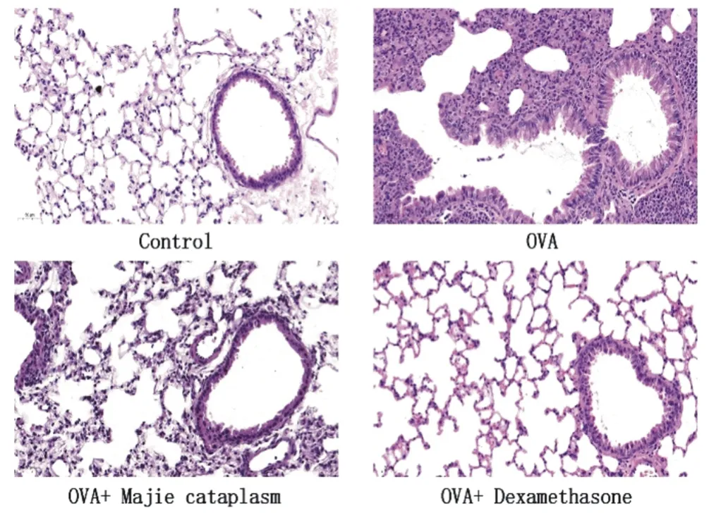
Figure 1 shows partial parenchyma of lung tissue in the asthma model group.With blood vessel hyperemia edema,alveoli and bronchiole wall appeared a large number of inflammatory cells,bronchial wall thickening.After treatment of Majie cataplasm,the pathological changes of lung tissues were relieved.The dexamethasone group recovered well.These results suggest that Majie cataplasm can reduce asthma inflammation caused by OVA.

Figure 1 H&E staining results of lung tissues in asthmatic mice(200X,scale=50μm,n=4)

Figure 2 The correlation of TSLP content in lung,skin and intestine in asthmatic mice
3.4 Effect of Majie cataplasm on TSLP content in lung,skin and intestine of allergic asthma model mice
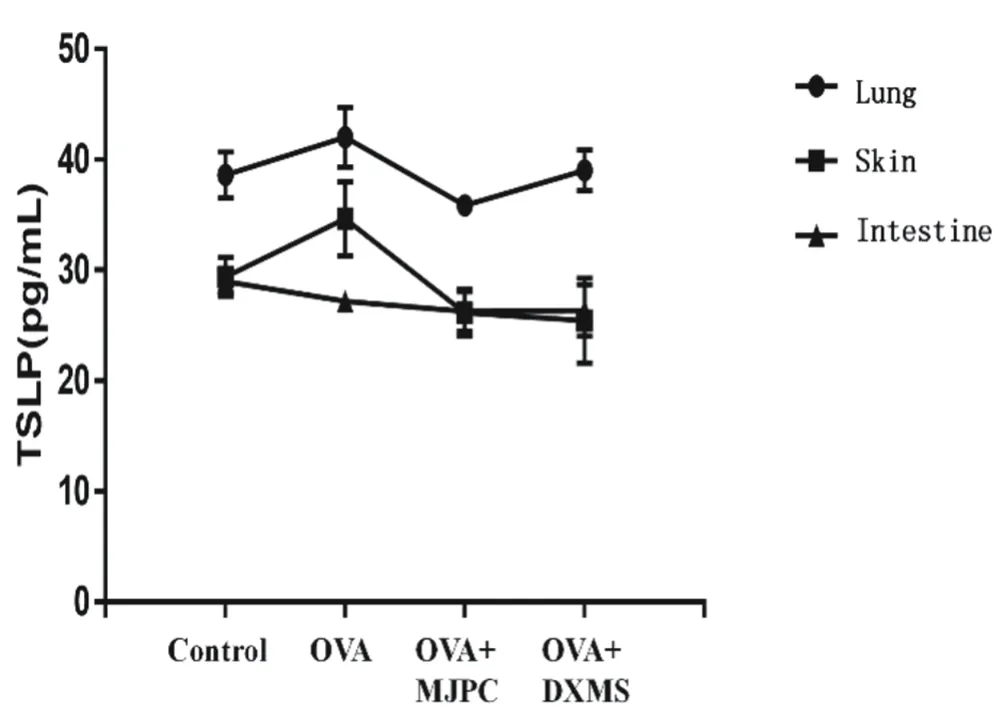
In lung tissue,there was a difference in TSLP between asthma model group and control group (P<0.05).Majie cataplasm decreased the content of TSLP (P<0.01),and dexamethasone had no significant effect on the content of TSLP.In skin tissue,there was a difference in TSLP between asthma model group and control group (P<0.05).Dexamethasone and Majie cataplasm decreased the content of TSLP (P<0.01,P<0.01).In intestinal tissue,there was no significant difference in TSLP between asthma model group and control group.Dexamethasone and Majie cataplasm decreased the content of TSLP compared with control group (P<0.05,P<0.05),as shown in Table 4.
Table 4 The expression of TSLP in lung,skin and intestine in asthmatic mice (pg/mL,n=4,)
Note:Compared with control group,# P<0.0 5,##P<0.01,###P<0.001,####P<0.0001;Compared with asthma model group,*P<0.05,** P<0.01,***P<0.001,****P<0.0001.
As shown in Figure 2,the correlation r of TSLP content between lung tissue and skin tissue was 0.689 (P>0.05),between lung tissue and intestinal tissue was -0.163 (P>0.05),and between skin tissue and intestinal tissue was 0.422 (P>0.05).
3.5 Effects of Majie cataplasm on the expression of TSLP mRNA and IL-33 mRNA in skin and intestine of allergic asthma model mice
The expression of TSLP mRNA in skin tissue of asthma model group was different from that of control group (P<0.05).Dexamethasone could reduce the expression of TSLP mRNA(P<0.05),while Majie cataplasm increased the expression of TSLP mRNA (P<0.05).The expression of TSLP mRNA in intestinal tissue of asthma model group was higher than that of control group(P<0.01).Dexamethasone group and Majie cataplasm could inhibit the expression of TSLP mRNA (P<0.0001,P<0.0001),as shown in Table 5.
Table 5 The expression of TSLP mRNA in skin and intestine in asthmatic mice (n=4,)

Table 5 The expression of TSLP mRNA in skin and intestine in asthmatic mice (n=4,)
Note:Compared with control group,# P<0.0 5,##P<0.01,###P<0.001,####P<0.0001;Compared with asthma model group,*P<0.05,** P<0.01,***P<0.001,****P<0.0001.
The expression of IL-33 mRNA in skin tissue of asthma model group was higher than that of control group (P<0.01).Dexamethasone group and Majie cataplasm could inhibit the expression of IL-33 mRNA (P<0.01,P<0.01).There was no significant difference in the expression of IL-33 mRNA in intestinal tissue between the asthma model group and the control group,but dexamethasone group and Majie cataplasm could inhibit the expression of IL-33 mRNA (P<0.05,P<0.05),as shown in Table 6.
4.Discussion
"Fei He Pimao" was first seen in the Huangdi Neijing.Su Wen Wuzang Shengcheng:"Fei Zhi He Pi Ye,Qi Rong Mao Ye".The Fei transpire the essence of water and grain to the body surface to moistening and nourishing the skin,so that it can play its normal physiological functions,such as guarding muscle table,resisting external pathogens,regulating the opening and closing of sweat pores,regulating body temperature,etc;At the same time,Pimao can also regulate sweat pores,promote Fei qi,help breathing,and regulate water metabolism.Su Wen Weilun“Fei Zhu Shen Zhi Pimao……Gu Fei Re Ye Jiao,Ze Pimao Xuruo Jibao,Zhuo Ze Sheng Weibi Ye.”Su Wen Kelun“Pimao Zhe,Fei Zhi He Ye,Pimao Xian Shou Xieqi,Xieqi Yi Cong Qi He Ye.”Pathologically,the lung and skin also interact.Cross sectional and longitudinal studies show that the occurrence of allergic diseases follows a chronological order:from atopic dermatitis and food allergy in infancy to allergic asthma and allergic rhinitis in childhood.Anatomically,it follows the spatial evolution of skin-gastrointestinal-respiratory tract,which is defined as "Atopic process"[6].These all emphasize the close relationship between lung and skin.“Fei Yu Dachang Xiang Biao Li”was originally from Huangdi Neijing Ling Shu Ben Shu.“Fei He Dachang,Chang Zhe,Chuandao Zhi Fu.”Fei Qi downdraft and makes the essence of water and grain distributed inward and downward to moisten and nourish other viscera,pushing the large intestine to carry waste.It interacts with the work of large intestine conduction.It was found that OVA induced asthma model rats had the phenomenon of goblet cell reduction and inflammatory cell infiltration in intestinal mucosa[7].There is a close relationship between lung,skin and intestine in physiology and pathology,which suggests whether there is a "lung-skin-intestine" axis,so that the pharmacodynamic effect acting on one viscera can affect other viscera on the axis,so as to give full play to the advantages of overall regulation.By studying the effect of Majie cataplasm on epithelial derived cytokines,the common substance of the "lungskin-intestine" axis,this experiment not only reveals the biological basis of its "percutaneous treatment of the lung",but also further explores the mechanism linkage of the "lung-skin-intestine" axis in the disease,so as to provide a new idea for the treatment of asthma.
Epithelial cells exist in the skin,respiratory tract and gastrointestinal tract,which can protect us from external damage.Epithelium not only acts as a physical barrier,but also initiates and drives type 2 immune response by releasing epithelial cytokines to respond positively to stimuli such as allergens,pollution or microorganisms.Dysregulated epithelial barrier activity in allergic diseases increases the possibility of allergen and microbial penetration and subsequent allergy.
TSLP belongs to the alarmins produced by epithelial cells on the surface of lung,skin and gastrointestinal mucosa.It plays an important role in regulating type 2 immune response in airway.TSLP is an effective innate immune activator and a precursor of adaptive and allergen specific Th2 response.In the mouse model,TSLP promotes type 2 immune response by activating conventional dendritic cells and group 2 innate lymphocytes[8].Interstitial macrophages expressing TSLP receptors may also spread TSLP mediated acute airway inflammation[9].TSLP gene variation and high level of TSLP expression are related to atopic diseases such as AD,asthma,allergic rhinoconjunctivitis and EOE[10].More and more literatures show that TSLP can activate a subset of sensory neurons to drive pruritus in allergic diseases such as AD[11].Overexpression of TSLP in keratinocytes aggravates airway inflammation in OVA asthma model mice[12].A recent study showed that when the skin barrier is damaged,TSLP induced basophils can promote skin surface allergy to food allergens and subsequent IgE mediated food allergy through IL-4[13].However,in the intestine,TSLP and IL-33 cytokines seem to have a protective effect on ulcerative colitis[14].Studies have shown that TSLPR -/ -mice exhibit overexpression of proinflammatory cytokines and severe intestinal inflammation in dextran sodium sulfate (DSS) -induced colitis[15].
IL-33 may play a wide role in the process from early immune development to the deterioration of atopic diseases.Genetic studies have repeatedly proved that there is a significant correlation between IL-33 and IL1RL1 gene variants and human asthma[16].Genetic variation of IL1RL1 gene is also associated with the risk of AD,and genetic variation of IL-33 and IL1RL1 loci is associated with the risk of EOE[17].Like TSLP,IL-33 has also been shown to mediate pruritus by activating sensory neurons[18].IL-33 may have different functions according to the tissue environment.IL-33 and its fragments aggravate the pro-inflammatory circuit and enhance the cytotoxic activity of CD8+T cells in the pathology of celiac disease[19].
In this experiment,the asthma model induced by OVA was evaluated by spleen index,serum IgE content and HE staining.The results showed that the spleen index and serum IgE content increased,bronchial wall thickening and inflammatory cell infiltration in the asthma model group,indicating that the asthma model was successfully established.The content of TSLP in lung,skin and intestine was detected.It was found that Majie cataplasm could reduce the content of TSLP in lung,skin and intestine,lower the expression of IL-33 mRNA in skin tissue and the expression of TSLP mRNA and IL-33 mRNA in intestine,but increase the expression of TSLP mRNA in skin tissue.It shows that Majie cataplasm has a certain stimulating effect on the skin barrier,resulting in the increase of TSLP mRNA in the skin epithelium,but it has no pathological significance and does not need to start the subsequent over immune response.Therefore,the mRNA has not been further translated,and the detected TSLP content has not increased,but decreased.It is speculated that it may be caused by the overall effect of Majie cataplasm on the "lung-skin-intestine" axis.
The correlation of TSLP content between different tissues was compared.The results showed that the correlation r of TSLP content between lung tissue and skin tissue was 0.689,between lung tissue and intestinal tissue was -0.163,and between skin tissue and intestinal tissue was 0.422,which was not statistically significant,which may be related to the nonlinear trend.However,it can be speculated that the content change of lung TSLP in asthma model may have a certain impact on the skin,but not on the intestinal tissue,which may be related to the anti-inflammatory effect of TSLP in the intestinal tract.The change trend of skin TSLP and intestinal TSLP may be related to the effect of Majie cataplasm on reducing the content of TSLP in skin and intestinal.
In a word,Majie cataplasm can alleviate asthma by affecting the TSLP produced by the epithelium of "lung-skin-intestine" and reducing the expression of IL-33,which provides a certain idea for the modern biological connotation of "Fei He Pimao" and "Fei Yu Dachang Xiang Biao Li".
Author's contribution
SHI Hanfen,Index detection and paper writing.FENG Tianyi,Animal modeling and drug intervention.ZHANG Shuang,Data analysis.JI Wenting,Data analysis.WANG Qingguo,Project design.WANG Xueqian,Project design and review.
 Journal of Hainan Medical College2022年1期
- Journal of Hainan Medical College的其它文章
- Study on the in vitro activity of Hehuan Yin aqueous extract against hepatitis C 2a virus
- Optimization of preparation process of cationic liposome nanoparticles containing Survivin-siRNA and osthol
- Intervention effect of Danbei Yifei formula on pulmonary fibrosis based on urine metabolomics by UHPLC-Q-Exactive
- Clinical characteristics,GRACE score,TIMI score and prognosis of patients with type 2 diabetes mellitus complicated with acute coronary syndrome
- Effect of intradermal needle at five-zang Back-Shu points on treatment of chronic fatigue syndrome
- Clinical effect of Chinese medicine aerosol fumigation on demodex infection related meibomian gland dysfunction
