
Six New 3,5-Dimethylcoumarins from Chelonopsis praecox,Chelonopsis odontochila and Chelonopsis pseudobracteata
2021-12-16 12:29:08ChangAnGengZhenTaoDengQianHuangChunLeiXiangJiJunChen
Chang-An Geng·Zhen-Tao Deng·Qian Huang·Chun-Lei Xiang·Ji-Jun Chen
Abstract Ten 3,5-dimethylcoumarins ( 1– 6 and 8-11) involving six new ones ( 1– 6), together with a known 3-methylcoumarin ( 7), were isolated from the aerial parts of three Chelonopsis plants,C.praecox,C.odontochila, and C.pseudobracteata.The structures of the new compounds were determined by extensive HRESIMS, 1D and 2D NMR spectroscopic analyses.According to the substitution at C-5, these coumarins were classifi ed into 5-methyl, 5-hydroxymethyl, 5-formyl, and 5- nor types.All the isolates were assayed for their inhibition on α-glucosidase, protein tyrosine phosphatase 1B, and T-cell protein tyrosine phosphatase in vitro.
Keywords 3,5-Dimethylcoumarins·3-Methylcoumarin·Chelonopsis ·Enzyme inhibiti on
1 Introduction
Coumarins with the benzo-α-pyrone core are widely distributed in plant kingdom and show a wide range of biological activities, including antimicrobial, antiviral, antidiabetic antiinfl ammatory, and antihypertensive activities,etc.[1].Structurally, coumarins can be divided into simple coumarins, C-substituted coumarins, miscellaneous coumarins, biscoumarins, and triscoumarins.Besides hydroxy and methoxy groups, isopentenyl related C5-groups are the most common substituents present in coumarins, which are generally located at C-3, C-6, or C-8 positions by C–C linkage [2– 4].The methyl substituent in coumarin is very unusual, and only limited coumarins with the methylation at C-3, C-5, or C-6 positions have been reported.Currently, tens of 3,5-dimethylcoumarins have been isolated fromClutia lanceolata[5],Clutia abyssinica[6],Juniperus sabina[7],Leucas infl ata[8], andSideritis pullulans[9], but never fromChelonopsisplants.Our previous investigation onChelonopsisplants yielded a series of diterpenoids withα-glucosidase inhibitory activity,i.e., tenent-kauranes fromC.praecox[10], and 13ent-labdanes and 11ent-kauranes fromC.odontochila[11].As a continuous search for antidiabetic candidates from natural sources [12– 16], ten 3,5-dimethylcoumarins ( 1– 6 and 8 -11) involving six new ones and one known 3-methylcoumarin ( 7) were fi rst isolated from threeChelonopsisplants (Fig.1).Herein, we report their isolation, structural elucidation, and enzymatic inhibition onα-glucosidase, protein tyrosine phosphatase 1B (PTP1B), and T-cell protein tyrosine phosphatase (TCPTP).
2 Results and Discussion
2.1 Structural Elucidation
Compound 1 had a chemical composition of C12H12O4deduced by the [M + H] + ion atm/z221.0809, accounting for seven indices of hydrogen defi ciency.The UV spectrum showed characteristic absorption atλmax321 nm for coumarins.The IR absorptions at 3204, 1681, 1610, and 1454 cm −1 were indicative for the presence of hydroxyl, carbonyl, and aromatic functionalities.In the 1 H NMR spectrum, twometa-coupled aromatic protons atδH6.93 (J= 2.4 Hz) and 6.87 (J= 2.4 Hz), one methoxy atδH 3.64, and two singlet methyls atδH 2.56 and 2.10 were well recognized (Table 1).The 13 C NMR spectrum displayed 12 carbons comprising one carbonyl carbon, eight olefi nic carbons, one methoxy, and two methyls.The 1 H and 13 C NMR data of 1 showed high resemblance with 6-hydroxy-3,5-dimethyl-4,7-dimethoxycoumarin ( 9) [5] except for the absence of a methoxy and an oxygenated methine in 9 being changed to be a methine in 1.In the HMBC spectrum, the correlations from the methyl (δH2.10) to C-2 (δC164.0) and C-4 (δC166.9), and from the methoxy (δH3.64) to C-4 (δC166.9) affi rmed the 3-methyl and 4-methoxy substitution.Taking the ROESY correlations of Me-3/OMe-4/Me-5/H-6 into consideration, this compound was characterized to be 7-hydroxy-4-methoxy-3,5-dimethylcoumarin ( 1).
The molecular formula of 2 was assigned to be C13H14O5by the positive HRESIMS ion atm/z251.0897 ([M + H] + , calcd.for 251.0914).By comparing its 1 H and 13 C NMR data with those of 1, the 5-methyl in 1 was changed to be a hydroxymethyl (δH4.89,δC56.2) in 2, as well as an additional methoxyl group.The substitution of 5-hydroxymethyl and 7-methoxyl was confi rmed by the HMBC correlations from H-10 (δH4.89) to C-6 and C-4a, and from OMe-7 (δH3.86) to C-7, as well as the ROESY correlations of H 3 -9/OMe-4/H 2 -10/H-6/OMe-7/H-8 (Fig.2).Hence, compound 2 was defi ned as 5-hydroxymethyl-4,7-dimethoxy-3-methylcoumarin.
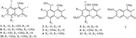
Fig.1 Chemical structures of compounds 1– 11

Table 1 1 H NMR data of compounds 1– 7 ( δ in ppm,J in Hz)
Compounds 3 and 4 were a pair of isomers with the same molecular formula of C14H16O 6 , indicating an additional CH2O moiety than 2.In their 1 H and 13 C NMR spectra, three methoxy groups (δH3.86, 3.93, 4.01 andδC56.2, 61.9, 62.1 for 3;δH3.95, 3.97, 3.98 andδC56.5, 61.67, 61.70 for 4) were obviously recognized (Table 2), suggesting compounds 3 and 4 should be the methoxylated derivatives of 2.The position of the additional methoxy in 3 and 4 were unambiguously determined by analyzing their ROESY experiments.In the ROESY spectrum of 3, the correlation peaks of H3-9/OMe-4/H2-10/OMe-6 and OMe-7/H-8 revealed the methoxy at C-6 position.Similarly, the ROESY signals of H3-9/OMe-4/H2-10/H-6/OMe-7 in 4 supported the methoxy at C-8 position.Thus, compounds 3 and 4 were concluded as 5-hydroxymethyl-4,6,7-trimethoxy-3-methylcoumarin ( 3) and 5-hydroxymethyl-4,7,8-trimethoxy-3-methylcoumarin ( 4), respectively.
Compound 5 had a chemical composition of C13H12O5according to the protonated ion atm/z249.0744 in the HRESIMS data.In the 1 H NMR spectrum, the presence of a formyl group atδH10.74, twometa-coupled aromatic protons atδH7.24 (J= 2.7 Hz) and 6.99 (J= 2.7 Hz), two methoxys atδH3.91 and 3.89, and a methyl atδH2.19 were easily recognized.By comparing with 2, compound 5 had an additional formyl group atδH10.74 andδC192.3, but with the absence of a hydroxymethyl group (δH4.89 andδC64.6), indicating the dehydrogenated derivative of 2.The formyl group was assigned at C-5 by the ROESY correlations of H3-9/OMe-4/H-10, and HMBC correlation from H-6 to C-10 and from H-10 to C-5, C-6 and C-4a.Consequently, compound 5 was defi ned as 5-formyl-4,7-dimethoxy-3-methylcoumarin.
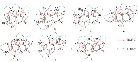
Fig.2 Selected 2D NMR correlations of compounds 1– 7
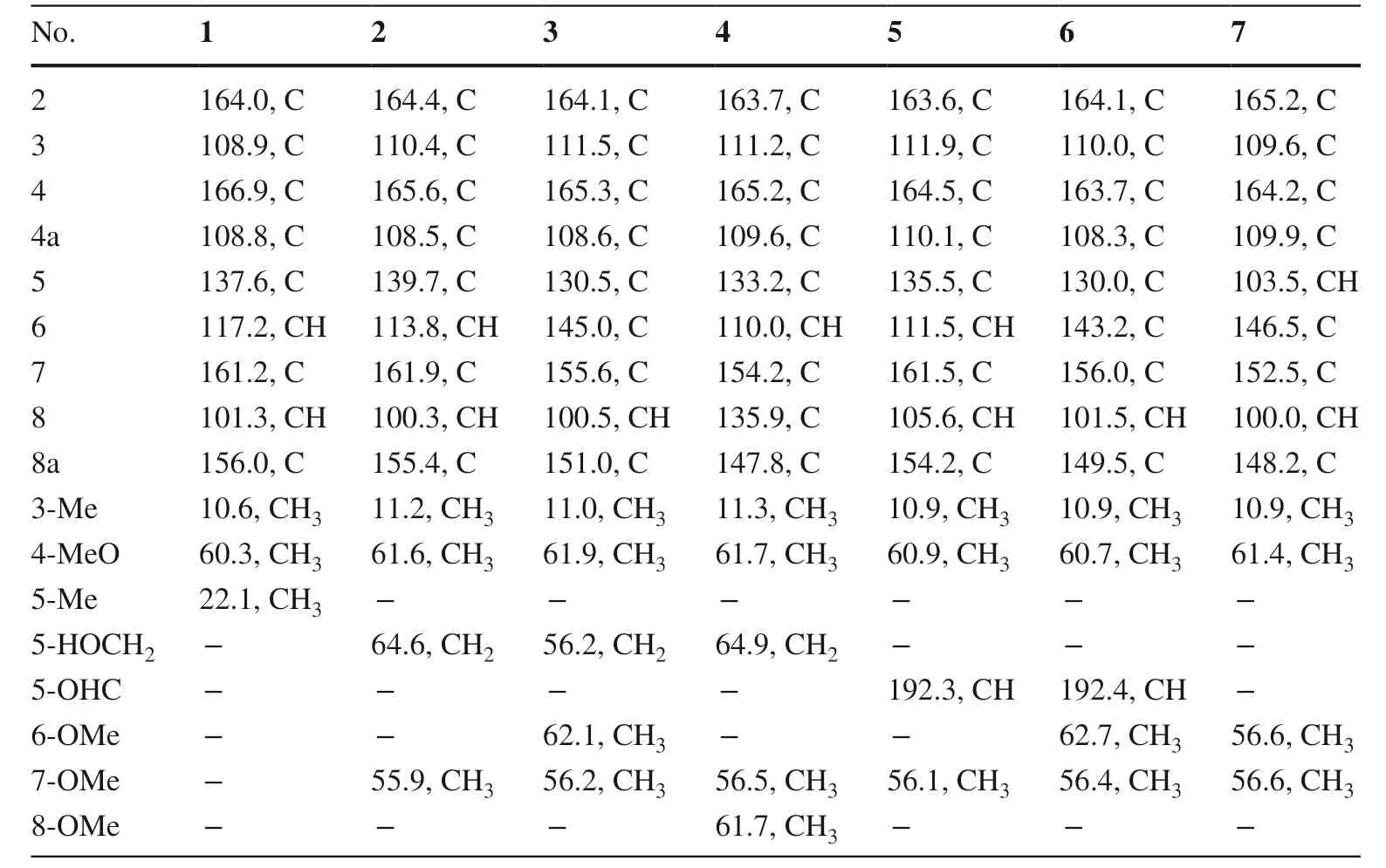
Table 2 13 C NMR data of compounds 1– 7 ( δ in ppm,J in Hz)
The molecular formula of 6 was assigned as C14H14O6by the [M + H]+ion atm/z279.0906 in positive HRESIMS spectrum.In the1H NMR spectrum, one formal atδH10.44, one aromatic singlet atδH6.89, three methoxy groups atδH3.81, 3.82, and 3.93, and one methyl group atδH2.15 were observed, showing an extra methoxy than 5.The above deduction was consistent with that twometa-coupled protons atδH7.24 and 6.99 in 5 was changed to be an aromatic singlet atδH6.89 in 6.By analyzing the ROESY experiment, the correlations of H3-9/OMe-4/H-10/OMe-6 and OMe-7/H-8 demonstrated the structure of 5-formyl-4,6,7-trimethoxy-3-methylcoumarin ( 6).
Compound 7 was assigned with the chemical formula of C13H14O5by the [M + H]+ion atm/z251.0898 in positive HRESIMS spectrum.In the 1 H NMR spectrum, two aromatic protons atδH7.07 and 6.85, three methoxy groups atδH4.01, 3.95, and 3.94, and a methyl group atδH2.17, were recognized.Compared with 4,6,7-trimethoxy-3,5-dimethylcoumarin ( 10) [6], the 5-methyl in 10 was absent in 7 but with an extra aromatic singlet atδH7.07.This proton (δH7.07) was assigned to be H-5 by the HMBC correlation from H-5 to C-4, and ROESY correlations of H-5/OMe-6 and H-8/OMe-7.Thus, compound 7 was deduced as 4,6,7-trimethoxy-3-methylcoumarin, the demethylated derivative of 10.Although this compound has been synthesized by methylation of 4-hydroxy-6,7-dimethoxy-3-methylcoumarin in 1949 [17], it is the fi rst report of its natural occurrence and NMR spectroscopic data.
The known coumarins were determined to be 4,7,8-trimethoxy-3,5-dimethylcoumarin ( 8) [7], 6-hydroxy-4,7-dimethoxy-3,5-dimethylcoumarin ( 9) [5], 4,6,7-trimethoxy-3,5-dimethylcoumarin ( 10) [6], and 5-formyl-4,7,8-trimethoxy-3-methylcoumarin ( 11) [8] by comparing their 1 H and 13 C NMR data with those in the literatures.
In order to evaluate their antidiabetic potency, all the coumarins were assayed for their inhibitory activity onα-glucosidase, PTP1B, and TCPTP.As shown in Table 3, all the compounds showed only weak or no inhibition to three enzymes at the concentration of 200 μM.According to the previous study [5], this type of coumarins could enhance the glucose-triggered secretion of insulin from murine islets.Thus, further studies will be needed to reveal their targets and mechanisms in exerting hypoglycemic eff ects.
3 Experimental Section
3.1 General Experimental Procedures
A Jasco model 1020 digital polarimeter (Jasco Corp., Tokyo, Japan) was used to measure optical rotations.UV and IR data were obtained using a Shimadzu UV2401PC spectrophotometer (Shimadzu, Kyoto, Japan) and a Nicolet iS10 spectrometer (Thermo Fisher Scientific, Madison, WI, USA), respectively.A Waters AutoSpec Premier P776 mass spectrometer (Waters, Milford, MA, USA) or aShimadzu LCMS-IT-TOF mass spectrometer (Shimadzu, Kyoto, Japan) was used to acquire the high-resolution mass spectra.ECD spectra were recorded on an Applied Photophysics Chirascan apparatus (Applied Photophysics, Surrey, UK).NMR spectra were obtained by using DRX-500, Avance III-600, and Ascend™ 800 MHz spectrometers (Bruker, Karlsruhe, Germany).TLC detection was run on silica gel plates (60 F254).Silica gel (200–300 mesh, Qingdao Makall group Co.Ltd., Qingdao, China) and Sephadex LH-20 (GE Healthcare Bio-Sciences AB, Uppsala, Sweden) were used for column chromatography.A Dr-Flash II apparatus was applied to accomplish the MPLC separations.HPLC purifications were conducted on a Shimadzu LC-CBM-20 system (Shimadzu, Kyoto, Japan), equipped with an Agilent Eclipse XDB-C18column (5 μm, 9.4 × 250 mm).
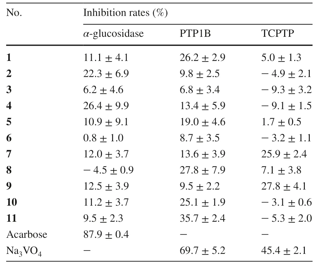
Table 3 Inhibitory rates of the isolates (200 μM) on α-glucosidase, PTP1B, and TCPTP
3.2 Plant Materials
The aerial parts of threeChelonopsisplants were collected in October 2016 from Lijiang, Yunnan Province of China, which were authenticated to beChelonopsis odontochilaDiels,Chelonopsis pseudobracteataC.Y.Wu et H.W.Li, andChelonopsis praecoxWeckerle and F.Huber by Dr.Chun-Lei Xiang.Voucher specimens (Nos.2016102101, 2016102102, 2016102103) were deposited in the Laboratory of Anti-virus and Natural Medicinal Chemistry, Kunming Institute of Botany, Chinese Academy of Sciences, China.
3.3 Extraction and Isolation
The air-dried plants ofC.pseudobracteata(6 kg) were powdered and extracted three times with 90% aqueous EtOH (25 L × 3) at room temperature.The extract was evaporated under reduced pressure, and the residue was suspended in H2O and partitioned with CHCl3.The CHCl3extraction (95 g) was subjected to silica gel column chromatography (Si CC) and eluted with an acetone-petroleum ether solvent system (from 10:90 to 50:50,v/v) to aff ord seven fractions (A–G).Fraction C (19.8 g) was subjected over MCI gel CHP 20P column (H2O–MeOH, 50:50–0:100) to provide fi ve fractions, Frs.C1–C5.Fr.C3(3.4 g) was purifi ed via Si CC (EtOAc-petroleum ether, 10:90–50:50) to give three fractions, Frs.C3-1–C3-3.Fr.C3-1 (600 mg) was purifi ed by Sephadex LH-20 CC (MeOH–CHCl3, 50:50) and semi-preparative HPLC (H2O–MeCN, 36:64) to give compounds 1 (16 mg) and 3 (18 mg).Compounds 2 (25 mg) and 4 (38 mg) were obtained from Fr.C3-2 (750 mg) by Sephadex LH-20 CC (MeOH–CHCl 3 , 50:50) and semi-preparative HPLC (H2O–MeCN, 50:50).
The air-dried plants ofC.praecox(25 kg) were powdered and extracted three times with 90% aqueous EtOH (100 L × 3) at room temperature.The combined EtOH extract was concentrated and partitioned between H2Oand CHCl3.The CHCl3extraction (380 g) was subjected to Si CC (4.0 kg, 30 × 100 cm), using a gradient elution of EtOAc-petroleum ether (from 10:90 to 100:0) to aff ord seven fractions (A–G).MPLC separation of Fr.D (46 g) by using a CHP20P MCI gel column (H2O-MeOH, from 50:50 to 0:100) provided fi ve fractions, Frs.D1–D5.Fr.D3 (1.3 g) was separated by Si CC (EtOAc-CHCl 3 , 2:98–50:50) to aff ord fi ve fractions, Frs.D3-1–D3-5.Fr.D3-1 (265 mg) was purifi ed by Si CC (acetone-petroleum ether, 5:95), and semi-preparative HPLC (H2O–MeCN, 42:58) to yield compounds 8 (25 mg),5 (18 mg), and 6 (18 mg).Fr.E (17 g) was subjected to MPLC to give fi ve fractions, Frs.E1–E5.Compounds 9 (5 mg) and 7 (25 mg) were obtained from Fr.E3 after repeated Si CC (acetone-petroleum ether, 10:90) and Sephadex LH-20 (MeOH–CHCl3, 50:50), and semipreparative HPLC (MeCN–H2O, 40:60).
Air-dried and powdered plants ofC.odontochila(8.0 kg) were extracted with 90% aqueous EtOH (35 L × 3) at room temperature.The combined EtOH extract was concentrated and partitioned between H2O and CHCl 3 .The CHCl 3 extract (160 g) was chromatographed on a silica gel column (1.3 kg, 30 × 100 cm), and eluted with acetone-petroleum ether gradient (from 0:100 to 100:0) to yielded seven fractions, Frs.A–G.MPLC separation of Fr.C (15 g) with MCI gel CHP 20P column (H2O–MeOH, from 50:50 to 0:100) gave rise to fi ve fractions, Frs.C1–C5.Fraction C3 (2.8 g) was chromatographed on a silica gel column (acetone-CHCl3, from 10:90 to 100:0) to provide four fractions, Frs.C3-1–C3-4.Compound 11 (12 mg) was purifi ed from Fr.C3-2 (500 mg) by Sephadex LH-20 CC (MeOH–CHCl3, 50:50), and semipreparative HPLC (H2O–MeCN, 64:36).After repeated separation over Si CC (acetone-petroleum ether, 15:85), Sephadex LH-20 CC (MeOH–CHCl 3 , 50:50), and semi-preparative HPLC (H2O–MeCN, 60:40), compounds 10 (27 mg) and 8 (21 mg) were obtained from Fr.C3-3 (470 mg).
3.4 Spectroscopic Data of Compounds
3.4.1 7-Hydroxy-4-Methoxy-3,5-Dimethylcoumarin ( 1)
White amorphous powder; UV (MeOH)λmax(logε): 321 (3.99) nm; IR (KBr)νmax: 3204, 1681, 1610, 1567, 1454, 1378, 1358, 1344, 1257, 1154, 1102, 1074, 1016 cm −1 ; 1 H and 13 C NMR data, see Tables 1 and 2; positive HRESIMSm/z221.0809 [M + H] + (calcd.for C12H13O 4 , 221.0808).
3.4.2 5-Hydroxymethyl-4,7-Dimethoxy-3-Methylcoumarin( 2)
White amorphous powder; UV (MeOH)λmax(logε): 222 (3.91), 320 (3.90) nm; IR (KBr)νmax: 3398, 1663, 1616, 1592, 1558, 1453, 1432, 1369, 1336, 1251, 1197, 1151, 1083, 1047, 1012, 946 cm −1 ; 1 H and 13 C NMR data, see Tables 1 and 2; positive HRESIMSm/z251.0897 [M + H] + (calcd.for C13H15O5, 251.0914).
3.4.3 5-Hydroxymethyl-4,6,7-Trimethoxy-3-Methylcoumarin ( 3)
White amorphous powder; UV (MeOH)λmax(logε): 294 (3.75), 328 (4.00) nm; IR (KBr)νmax: 3434, 1683, 1603, 1562, 1452, 1418, 1366, 1331, 1262, 1224, 1161, 1130, 1081, 1061, 1004, 987 cm −1 ; 1 H and 13 C NMR data, see Tables 1 and 2; positive HRESIMSm/z281.1039 [M + H] + (calcd.for C14H17O6, 281.1020).
3.4.4 5-Hydroxymethyl-4,7,8-Trimethoxy-3-Methylcoumarin ( 4)
White amorphous powder; UV (MeOH)λmax (logε): 318 (4.07) nm; IR (KBr)νmax: 3435, 1687, 1595, 1571, 1457, 1421, 1337, 1274, 1136, 1096, 1052 1012 cm −1 ; 1 H and 13 C NMR data, see Tables 1 and 2; positive HRESIMSm/z281.1014 [M + H] + (calcd.for C14H17O6, 281.1020).
3.4.5 5-Formyl-4,7-Dimethoxy-3-Methylcoumarin ( 5)
White amorphous powder; UV (MeOH)λmax(logε): 218 (3.09), 291 (2.74), 328 (2.83) nm; IR (KBr)νmax: 3426, 1725, 1688, 1605, 1448, 1367, 1336, 1258, 1168, 1089, 953 cm −1 ; 1 H and 13 C NMR data, see Tables 1 and 2; positive HRESIMSm/z249.0744 [M + H] + (calcd.for C13H13O5, 249.0758).
3.4.6 5-Formyl-4,6,7-Trimethoxy-3-Methylcoumarin ( 6)
White amorphous powder; UV (MeOH)λmax(logε): 207 (3.67), 224 (3.63), 290 (3.20), 329 (3.45) nm; IR (KBr)νmax: 3420, 1721, 1693, 1603, 1455, 1388, 1369, 1266, 1226, 1078, 1006, 959 cm −1 ; 1 H and 13 C NMR data, see Tables 1 and 2; positive HRESIMSm/z279.0906 [M + H] + (calcd.for C14H15O6, 279.0863).
3.4.7 4,6,7-Trimethoxy-3-Methylcoumarin ( 7)
Colorless gum; UV (MeOH)λmax(logε): 208 (2.99), 222 (2.83), 287 (2.30), 333 (2.58) nm; IR (KBr)νmax: 3431, 1709, 1620, 1580, 1453, 1372, 1337, 1249, 1215, 1162, 1025, 994 cm −1 ; 1 H and 13 C NMR data, see Tables 1 and 2; positive HRESIMSm/z251.0898 [M + H] + (calcd.for C13H15O5, 251.0914).
3.5 In Vitro Enzyme Inhibition Assays
In this study, three enzymes closely related to diabetes, namelyα-glucosidase, PTP1B, and TCPTP, were applied to assess the antidiabetic potency of compounds.Enzyme inhibition was assayed in accordance with the previous reports [18,19].Acarbose (forα-glucosidase) and Na3VO4(for PTP1B and TCPTP) were used as the positive controls.
4 Supporting Information
1D and 2D NMR, HRMS, UV and IR spectra of compounds 1 − 7.
Supplementary InformationThe online version contains supplementary material available at https:// doi.org/ 10.1007/ s13659- 021- 00318-9.
AcknowledgementsThis work was supported by the Yunnan Wanren Project (YNWR-QNBJ-2018-061), the Natural Science Foundation of Yunnan Province (2019FI017), and the Reserve Talents of Young and Middle-Aged Academic and Technical Leaders in Yunnan Province.
Declarations
Conflict of interestThe authors declare that they have no competing fi nancial interest.
Open AccessThis article is licensed under a Creative Commons Attribution 4.0 International License, which permits use, sharing, adaptation, distribution and reproduction in any medium or format, as long as you give appropriate credit to the original author(s) and the source, provide a link to the Creative Commons licence, and indicate if changes were made.The images or other third party material in this article are included in the article's Creative Commons licence, unless indicated otherwise in a credit line to the material.If material is not included in the article's Creative Commons licence and your intended use is not permitted by statutory regulation or exceeds the permitted use, you will need to obtain permission directly from the copyright holder.To view a copy of this licence, visit http:// creat iveco mmons.org/ licen ses/ by/4.0/.
 Natural Products and Bioprospecting2021年6期
- Natural Products and Bioprospecting的其它文章
- Isoprenylated Flavonoids as Ca v3.1 Low Voltage-Gated Ca2+ Channel Inhibitors from Salvia digitaloides
- Scutellarin Reduces Cerebral Ischemia Reperfusion Injury Involving in Vascular Endothelium Protection and PKG Signal
- Furostanol Saponins from Asparagus cochinchinensis and Their Cytotoxicity
- Anticancer Properties and Mechanism of Action of Oblongifolin C, Guttiferone K and Related Polyprenylated Acylphloroglucinols
- Natural Products as Potential Lead Compounds for Drug Discovery Against SARS-CoV-2
- Advances in Research on Chemical Constituents and Their Biological Activities of the Genus Actinidia