
Descriptive histomorphological evaluation of the testis and caudal epididymis following treatment with rooibos (Aspalathus linearis), honeybush (Cyclopia intermedia) and sutherlandia (Lessertia frutescens) in healthy and streptozotocininduced diabetic rats
2021-08-17 09:14:14TemidayoOmolaoyeStefanduPlessis
Temidayo S. Omolaoye, Stefan S. du Plessis
1Department of Basic Sciences, College of Medicine, Mohammed Bin Rashid University of Medicine and Health Sciences, Dubai, United Arab Emirates
2Division of Medical Physiology, Faculty of Medicine and Health Sciences, Stellenbosch University, Tygerberg, South Africa
Supplementary Materials

Supplementary Figure 1. Classification for the seminiferous tubules (AB/PAS stain; A-C: the scale bar 20 µm; D: the scale bar 50 µm). A: Normal (complete spermatogenic phases, regular cellular organization, normal cellular association and regular interstitial spaces); B: Atrophic (epithelium shrinkage, few or absence of germ cells and cellular disorganization); C and D: Sloughy (presence of immature accumulation of cells in the lumen and absence of some or all of the spermatogenic phases). SG: spermatogonia, PS: primary spermatocytes, S: spermatids.
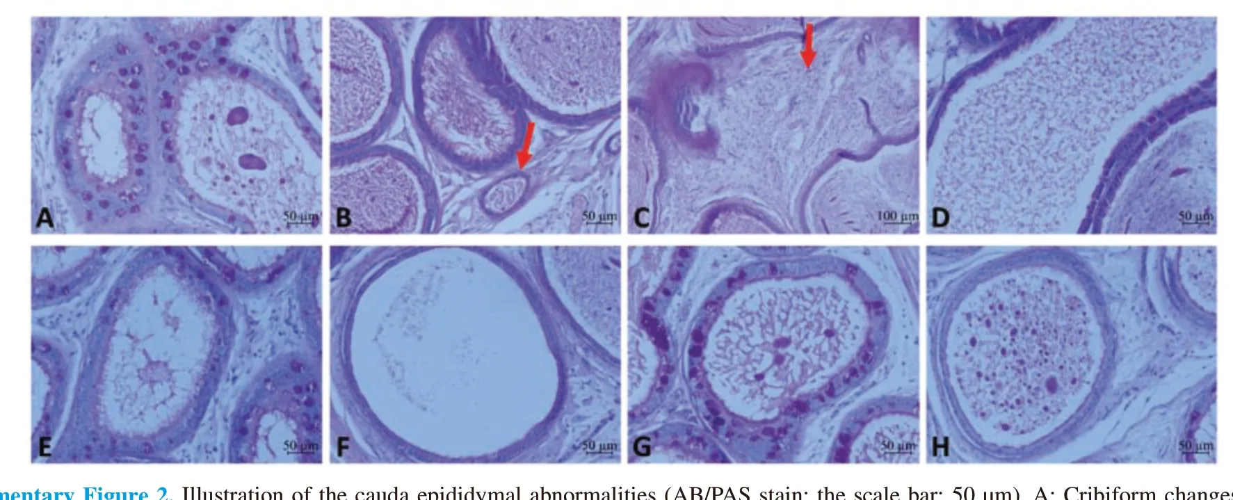
Supplementary Figure 2. Illustration of the cauda epididymal abnormalities (AB/PAS stain; the scale bar: 50 µm). A: Cribiform changes (hyperplastic modification of the epithelium, e.g. infolding of the epithelium); B and C: Inflammatory infiltrate (excessive accumulation of cells in the interstitium); D and E: Debris in the lumen (presence of cytoplasmic shedding in the lumen instead of spermatozoa); F: Clear lumen (no spermatozoa in the lumen); G: Epithelium disruption (altered epithelium organization); H: Cellular accumulation in the lume.
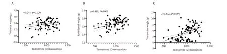
Supplementary Figure 3. Correlations between plasma testosterone concentration and testicular weight (A), epididymal weight (B), and visceral fat weight (C).
 Asian Pacific Journal of Reproduction2021年4期
- Asian Pacific Journal of Reproduction的其它文章
- Oral supplementation of selenium improves post-thaw sperm quality in Saanen bucks
- Taxifolin attenuates ischemia-reperfusion induced oxidative ovarian damage in rats
- Proposed age-stratified reference intervals of FSH derived from normozoospermic men
- Genetic association of rs7754840 and rs7756992 polymorphisms in the CDKAL1 gene and gestational diabetes mellitus in selected Filipino pregnant women
- Etiopathogenesis of reproductive tract infections and the emerging role of bitter taste receptors: A scoping review