Biomaterial-based platforms for cancer stem cell enrichment and study
Chunhua Luo*, Zhongjie Ding*, Yun Tu, Jiao Tan, Qing Luo, Guanbin Song
1Key Laboratory of Biorheological Science and Technology, Ministry of Education, College of Bioengineering, Chongqing University, Chongqing 400044, China; 2Institute of Pathology and Southwest Cancer Center, Southwest Hospital, Third Military Medical University (Army Medical University), Key Laboratory of Tumor Immunopathology, Ministry of Education of China, Chongqing 400038, China; 3School of Pharmacy, Chongqing Medical and Pharmaceutical College, Chongqing 401331,China
ABSTRACT Cancer stem cells (CSCs) are a relatively rare subpopulation of tumor cell with self-renewal and tumorigenesis capabilities. CSCs are associated with cancer recurrence, progression, and chemoradiotherapy resistance. Establishing a reliable platform for CSC enrichment and study is a prerequisite for understanding the characteristics of CSCs and discovering CSC-related therapeutic strategies. Certain strategies for CSC enrichment have been used in laboratory, particularly fluorescence-activated cell sorting (FACS)and mammosphere culture. However, these methods fail to recapitulate the in vivo chemical and physical conditions in tumors, thus potentially decreasing the malignancy of CSCs in culture and yielding unreliable research results. Accumulating research suggests the promise of a biomaterial-based three-dimensional (3D) strategy for CSC enrichment and study. This strategy has an advantage over conventional methods in simulating the tumor microenvironment, thus providing a more effective and predictive model for CSC laboratory research. In this review, we first briefly discuss the conventional methods for CSC enrichment and study. We then summarize the latest advances and challenges in biomaterial-based 3D CSC platforms. Design strategies for materials, morphology,and chemical and physical cues are highlighted to provide direction for the future construction of platforms for CSC enrichment and study.
KEYWORDS Cancer stem cell; biomaterial; CSC enrichment and study; three-dimensional culture platform
Introduction
Cancer stem cells (CSCs) are a relatively rare subpopulation of tumor cells with self-renewal and tumorigenesis capabilities. These cells have been correlated with cancer recurrence, progression, and chemoradiotherapy resistance1.Understanding the characteristics of CSCs and discovering CSC-related drugs have important implications in anticancer therapy. A research prerequisite is the establishment of stable and repeatable culture conditions for the enrichment and study of CSCs while maintaining their stemness propertiesin vitro.
The conventional laboratory cell culture platform is twodimensional (2D) and typically based on substrates optimized for the attachment of cells supplied with serum-rich medium,21% oxygen and 5% CO2. However, CSCs are poorly adherent within tumors, under the conditions of restricted nutrients and oxygen2. Under typical 2D culture conditions, CSCs are forced to adhere and polarize on the rigid bottoms of dishes and are subjected to excessive nutrition and oxygen3; these conditions clearly fail to reflect thein vivosituation of CSCs in tumors. Inappropriate culture conditions may decrease the malignancy of CSCs4and yield unreliable results in drug screening and mechanistic discovery investigations. To overcome the drawbacks of the 2D culture approach, three-dimensional (3D) culture models for CSC enrichment and study have been developed, among which fluorescence-activated cell sorting (FACS) and mammosphere culture models are most extensively used. Although these methods can establish 3D growth conditions for CSCs, they have limitations in their ability to represent the crosstalk between CSCs and environmental elements, such as the extracellular matrix (ECM),matrix stiffness, and other chemical and physical cues. Tumor slice culture, a 3D model originating decades ago, can retain the complexity of the original tumor microenvironment through culturing of tumor slices in selected conditions5.This model’s limitations include requirements for extensive cell manipulation and fresh tumor specimens, thus restricting its wide application.
In recent years, some biomaterial-based 3D strategies for CSC enrichment and study have been developed and provided advantages in simulating the tumor microenvironmentin vitro. Organoids, a novel stem cell study model, have been introduced in the study of CSCs through the embedding of CSCs and other cancer-related cells in Matrigel or ECM-like biomaterials6. This model not only supports the CSC enrichment but also recapitulates the histopathology and cellular heterogeneity of tumors7-10. Additionally, unlike conventional study systems based on cancer cell lines, organoids can be generated from specimens of individual patients,thereby enabling the development of personalized therapeutic regimens6. The utilization of organoids in CSC studies has been reviewed by Drost et al.6. However, because technical difficulties exist in constructing organoids, and clinical specimens are usually needed, organoid studies are not achievable in every laboratory studying CSCs. For this reason, CSCs enriched from cancer cell lines remain the mainstream method used in CSC studies. Thus, in this review, we emphasize 3D platforms constructed for CSC enrichment from cancer cell lines.
Compared with CSCs obtained from conventional mammosphere culture, those grown on 3D scaffolds exhibit enhanced CSC characteristics4,11, thus suggesting that the biomaterial-based 3D culture platform can better maintain malignancy. Therefore,in vitroinvestigations performed on biomaterial-based 3D culture platforms may provide more effective and predictive data for clinical use. Here, we provide an overview of the conventional methods for CSC enrichment and study and summarize the latest advances and challenges in biomaterial-based 3D platforms for CSC enrichment and study, with an emphasis on the design of materials, morphology, and chemical and physical cues involved in the construction of the platforms.
Conventional methods for CSC enrichment and study
CSCs make up a minority of cells in heterogeneous tumors1.To study CSCs in laboratory settings, methods for the isolation, enrichment, and culture of CSCs are required. According to the characteristics of CSCs-such as their nonadherence,CSC-related gene expression, and chemo- and radioresistance-strategies for CSC enrichment have been developed,including mammosphere culture, CSC marker-based sorting, chemo- and radioselection, and genetic reprogramming(Figure 1). In these strategies, CSC marker-based sorting using fluorescence-activated cell sorting (FACS) and mammosphere culture are the most extensively used techniques for CSC isolation and enrichment. However, mammosphere culture is the common use method for the further study of CSCs, because CSCs isolated by FACS, chemo- and radioselection, or genetic reprogramming must be enriched by mammosphere culture,and mechanistic investigations and antidrug screening are based on mammospheres.
FACS selection is a strategy using flow cytometry to isolate CSCs according to the specific markers that they express. Since Bonnet et al.12discovered CD34+CD38-leukemia stem cells in the hematopoietic system in 1997, CSCs have been observed in various solid tumors exhibiting a tissue-specific antigenic phenotype. To date, several specific markers for CSCs have been identified, such as CD133 for breast, pancreas, prostate,brain and liver cancers; aldehyde dehydrogenase 1 (ALDH1)for breast, lung, ovarian, and colon cancer; CD44 for breast,pancreatic, colon, and prostate cancer; epithelial cell adhesion molecule (EpCAM) for stomach, pancreatic, and liver cancer; and CD166 for lung and colon cancer12,13. Through labeling of cells with fluorescent or magnetic bead-conjugated antibodies, the CSC population can be separated from tumors or cancer cells through FACS or magnetic-activated cell sorting (MACS). In addition to sorting through the use of surface markers, studies have also suggested a FACS approach according to the stem cell featured side population, through the use of Hoechst 3334214,15. In practice, FACS is preferred over MACS because its selection is purer and more accurate.However, FACS relies on costly dedicated equipment and consumes large amounts of antibodies, thus making FACS expensive and not accessible or affordable for every laboratory. Moreover, owing to cost concerns, FACS separation is usually performed on the basis of single biomarkers; however,no unique biomarker is acceptable for CSC identification12.Moreover, because of the effect of laser scanning in flow cytometry, the number of viable cells obtained by FACS is usually lower than the theoretical value.

Figure 1 Schematic representation of conventional methods used for CSC enrichment and study. In laboratory settings, CSCs can be enriched from tumor or cancer cell lines. Mammosphere culture, also called suspension culture or spheroid culture, is the most extensively used method; this method aggregates CSCs spherically by culturing cells in suspension by using ultralow attachment plates, hanging drops,gyrator rotation and spinner flasks, or NASA rotary cell culture systems. Marker-based sorting is usually performed with FACS or MACS,according to the surface markers specifically expressed by CSCs. Chemo- and radioselection enriches CSCs by treating cells with anticancer drugs or radiation according to their chemo- and radioresistance characteristics. Genetic reprogramming is a novel method for CSC enrichment through epigenetic methods or genetic modifications. Regardless of the method through which CSCs are enriched, mammosphere culture is the common method for CSC subculture and study.
In mammosphere culture, pioneered by Sutherland and colleagues in 197016, and also called suspension culture or spheroid culture, cells are cultured in serum-free medium and kept in suspension through the use of an ultralow attachment plate, hanging drop, gyrator rotation and spinner flask, or NASA rotary cell culture system, which aggregates cells spherically and simulates nonadherent conditionsin vivo17. Under suspension conditions, only CSCs, but not differentiated cells,such as cancer cells and stromal cells in tumors, survive and form floating colonies. Compared with FACS, mammosphere isolation of CSCs is technically more practical for laboratories lacking dedicated facilities and is feasible for the enrichment of multiple types of CSCs. However, interestingly, a recent study has suggested that mammosphere culture induces CSC enrichment in a cell line-dependent manner18.
In addition to FACS and mammosphere culture, some novel approaches for CSC enrichment have been elucidated.Because of the resistance of CSCs to chemotherapeutic agents and radiation, only CSCs are believed to be able to survive after treatment with chemotherapeutic drugs or radiotherapy.Studies have shown that cells expressing CSC surface markers are enriched after treatment with chemotherapeutic agents or radiation19-21, thereby suggesting a novel chemo- and radioselection approach for CSC enrichment.
According to the different expression profiles between CSCs and cancer cells, some studies have enriched CSCs by reprogramming cancer cells through epigenetic methods or genetic modification. Ikegaki et al.22have reported a method of enriching stable neuroblastoma stem cells with tumor-initiating ability from neuroblastoma cells by treatment with epigenetic modifiers, DNA methylation (5AdC),and/or histone deacetylase (4-phenylbutyrate, or 4PB).Likewise, Mani et al.23have generated CSCs by ectopically expressing factors involved in epithelial to mesenchymal transition (EMT) and have suggested that EMT is associated with the stem-like phenotype of cells23. However, these cells are genetically modified. Consequently, it will be interesting to directly compare these cells with those selected by FACS or mammosphere culture to ensure their reliability in preclinical research.
Biomaterial-based CSC platforms
To maintain stemnessin vivo, CSCs require the proper niche,including the ECM, cancer-associated fibroblasts, hypoxia,growth factors/cytokines, immunocytes, and other components1. In addition, biophysical cues in the tumor microenvironment, such as stiffness, porosity, topography, stretch,interstitial fluid flow, and compression, are crucial factors regulating the stem cell state of CSCs24. These chemical and physical elements maintain the malignant characteristics of CSCs, such as rapid proliferation, metastasis, and anticancer drug resistance.
Although mammosphere culture, the most commonly used conventional method to date, can mimic thein vivononadherent 3D growth morphology of CSCs to some extent by forming multicellular tumor spheroidsin vitro, it cannot reestablish the interaction of CSCs with biochemical and biophysical cues in the niche. These drawbacks not only decrease the enrichment efficiencyin vitrobut also, more importantly, potentially provide less reliable data in the study of pathological mechanisms and drug screening4. Given the weaknesses of conventional methods, in recent years, some novel 3D CSC platforms based on natural and synthetic biomaterials have been developed for enrichment and study; these platforms have advantages in mimicking biochemical and biophysical elements in the tumor microenvironment and in allowing them to interact with cells.
Biomaterials for platform construction
The ECM is the major component in the tumor niche, providing structural and biochemical support for the stemness phenotype of CSCs and regulating their fates1,25. The components of tumor ECM have been studied, mainly proteins, glycoproteins, proteoglycans, and polysaccharides1,25. According to these discoveries, biomaterials based on the natural components of the ECM or their analogs are generally used to construct platforms for CSC enrichment, thus not only providing 3D structural support for growth but also reestablishing the ECM-CSC interaction.
To simulate ECM-cell interaction, a decellularized amnion membrane scaffold containing the main components of the ECM has been proposed26. Likewise, commercially available Matrigel, a solubilized basement membrane matrix composed of certain ECM proteins, such as laminin and collagen IV, has been demonstrated to promote the enrichment of CSCs from adenocarcinoma cells, breast cancer cells, and prostate cancer cells27. Although these scaffolds have similar compositions to that of the ECM, potential variation may exist among scaffolds from different sources, and the exact components of scaffolds are usually unclear. To avoid potential variation and to generate a more controllable and repeatable research system, scaffolds with defined components are favored by researchers.
Hyaluronic acid (HA), a major glycosaminoglycan in the tumor ECM, has been demonstrated to be correlated with tumor angiogenesis, invasion, and metastasis28. However,although HA has good biodegradability and biocompatibility,its low mechanical strength and rapid bioabsorption rate have limited its use as the sole component in platform construction29,30. To exploit its advantages and bypass its disadvantages,HA is usually mixed with biopolymers with good mechanical properties, such as chitosan, to construct CSC platforms29,30or is used as a modifier to improve enrichment efficiency, taking advantage of its ability to activate chemical signaling in cells31.
Collagens are the main structural element of the tumor ECM, and multiple collagen subtypes (e.g., collagen I, collagen III, and collagen IV) have been demonstrated to be associated with tumor initiation, EMT, drug resistance, and CSC self-renewal25. Platforms based on collagen I have been explored for the enrichment of liver CSCs32, colorectal CSCs33, and breast CSCs34.
Compared with costly natural ECM-based hydrogels or scaffolds, some naturally occurring polymers sharing similar molecular structures with tumor ECM components, such as alginate and chitosan, have attracted much attention17,31,35-40.Keratin, an inexpensive ECM-like protein with integrin-mediated cellular interactions, has been found to perform comparably to collagen I in facilitating tumor spheroid formation and CSC enrichment, thus suggesting that it is an economical material for CSC enrichment and study33. Alginate has a similar molecular structure to that of glycosaminoglycan in the ECM; is easily gelated in divalent cation solutions, such calcium chloride; and has good biocompatibility and low immunogenicity41. When used in cell culture, alginate is usually covalently modified by cell-binding ligands or peptides, such as RGD and cadherins, to facilitate cell adhesion, owing to its lack of a cell adhesion domain41. Rigid alginate hydrogels can be easily fabricated by modifying the concentration of alginate or calcium chloride38,42, thus providing a tool for exploring the effects of matrix stiffness on the biological behaviors of CSCs. Because of these merits, alginate hydrogels are extensively used in constructing platforms for CSC enrichment and study17,31,35-40.
Some synthetic materials are also used for CSC platforms,including poly(ε-caprolactone) (PCL)43, polydimethylsiloxane(PDMS)44, polyethylene glycol diacrylate (PEGDA)45, poly(allylamine hydrochloride) (PAH)46, and poly(2,4,6,8-tetravinyl-2,4,6,8-tetramethyl cyclotetrasiloxane) (pV4D4)47.Compared with natural biomaterials, synthetic materials enable experimental reproducibility by avoiding variations in the origin and purification of materials. Because they allow less chemical signaling, synthetic materials have advantages in investigating the influence of structural and physical cues by avoiding chemical crosstalk between cells and the chemical components of materials. Another merit of synthetic materials is that they are easier to functionalize with chemical groups,growth factors, drugs, and peptides, thereby enabling study of the role of chemical cues in a 3D microenvironment. Certain synthetic materials have been used in the study of cancer cells,such as polyethylene glycol (PEG), PDMS, PCL, poly(D,L-lactide-coglycolide) (PLG), and PLGA/polylactic acid (PLA)48,49,thus offering several candidates for the development of CSC platforms. However, whether these materials are qualified for enrichment and study of CSCs remains unclarified or controversial. For example, Palomeras et al.43have demonstrated that PCL scaffolds improve the enrichment of breast CSCs, whereas Kievit et al.40have reported that tumor spheroids cannot form on either PCL or chitosan-alginate scaffolds coated with PCL,thus suggesting that PCL is not suitable for the growth of glioblastoma CSCs.
Generally, on these platforms based on natural or synthetic materials, the proportion of cells expressing CSCspecific markers and the expression of stemness-related genes increases, and the chemo- and radioresistance of cells is improved over that of cells cultured on 2D substrate or 3D mammosphere culture conditions. This improvement is due to not only to the 3D structure constructed by materials but also to the chemical signaling provided by the chemical components of materials, as discussed in the following section. The reported natural and synthetic materials used to construct a 3D platform for CSC enrichment and study are summarized in Table 1.
Morphology of biomaterial-based platforms
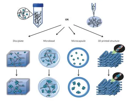
To construct a platform for CSC enrichment and study, materials are usually structured into discs/plates or microcapsules/microbeads. Through simple seeding of cells onto hydrogel or polymer thin films predeposited on cell culture dishes31,33,34,47or sectioned presynthesized scaffolds29,30,39,40, 3D platforms for CSC culture and study can be constructed. However, in practice, the penetration depth of cell suspensions is limited by the surface properties of the material and the air pre-existing in the scaffolds, thus potentially resulting in the heterogeneous seeding of cells and the wasting of scaffolds. An alternative method is gelating cells with hydrogels to achieve a uniform distribution of cells27,32,50. In designing such cell-laden hydrogels, the pore size is a crucial issue that must be considered,because tumor spheroids usually grow into spheres larger than 100 μm in diameter, whereas hydrogels with smaller pore sizes may restrict the growth of CSCs. Additionally, nutrients may be adequately supplied for cells embedded in hydrogel35.
Alginate is an extensively explored biomaterial in constructing CSC platforms, because of its low cost and ECM-like properties. In the laboratory, alginate is usually mixed with cells,and cell-laden alginate microbeads are generated by extrusion of the mixture into calcium chloride solution with an electrostatic droplet generator or a syringe37,38. As discussed previously, pore size restriction and nutrient insufficiency may potentially affect the growth of CSCs. Rao et al.17have developed a miniaturized 3D liquid core of microcapsules with an alginate hydrogel shell by using 2 syringes that push the core fluid with cancer cells and the shell fluid of sodium alginate.This microcapsule has been found to provide human prostate cancer cells with sufficient space and nutrients for the formation of tumor spheroids, and to allow the culture time to be shortened to 2 days while yielding CSC aggregates with better quality17. These findings have been supported by a direct comparison of microcapsules and microbeads by using embryonic stem cells, which has indicated a higher enrichment efficiency of microcapsules than microbeads. Sakai et al.36have further improved the homogeneity of these microcapsules by templating the size and shape of cavities by using gelatin microparticles, which may improve the reproducibility of results obtained from the microcapsule platform. However, owing to the lack of physical attachment to the structural material,microcapsules do not support the investigation of cell-matrix interactions, such as the effect of matrix stiffness on the biological behaviors of CSCs.

Table 1 Reported biomaterial-based platforms for CSC enrichment and study
A reproducible platform is a crucial premise for generating reproducible results in studies. The 3D bioprinting technique has advantages in constructing reproducible platforms, because it can precisely position biological materials, biochemicals,and living cells layer by layer, and enable spatial control of the placement of functional components51. Some 3D-bioprinted CSC platforms have been developed by using alginate and PCL35,43, and the effect of the scaffold angle on the enrichment of CSCs has been investigated43. However, the current platforms have not taken full advantage of 3D bioprinting techniques in generating 3D structures with well-defined geometries. For example, through precise design, an engineered 3D tumor microenvironment can be constructed by mimicking thein vivodistribution of tumor/nontumor cells, blood vessels, chemical components, and matrix rigidity. In this sense,3D-bioprinted platforms have broad application potential in tumor studies by providing an engineered microenvironment combining various cellular and physicochemical factors.Additionally, because matrix topography has been demonstrated to determine the stem cell fate and EMT of cancer cells52,53, the inner structure of scaffolds or hydrogels can be designed with 3D bioprinting, which enables the study of the effect of topography in a 3D microenvironment.
The morphology of biomaterial-based platforms for CSC enrichment and study is summarized in Figure 2.
Chemical cues of biomaterial-based platforms
Biomaterial-based platforms are more than 3D structures supporting CSCs for growth; in fact, some biomaterials facilitate the enrichment of CSCs. On biomaterial-based platforms, CSC enrichment can be achieved without supplementation with the cytokines regularly used in conventional mammosphere culture (Table 1), such as epidermal growth factor (EGF),basic fibroblast growth factor (bFGF), N2, and B27, probably not only because of the 3D structural properties of the platform but also because of the chemical characteristics of materials in promoting CSC self-renewal, proliferation, and EMT40.The addition of regular cytokines in biomaterial-based culture further shortens the enrichment time17. Several natural ECM components have been demonstrated to be associated with the activation of signaling pathways regulating the stem cell state,self-renewal, and proliferation of CSCs24,25,51,54, thus indicating the merits of using natural ECM polymers in constructing CSC platforms. The signaling pathways correlated with the natural ECM polymers are summarized in Figure 3.

Figure 2 Schematic representation of the morphological design of the biomaterial-based platform for CSC enrichment and study. To construct CSC platforms, cell-laden hydrogels or presynthesized hydrogels/scaffolds are used to form discs/plates, microcapsules, microbeads,or defined 3D printed morphologies.

Figure 3 Signaling pathways correlating with the natural ECM components that regulate the stem cell state, self-renewal, and proliferation of CSCs. ILK: integrin-linked kinase; NF-κB: nuclear factor kappa B; GSK-3β: glycogen synthase kinase-3β; LEF1: lymphoid enhancer factor 1;FAK: focal adhesion kinase; AKT: also known as protein kinase B (PKB); ERK: extracellular signal-regulated kinase; β-cat: β-catenin; TAZ: transcriptional coactivator with PDZ-binding motif.
HA is one of the most notable biomaterials in CSC studies, because of its ability to bind the cellular receptor CD44,which activates downstream signaling pathways associated with cancer stemness, motility, and EMT25,54,55. Although the signaling molecule downstream of the HA-CD44 interaction has been poorly investigated in CSCs, studies nonetheless strongly suggest a role of HA in regulating CSC stemness and proliferation. Moreover, HA’s specific binding to CD44 further promotes the selection and enrichment of CSCs expressing CD44, such as liver, breast, prostate, ovarian, gastric, head,and neck CSCs56. Therefore, HA is an ideal candidate biomaterial for CSC enrichment or a modification material that can be combined with structural materials to improve the enrichment efficiency31. The advantage of HA in the enrichment of CD44-expressing CSCs suggests the possibility of using ligand-modified material in the exploitation of platforms,and accelerating CSC enrichment by targeting CSC-specific markers.
The structural material of the platform can be modified and functionalized with chemical groups, cytokines, growth factors,and peptides, depending on the culture requirements or study interests. Qiao et al.31have immobilized EGF and bFGFcytokines required for CSC enrichment in mammosphere culture-on alginate hydrogel, thus improving the enrichment efficiency of breast CSCs by prolonging the biological activities of these cytokines. Therefore, this method may provide a more economical culture strategy for CSCs. Likewise, some growth factors or cytokines, such as vascular endothelial growth factor(VEGF), a crucial factor in angiogenesis, can be engineered on scaffolds, thereby enabling study of angiogenesis in the tumor microenvironment. The immobilization of chemical reagents on materials, such as cytokines, growth factors, and proteases,might possibly simulate a microenvironment with cytokine or growth factor gradients in tumors.
Better hydrophilicity has been shown to facilitate the enrichment and stemness of prostate CSCs57, thus suggesting that the enrichment efficiency can be improved by modifying the surface properties of materials. This modification can be achieved through use of synthetic materials that can be easily functionalized with hydrophilic groups. Additionally, the covalent linking of the scaffold polymer to chemical groups or peptides enables the study of their functions in a 3D microenvironment. Synthetic materials have advantages in constructing engineered 3D platforms, owing to their characteristics of easy modification and low chemical crosstalk with cells.
Physical cues of biomaterial-based platforms
Matrix stiffness in the tumor microenvironment is a major physical cue affecting CSCs. Tumors are stiffer than paracarcinoma tissues, because of the increased deposition of ECM components, whereas the stiffness distribution in tumors is heterogeneous24. Studies based on 2D substrates with determined rigidity have shown that matrix stiffness plays crucial roles in regulating the malignancy of cancer cells58,59.However, CSCs are low-adherent cells, and their connection to the adherent surface is unstable, thus possibly preventing CSCs from sufficiently sensing the physical properties of the substrate. Comparatively, 3D culture models allow for more physical contacts and interactions by embedding CSCs in hydrogels or scaffolds, which are superior to 2D substrates,particularly in the study of the response of CSCs to the physical microenvironment.
Alginate hydrogels are commonly used to construct 3D structures with different stiffnesses, ranging from hundreds to thousands of Pascals, by modulation of the concentration of alginate31,38or the concentration of calcium chloride solution for cross-linking of alginate hydrogels42. Other natural or synthetic polymers, such as fibrin50and polyethylene glycol diacrylate (PEGDA)45, have also been used to establish 3D structures with different stiffness. These models have revealed that an appropriate stiffness facilitates better enrichment and stemness of CSCs31,38,45,50, thus indicating that a more reliable and persuasive drug screening and mechanistic studies should be performed on hydrogels or scaffolds with appropriate stiffness according to thein vivomicroenvironment.
However, to some extent, none of these biomaterials are ideal or satisfactory materials for fabricating different stiffnesses. First, as mentioned previously, chemical crosstalk exists between cells and the chemical components of biomaterials, particularly hydrogels or scaffolds constructed from natural ECM or ECM-like components. Hydrogels with different stiffnesses are usually generated by altering the concentrations of biomaterials31,38,50; however, in these research systems, the chemical effects of scaffold components cannot be excluded, because variations in concentration may result in different cell responses31. The chemical interaction between cells and material components could be avoided by the use of materials enabling fewer biochemical effects, such as synthetic materials. Second, together with changes in stiffness,other structural properties of scaffolds may also be altered,such as pore size, density, complexity, and roughness38, thus potentially affecting cell fate60. Therefore, owing to the chemical and structural variations in the construction of hydrogels or scaffolds with different stiffnesses, stiffness is not the sole factor responsible for the changes in cell behaviors. To minimize the concurrent adverse effects along with the change in stiffness, Liang et al.32have developed a stiffness-controlled collagen-PEG gel and controlled the elastic modulus of the collagen hydrogel by incorporating varying amounts of PEGdiNHS into a pregel solution of collagen, thus allowing for the control of stiffness without significantly changing the permeability and number of cell adhesion motifs. By adjusting the concentration of Ca2+in alginate, Lin et al.42have fabricated hydrogels with different stiffnesses without-a significant change in inner structures.
The stiffness of the tumor microenvironment is one of the major physical cues affecting CSC fate, and therefore has been a major focus in recent 3D biomaterial-based studies. Instead of biomaterial platforms with uniform mechanical properties,a recent study has presented a system constructed with alginate and near-infrared (NIR) light-triggered liposomes, which allows for both spatial and temporal control of stiffness by light61. Because mechanical heterogeneity of the ECM exists in tumors62, this system better mimics the mechanical gradient in tumors.
In addition to matrix rigidity, other physical factors control the biological behaviors of CSCs, such as shear stress in blood vessels and interstitial fluid, compression, and matrix topography24. To date, the influences of these physical factors on CSCs have been investigated in only 2D conditions; 3D engineered models enabling the study of these physical factors have rarely been reported.
Future prospects
CSCs are a rare tumor cell subpopulation responsible for tumor progression, metastasis, drug resistance, and recurrence. Thein vivotumor microenvironment is complex,comprising various cellular and chemicophysical elements.To obtain reliable and credible laboratory data, and to bridge the gap from laboratory studies to clinical use, a CSC enrichment and study platform should be constructed by simulating the tumor microenvironment to the greatest extent possible.On the basis of the understanding of the tumor microenvironment, and the development of biomaterials and their related fabrication techniques, composite platforms could be constructed by considering multiple factors, including blood vessels, hypoxia, cancer/noncancer cells, growth factors/cytokines, and mechanical cues (Figure 4).

Figure 4 Schematic representation of the composite biomaterial-based platform for CSC enrichment and study. To better mimic the in vivo microenvironment of tumors and obtain valid, reliable, and credible research data, prospective CSC studies should be based on a composite platform, considering multiple cellular and chemicophysical elements.
The construction of a CSC platform that better mimics the characteristics of these elements in tumors requires the development of biomaterial technology, which could potentially enable the establishment of spatially and temporally designed distributions of biomechanical and biochemical elements in a 3D platform61and allow for vascularization63. Moreover, with techniques for measuring and simulating cell-ECM interactions, the interactions between CSCs and the surrounding matrices could be investigated at the cellular and subcellular levels, over biologically relevant time scales64,65. The methodological improvement of biomaterials would facilitate better mimicry of the tumor microenvironment in the laboratory and greatly improve the outcomes of drug screening and the understanding of tumor development and heterogeneity.
Grant support
This work was supported by grants from the National Natural Science Foundation of China (Grant Nos. 11832008 and 81703012), the Program of the Postgraduate Tutor Team of Chongqing Education Commission (2018), the Fundamental Research Funds for the Central Universities (Grant No.2019CDXYSG0004), and the Visiting Scholar Foundation of the Key Laboratory of Biorheological Science and Technology (Chongqing University), Ministry of Education(Grant No. CQKLBST-2017-005).
Conflict of interest statement
No potential conflicts of interest are disclosed.
 Cancer Biology & Medicine2021年2期
- Cancer Biology & Medicine的其它文章
- A novel expressed prostatic secretion (EPS)-urine metabolomic signature for the diagnosis of clinically significant prostate cancer
- The relationship between treatment-induced hypertension and efficacy of anlotinib in recurrent or metastatic esophageal squamous cell carcinoma
- A nomogram-based immune-serum scoring system predicts overall survival in patients with lung adenocarcinoma
- BRCA mutation rate and characteristics of prostate tumor in breast and ovarian cancer families: analysis of 6,591 Italian pedigrees
- Epithelial-mesenchymal transition-related circular RNAs in lung carcinoma
- The biological role of the CXCL12/CXCR4 axis in esophageal squamous cell carcinoma
