
Inhibitory effects of oxyresveratrol on ERK and Smad1/2 phosphorylation and HSC activation in preventing carbon tetrachloride-induced rat liver fibrosis
2021-05-19 05:21:38GulingYngJinfengZhnYiwenYngLiYunPeileiWngChiTngHoShimingLiHueiKeyLortoryofEFGIRHunggngNormlUniversityHunggngHueiChin
食品科学与人类健康(英文) 2021年1期
Guling Yng,Jinfeng Zhn,∗,Yiwen Yng,Li Yun,Peilei Wng,Chi-Tng Ho,Shiming Li,,∗Huei Key Lortory of EFGIR,Hunggng Norml University,Hunggng,Huei,Chin
bDepartment of Food Science,Rutgers University,New Brunswick,NJ,USA
Keywords:
Oxyresveratrol
Hepatic fibrosis
Oxidative stress
TGF-β/Smad/ERK signaling pathway
ABSTRACT
Oxyresveratrol(ORes,trans-2,4,3',5'-tetrahydroxy stilbene)naturally exists in mulberry,grapes,peanuts and other plants.It belongs to stilbene polyphenolic family and has an extra hydroxyl group at 2-position comparing with resveratrol(Res).Hence,ORes has stronger antioxidant activity than resveratrol.In present study,we employed a rat hepatic fibrosis model induced by carbon tetrachloride(CCl4)and administrated ORes via gavage feeding to study the protective effects and potential mechanisms of ORes against hepatic fibrosis.We demonstrated that rat liver oxidative damage induced by CCl4was significantly alleviated after ORes feeding.Furthermore,the mRNA transcription levels of α-smooth muscle actinn(α-SMA),desmin,and two MMPs(MMP2 and MMP9)were reduced and the expression levels of transforming growth factor β1(TGF-β1),p-small mother against decapen-taplegic protein(Smad)1/2 and p-extracellular signal-regulated kinases(ERK)1/2 in the liver tissue down-regulated dramatically.In a parallel study with Res,ORes showed more efficacious protective effect than Res against rat liver fibrosis,which is attributed to extended conjugation system due to the extra hydroxyl group at 2-position on ORes making it more electron-rich and susceptible to oxidation than Res.Therefore,dietary consumption of mulberry and other fruits containing ORes may be beneficial in the prevention of liver fibrosis.
1.Introduction
Oxyresveratrol(ORes),trans-2,4,3',5'-tetrahydroxy stilbene,exists in mulberry and other moraceae family and also in grapes,peanuts and other plants[1].Comparing with resveratrol(Res),it has an extra hydroxyl group at 2-position in a 1,2-diphenylethylene skeleton(stilbene),which extends the electron-rich conjugation system and makes it more susceptible to oxidation than that of Res.Hence,ORes has stronger antioxidant activity than Res[2].The antioxidant and radical scavenging activity of ORes was demonstrated in a hydrogen peroxide induced oxidative stress and apoptosis in human lens epithelial cells(HLECs),in which ORes effectively inhibited the activation of protein kinase B(Akt)/heme oxygense-1(HO-1)pathway,thereby protected H2O2-triggered cataract[3].ORes also demonstrated inhibitory effects of inflammation in rodents[4,5]and antitumor activity against hepatocellular carcinoma through angiogenesis and lymph node metastasis[6].There were several studies concerning the hepatoprotective effects of ORes,such as amelioration of nonalcoholic fatty liver disease by inhibiting hepatic lipogenesis through AMPK pathway[7],abrogation of oxidative stress in tert-butyl hydrogen peroxide(tBHP)-induced HepG2 cells and preventive effects of acute livery injury in mice induced by carbon tetrachloride(CCl4)[8],and protection of lipopolysaccharides(LPS)or D-galactosamine(DGaIN)induced acute livery injuryviainhibiting of the pathway of nuclear factor kappa B(NF-κB)and the phosphorylation of mitogen activated protein kinase(MAPK),and up-regulating the expression levels of antioxidation enzymes,nuclear factor erythroid 2-related factor 2(Nrf2),NADH qinone oxidoreductase(NQO1)and HO-1 in mice[9].
Hepatic fibrosis is a preliminary liver disease that is a spontaneous repairing process of liver responding to the injury resulting from various stimuli,i.e.alcohol abuse,hepatic infection,high fat diet and other unhealthy lifestyle.Following liver injury,hepatic stellate cells(HSCs)will be activated to enhance migration and deposition of extracellular matrix(ECM)components due to its unbalanced fast synthesis and slow decomposition.Simultaneously,hepatic injury will trigger prompt over-expression of matrix metalloproteinases(MMPs)of the activated HSCs,leading to an excessive deposition of ECM in the liver due to its unbalanced fast synthesis and slow decomposition[10].Activation of HSCs is accompanied with the over-expression of transforming growth factor-β1(TGF-β1),connective tissue growth factor(CTGF)and other fibrogenic cytokines,which subsequently promotes the occurrence of hepatic fibrosis[10].Untreated liver fibrosis often leads to cirrhosis or even liver cancer,a serious adverse consequence resulting in mortality in high occurrence[11,12].Fortunately,there are evidences revealing that hepatic fibrosis can be reversed completely[13,14].Consequently,elimination of pathogenic factors and alleviation of HSCs activation are current main strategies to prevent and reverse hepatic fibrogenesis[15].
CCl4-induced hepatic fibrosis animal model is a commonly used method in rodents.Previous study has revealed that the close family of ORes,Res and another dietary stilbene compounds has demonstrated the inhibitory effects of hepatic fibrosis in rats induced by CCl4[16,17].Res effectively prevented the activation of NF-KB and the increase of transforming growth factor-β1(TGF-β1) in chronic treated rats[16];trans-2,3,5,4'-tetrahydroxystilbene 2-O-β-D-glucopyranoside,a dominant stilbene phytochemiscal in herbal plantPolygonum multi florum,alleviated liver fibrosis in rats induced by CCl4through reducing the level of HSC activation due to its inhibitory effect on α-smooth muscle actin(α-SMA),TGF-β1[17].However,to the best of our knowledge,the efficacy and mechanism ORes,in particular its direct comparison with Res in preventing liver fibrosis remains unclear.Hence,the present study is to investigate the preventive effects and molecular mechanisms of ORes against hepatic fibrosis in rats induced by CCl4,and to compare the anti-fibrotic activities of ORes and Res.
2.Materials and methods
2.1.Materials
ORes and Res were purchased from Nanjing Spring&Autumn Biological Engineering Company(Nanjing,Jiangsu,China).The purity of ORes and Res was 98% or above measured by in-house reversed phase high performance liquid chromatography(HPLC,Agilent Technologies,Santa Clara,California,USA)with established analysis method using standard compounds(ORes and Res),for which the identity and purity were characterized by HPLC-MS(mass spectroscopy)and proton NMR(nuclear magnetic resonance).
2.2.Animals and treatment
Fifty-six of Sprague Dawley(SD)female rats,5–6weeks old with an average body weight of 200–250g,were purchased from Hubei Center for Disease Control and Prevention(Wuhan city,Hubei,China).The animals were housed in a controlled temperature maintained at(25±1)◦C with 50% relative humidity and 12h of light/dark cycle and had free access to water and commercial rodent diet 5001.Animal care followed procedures in accordance with the Guidelines for Care and Use of Laboratory Animals of Huanggang Normal University.The animal experiments(refer to the methods we used previously[17])were approved by the Animal Ethics Committee of Huanggang Normal University,Huanggang,Hubei province(Approval No.:201708102),China.Bodyweight(BW)was measured and recorded once every 7 days,and physical condition and clinical symptoms of the animals were observed.Experiments began after one week of adaptation. The rats were randomly divided to 7 groups with 8 rats per group: normal group(CON),CCl4-treated model group(CCl4),silymarin treated group(CCl4+Silymarin),100 or 300mg/kg BW of ORes(CCl4+ORes100;CCl4+ORes300)and Res(CCl4+Res100;CCl4+Res300).All rats except of CON group were injectedviaIP of 40%CCl4(0.1mL/100g BW of rats)twice per week on Wednesdays and Saturdays for a cycle of 8 weeks.Rats of CON group and hepatic fibrosis model group(CCl4)received an intraperitoneal(IP)injection of corn oil at a volume of 1mL daily continuously for 8 weeks.Rats in silymarin-treated group(CCl4+Silymarin)were given intragastrical administration(IG)with silymarin dissolved in corn oil(200mg/kg BW)daily.Direct IG feeding of Res and ORes were applied to the rest groups of rats(100 or 300mg/kg BW)daily.All treatments were sustained for 8 weeks.At the end of the experiment,all rats were fasted overnight and sacrificed by CO2asphyxiation and the blood and liver tissue were immediately collected.
2.3.Histopathological analyses
Liver tissue samples were taken from each rat and then fixed in 10%(V/V)neutral formalin for more than 24h,dehydrated with different concentrations of alcohol,embedded in paraffin,then cut into pathological sections.The obtained sections were stained with hematoxylin and eosin(H&E)or0.2% Sirius red and examined at 200×magnification under a BH2 optical microscope(Olympus,Hino,Tokyo,Japan).Fibrosis degree of the liver was independently evaluated by two experienced pathologists referencing to the METAVIR scoring system as the following:S0=no fibrosis observed;S1=perisinusoidal or portal fibrosis;S2=fiber septum formation and hepatic lobular structure preservation;S3= fiber septum formation and hepatic lobular structure destruction;and S4=early cirrhosis[15].
2.4.Biochemical analysis
Blood samples were kept at room temperature for 1 h,then the serum were separated after centrifuged at 1000r/min for 10min.Serum levels of alanine aminotransferase(ALT,cat#C009-1),aspartate aminotransferase(AST,cat#C010-1),glutathione(GSH,cat#A006-1),superoxide dismutase(SOD,cat#A001-1)and malondialdehyde(MDA,cat#A003-1)were determined using appropriate commercial assay kits(Jiancheng Bioengineering Institute Nanjing,Jiangsu,China),respectively.Absorbance value was measured on a MK3 microplate reader(Thermo Fisher,Waltham,MA,USA).
2.5.Real-time PCR
Total RNA was extracted from homogenized liver tissues with TRIzol reagent(cat#:15596026,Ambion,TX,USA),then mRNA was transcribed into cDNA with a commercial mRNA reverse transcription kit(Advantage RT-for-PCR Kit,cat#:639505,Clontech,Palo Alto,CA,USA).Quantitative real-time PCR(RT-PCR)was performed with SYBR Green Master Mix(cat#:KM4101,Kapa Biosystems,Wilmington,MA,USA)under 39 cycles of amplification in Bio-Rad Real-Time PCR System(Bio-Rad,Hercules,CA,USA).Primers of RT-PCR were synthesized by GenScript Biotech Corp(Nanjing,Jiangsu,China):MMP2-F:5'-ATCGGTTTATTTGGC-3'and MMP2-R:5'-TACTCATTCCCTGCG-3',MMP9-F:5'-GACACATAAAAGGCA-3'and MMP9-R:5'-CACATAGTGGGAGGAG-3'.The mRNA transcription level in each sample was normalized against GAPDH gene(F:5'-CAAGTTCAACGGCACAG-3'and R:5'-CCAGTAGACTCCACGACAT-3').The relative transcription level was calculated using the 2−ΔΔCtmethod.
2.6.Enzyme-linked immunosorbent assays(ELISA)
Serum was separated after the blood samples were centrifuged at 1500r/min for 10min.The contents of tumor necrosis factor α(TNF-α)(cat#:RA20035),TGF-β (cat#:RA20080)and IL-17(cat#:RA20117)were detected according to the operation manual of corresponding ELISA kit(Bio-Swamp,Wuhan,Hubei,China),separately.The optical densit(OD)at 450nm was measured using a MK3-Microplate Reader(LabSystems,Helsinki,Finland).
2.7.Protein extraction and Western blot analysis
The liver tissue was cut into small pieces,and homogenized completely in a tissue homogenizer with the addition of a proportion of radio-immunoprecipitation assay(RIPA)lysis buffer(150−250μL RIPA lysate buffer(cat#:2114,BioVision Inc.,Milpitas,CA,USA)per 20mg tissue).The lysed samples were centrifuged for at 4◦C and 12000r/min 15min.The supernatant was collected and total protein was measured using a Bio-Rad protein assay kit(cat#:5000002,Hercules,CA,USA).Western blot analysis was performed according to our previously report[18].Primary monoclonal antibodies of p-extracellular signal-regulated kinases(ERK)1/2(cat#:4376S),HO-1(cat#:43966),β-actin(cat#:4970)and GAPDH(cat#:5174)were purchased from Cell Signaling Technology(Danvers,MA,USA).Primary polyclonal antibodies of α-SMA(cat#:ab5694),TGF-β (cat#:ab92486),ERK1/2(cat#:ab17942),small mother against decapen-taplegic protein(Smad)1(cat#:ab63356),p-Smad1(cat#:ab73211),monoclonal antibodies of Smad2(cat#:ab40855),p-Smad2(cat#:ab40855)and secondary horseradish peroxidase(HRP)-conjugated antibodies(cat#:ab6721)were purchased from Abcam(Cambridge,MA,USA).
2.8.Statistical analysis
One-way analysis of variance(one-way ANOVA)was performed for variance analysis of more than three sets of data and Duncan’s multiple-range comparison was used for multiple comparisons.Two-tailed Student’st-test was used for the statistical analysis of three sets of data.SPSS software 17.0(SPSS Inc.,Chicago,IL,USA)was used for statistical analysis,and the graphs were plotted with Excel 2007.All results were presented as mean±SEM.P<0.01 was considered to indicate statistical significance.
3.Results and discussion
ORes and Res are stilbene compounds that have attracted broad attention due to their strong antioxidant and other biological activities[2,19].ORes has an extra hydroxyl group at C-2'position of a 1,2-diphenylethylene skeleton, thus possess stronger antioxidative activity due to its more electrophilic property than Res.Protective effects of chronic liver injury by other stilbene compounds such as Res and pterostilbene were studied and effective.The protection mechanism of ORes in acute liver injury was firstly elucidated by Choi et al.[8].In present study,we investigated the protective effects of ORes against liver fibrogenesis in the process of chronic liver injury.In the elucidation of anti-liver fibrosis of ORes,we performed anin vivoexperiment in rats treated with CCl4and fed also with Res as a direct comparison at two dosages of 100 and 300mg/kg BW.
3.1.ORes prevented CCl4-induced progression of hepatic fibrosis
To investigate whether ORes possesses anti-fibrotic effect or not,we used H&E staining and Sirius staining assays to examine liver fibrogenesis pathologically.Results observed from H&E staining(Fig.1A)and Sirius staining(Fig.1B)demonstrated that IP injection of CCl4-induced severe liver injury,manifested by the inflammation,degeneration and necrosis of liver cells.There was also severe collagen accumulation detected in liver tissue.However,the markers of liver fibrosis,for instance,necrosis and infiltration of inflammatory cells macrovascular steatosis and regenerating nodules of hepatocytes,were effectively ameliorated in the treatment groups of rats with silymarin,ORes or Res.As illustrated in Fig.1B, the degree of liver fibrous septum was reduced dramatically with the treatment of silymarin,ORes or Res compared to CCl4-treated rats,which is consistent with the H&E results.Results from METAVIR grading evaluation demonstrated a dramatic decrease of the S3 phase of hepatic fibrosis and an increase of S2 phase after the treatment with silymarin,ORes or Res,compared with the CCl4treatment group(Fig.1C).Thickened liver septal fibrosis of CCl4-treated rats became thinner and collagen deposition was remarkably alleviated with the cotreatment of silymarin,ORes or Resviaanalyzing the imaging results of Sirius red staining(Fig.1D).In summary,collagen secretion in rat liver tissue after CCl4treatment was remarkably increased compared to the CON group,but co-treatment with ORes or Res significantly decreased collagen accumulation in the liver.Therefore,treatment with silymarin,ORes or Res effectively prevented liver fibrosis.
3.2.ORes improved liver function and attenuated oxidative stress
Reducing oxidative stress has been attested to have heptaprotective effects by the extracts of Pu-erh tea[20],andAllium cepa[21]among others.To address if ORes could improve liver functions and have preventive effects for liver injury induced by CCl4,we next performed serum enzymological tests.To evaluate the liver function of experimental rats,the levels of ALT and AST were measured using corresponding assay kits.As shown in Fig.2A,serum ALT and AST activities were significantly increased in CCl4-treated rats,compared with those in CON group.However,these increased levels of ALT and AST were remarkably attenuated by the intervention of silymarin,ORes or Res.
The oxidative status of hepatic fibrosis rats was also ameliorated serum levels of MDA,GSH and SOD,typical biomarkers re flecting the degree of oxidative damagein vivo.Rats treated with CCl4had reduced GSH content and SOD enzyme activity,whereas the application of ORes,Res or silymarin effectively revived the defense antioxidant indicators such as GSH and SOD close to normal levels(Fig.2B).Lipids in liver tissue are peroxidized due to the abstraction of lipid hydrogen atoms from trichloromethyl peroxyl radical(–OOCCl3)in vivoafter CCl4metabolization by cytochrome P450 system.Subsequently, large amount of ROS are produced[22].MDA is therefore a product resulted from lipid peroxidation and a liver MDA level increase re flects an elevated lipid peroxidation.As illustrated in Fig.2A,the concentration of serum MDA in the rats of CCl4-treated group was increased by 14-fold comparing with CON group.However,MDA levels were significantly decreased in the treatment groups of ORes or Res with dosages of 100 or 300mg/kg,and silymarin at 200mg/kg.
Heme oxygenase 1(HO-1),an inducible form of HO and powerful free radical scavenger,plays an important role in protecting against oxidative stress-mediated injury[23].CCl4stimulation of rats increased the relative expression level of HO-1 protein by 3.23-fold comparing with that in CON group(Fig.2C),demonstrating that CCl4stimulated oxidative damage of rat liver tissue.Treatment with ORes or Resviaoral gavage(PO)of 300mg/kg BW to the CCl4-treated rats,however,the values of HO-1 reduced to 0.68 and 0.51-times,respectively,compared with CCl4-treated group(Fig.2C,D),which manifesting that both ORes and Res play an important protective role of liver by alleviating CCl4-induced liver oxidative injury.It was also demonstrated that ORes is a stronger inhibitor of HO-1,i.e.better antioxidant than Res in protecting CCl4-induced oxidative liver damage.

Fig.1.H&E staining and Sirius red staining on liver tissue:(A)hematoxylin and eosin staining;(B)Sirius red staining;(C)HE staining METAVIR score was evaluated and calculated;(D)quantitative analysis of Sirius red staining was carried out by using Image J.All slides are based on the examination of five different fields,200×magnifications.Results are expressed as the mean±SEM(n=3 per group);The scale bar represents 200μm.Different letters(a–d)above the columns represent significant differences of the same indexes for different groups when P<0.01.
3.3.ORes inhibited pro-inflammation cytokines
CCl4-treatment induced oxidative damage of rat liver tissue.In response to the induced oxidative stress,the injured liver secretes number of pro-inflammation cytokines such as TGF-β1,TNF-α and IL-17.The released pro-inflammation cytokines from injured hepatocytes and macrophage Kupffer cells play significant roles in driving fibrogenesis.Hence,downregulation of related cytokine activity can inhibit hepatic fibrogenesis[24].Because in particular,TGF-β1 plays a crucial role in activating HSCs,inhibition of TGF-β1 is considered an effective strategy in combating liver fibrosis[25,26].After determined the antioxidant activity of ORes in protection of liver function,we further measured the levels of serum proinflammatory cytokines TNF-α,IL-17 and TGF-β with ELISA assays(Fig.3).Levels of TNF-α,IL-17 and TGF-β were increased severely by 5.97,3.59 and 3.57-folds,respectively,after CCl4challenge comparing to CON group.However,the levels of these three cytokines were reduced significantly after gavage feeding of ORes and Res.Particularly at high dose(300mg/kg)of ORes and Res,the three serum cytokines were reduced to the levels close to those rats in silymarin or normal chow diet group.It is therefore that ORes and Res have suppressed liver inflammation induced by CCl4and prevented the formation of hepatic fibrosis.

Fig.2.The serum content of ALT(U/L),AST(U/L),MDA(nmol/gprot)(A)and GSH(mg/gprot),SOD(U/gprot)(B),and the relative expression level HO-1 protein(C,D)in liver homogenate was measured(n=3,mean±SEM.The unit of dosage is mg/kg bw.Different letters(a–e)above the columns represent significant differences of the same indexes for different groups when P<0.01).
3.4.ORes inhibited HSCs activation
In response to oxidative stress in liver injury,HSCs are activated for proliferation and myofibroblastic transformation and increase fibrillar collagen production.ECM,such as collagen and glycopro tein released from the activation of HSCs, participates hepatic fibrosis formation and intrahepatic structure reconstruction[27].In present study,we examined the CCl4-induced activation of HSCs and the suppression of ORes and Res on HSCs activation.Results in Fig.4A and B showed that the relative expression levels of α-SMA in co-treated groups with ORes or Res at dosage of 300mg/kg BW(CCl4+ORes 300;CCl4+Res300)were36.5% and32.2%,respectively,compared to that in CCl4-treated group.Furthermore,desmin is also considered another characteristic feature of HSCs activation.The values of desmin fluorescent intensity(Fig.4C,D)of CON,CCl4,CCl4+Silmarin,CCl4+ORes300 and CCl4+Res300 groups are 0.53%,1.60%,0.82%,1.01% and 0.85%,respectively,attesting that ORes inhibited desmin expression effectively in HSCs.It also demonstrated that ORes is amuch stronger inhibitor of desmin than Res.
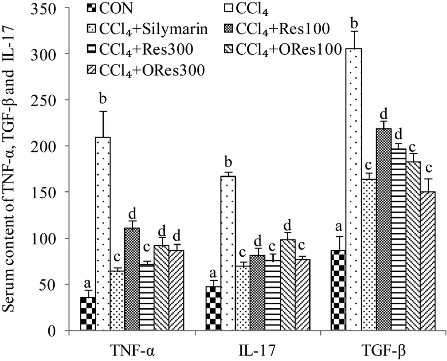
Fig.3.TNF-α,IL-17 and TGF-β in serum was measured by ELISA assays(n=3,mean±SEM.Different letters(a–e)above the columns represent significant differences of the same indexes for different groups when P<0.01).
Furthermore,ORes and Res substantially reduced the mRNA expression levels of MMP-2 and MMP-9(Fig.4E),comparing a sharp increase in CCl4treated rats,demonstrating that ORes and Res are efficient inhibitors of HSCs activation.
Production of matrix metalloproteinases(MMPs)from activated HSCs are directly involved in the process of ECM degradation and accumulation.MMP-2 and MMP-9 excessive expression level marks an early signal response from the liver tissue to hepatic inflammation[12].The activity of MMP-2 and MMP-9 was increased in activated HSCs in rodent liver fibrosis models[28].The increased transcriptional levels of MMP-2 and MMP-9 in the liver tissue were significantly increased in the CCl4-treated group of rats as illustrated in qPCR results(Fig.4E).Co-treatment with ORes or Res significantly lowered the mRNA transcriptional levels of these two genes.Activated HSCs stimulates the up-regulation of fibrotic proteins such as desmin and α-SMA[29].As demonstrated from immunofluorescence and western blot clearly,the expression of desmin and α-SMA was upregulated significantly in the liver tissue of CCl4-treated group,but co-treatment with silymarin,ORes or Res decreased the expression levels remarkably.Therefore,ORes and Res effectively inhibited the activation of HSCs in CCl4-induced liver fibrosis.
3.5.ORes downregulated the phosphorylation level of Smad and ERK
The signaling pathway of TGF-β1/Smad/ERK is closely associated with the progression of liver fibrosis.High expression level of TGF-β1 leads to the phosphorylation of Smad and ERK proteins.We demonstrated that in CCl4-induced liver fibrosis,increased expression of TGF-β activated the phosphorylation of ERK1/2 during the process of liver fibrogenesis[30].We therefore further investigated whether ORes alleviating hepatic fibrosis is associated with the phosphorylation of ERK and Smad.As illustrated in Fig.5A,western blot results demonstrated that after CCl4treatment,the relative expression values of TGF-β, p-ERK1/ERK1,p-ERK2/ERK2,p-Smad1/Smad1,p-Smad2/Smad2 proteins in liver tissues increased by 3.70-,3.87-,2.30-,7.57-,and 6.86-folds comparing to those in CON group(Fig.5A,B).Delightfully,co-administration with ORes or Res significantly down-regulated TGF-β expression and decreased the phosphorylation levels of Smad1/2 and ERK1/2 in liver tissues compared with those in CCl4-treated rats.The expression levels of these proteins were significantly attenuated after treatment with ORes or Res,revealing that ORes and Res can reduce the formation of collagen and alleviate the accumulation of ECM in liver tissue through the TGF-β1/Smad/ERK pathway,thus mitigating the degree of liver fibrosis.It has been demonstrated by many studies[31,32] that silymarin, a major bioactive component from milk thistle seeds, has strong preventive effects of liver fibrosis and has been used as a treatment control in majority of the liver disease related studies.In this study,we compared ORes and Res with silymarin in preventing liver fibrosis.The experimental results showed that 300mg/kg ORes or Res had considerable vitality compared with silymarin of 200mg/kg in terms of prevention of ROS formation and inhibitory effects on inflammatory signaling pathway,and there was no significant difference among them.

Fig.4.Stilbenes attenuates the activation of HSCs in liver:the relative expression level of α-SMA was determined by western blot against β-actin(A,B),desmin was detected by immunofluorescence(C)and then qualified by the fluorescent intensity in every field(D),and relative expressions of hepatic MMP-2 and MMP-9 mRNA levels were quantified by Real-Time PCR(E).Values are expressed as mean±SEM,n=3.The scale bar represents 200μm.The unit of dosage is mg/kg bw.Different letters(a–d)above the columns represent significant differences of the same indexes for different groups when P<0.01.
4.Conclusion
In summary,oxyresveratrol has demonstrated the biological effects of reducing liver oxidative damage and alleviating fibrosis induced by CCl4.It is attested that ORes has a stronger reducing effects against oxidative stress than Res.ORes achieved the preventive effect of liver fibrosis by suppression of excessive expression of TGF-β1,p-Smad1/2 and p-ERK1/2,activation of HSCs and accumulation of collagen in liver tissues.Hence we can conclude that dietary consumption of fruits or food products containing oxyresveratrol,mulberry in particular,has preventive beneficial effects against liver fibrosis.However,human study is warranted in the near future to have a comprehensive understanding of the cellular and molecular mechanisms of oxyresveratrol reversing the form of liver fibrosis and providing guidance of food products using oxyresveratrol as a bioactive marker.

Fig.5.The expression levels of TGF-β,p-Smad 1/2,Smad1/2,p-ERK1/2 and ERK1/2in liver were detected by(A)western blot and(B)relative to GAPDH in three independent experiments.The unit of dosage is mg/kg bw.Different letters(a–d)above the columns represent significant differences of the same indexes for different groups when P<0.01.
Declaration of Competing Interest
On behalf of all authors,we declare that there is no conflict of interests.
Acknowledgement
Grant from Hubei Province,China (GRANT number 2019ABA100)
- 食品科学与人类健康(英文)的其它文章
- Effect of xanthan gum on the quality of low sodium salted beef and property of myofibril proteins
- Peptide fraction from sturgeon muscle by pepsin hydrolysis exerts anti-inflammatory effects in LPS-stimulated RAW264.7 macrophages via MAPK and NF-κB pathways
- Chronic consumption of thermally processed palm oil or canola oil modified gut micro flora of rats
- Hypoglycemic polysaccharides from Auricularia auricula and Auricularia polytricha inhibit oxidative stress,NF-κB signaling and proinflammatory cytokine production in streptozotocin-induced diabetic mice
- Simultaneous determination of 15 pesticide residues in Chinese cabbage and cucumber by liquid chromatography-tandem mass spectrometry utilizing online turbulent flow chromatography
- Characterization of spoilage bacterial communities in chilled duck meat treated by kojic acid