“非压即黑”眼底多模式影像展示报告
2021-04-07 08:41:04林鹏耀
中华眼视光学与视觉科学杂志 2021年3期
林鹏耀
作者单位:宁波市第一医院眼科 315010
病例1:患者,男,34 岁,因“右眼视网膜脱离玻璃体切割术后1年复查”于2019年5月8日来宁波市第一医院就诊,发现眼底异常改变,无不适症状。专科检查:双眼视力1.0,眼压正常,双眼前节无异常,左眼底无异常改变。右眼底见中周部视网膜呈斑片状且颜色加深,与正常视网膜有明显分界线,透见脉络膜血管,炫彩照可见相应改变区呈暗红色斑片状(见图1)。炫彩光学相干断层扫描(OCT)可见暗红色区域的椭圆体带呈低反射信号(见图2)。自发荧光呈低荧光改变,眼底荧光血管造影(FFA)联合吲哚菁绿血管造影(Indocyanine green angiography,ICGA)晚期未见异常荧光渗漏(见图3─4)。30-2中央视野见右眼生理盲点扩大,60-4周边视野见右眼鼻下方周边暗点(对应颞上方原视网膜裂孔);双眼视网膜电图(ERG)检查示:右眼与正常左眼相比无明显改变(见图5)。随访半年,眼底无明显改变(见图6)。
病例2:患者,女,28岁,“左眼结膜炎”于2019年7月7日来宁波市第一医院就诊,发现眼底异常改变,无不适症状。专科检查:双眼视力1.0,眼压正常,双眼前节无异常,右眼底无异常改变。左眼底见中周部视网膜呈斑片状且颜色加深,与正常视网膜无明显分界线(见图7)。炫彩照可见相应改变区呈暗红色斑片状(见图7)。炫彩OCT可见暗红色区椭圆体带低反射信号(见图8)。半年后电话随访,患者无视力下降主诉。
讨论:
“非压即黑”首次报道于1975年[1],表现为发生于外层视网膜的无症状的眼底病变,多为眼底检查时无意中发现。组织病理学改变不清。近几年随着多模式影像检查设备的普及,这类病变越来越多被发现和认识[2],OCT检查发现这类病变均位于视网膜外层,表现为椭圆体带呈低反射信号。炫彩成像采用不同波段激光扫描成像,一次扫描同时获得基于488 nm蓝光反射、515 nm绿光反射、820 nm红外光反射成像,合成炫彩图像,分别呈现玻璃体视网膜交界面、视网膜血管层、视网膜深层及脉络膜结构。因此较传统眼底彩照更能清晰显示病变范围及位置,已经在中心性浆液性脉络膜视网膜病变、糖尿病视网膜病变、黄斑前膜等疾病的检查中体现出明显的优势[3-5]。由于“非压即黑”主要表现为视网膜外层椭圆体带的异常信号,所以在炫彩的绿光发射成像较红光反射和蓝光反射成像更清晰(见图9)。因这种眼底改变对患者视功能无影响,故定期随访观察。
利益冲突申明本研究无任何利益冲突
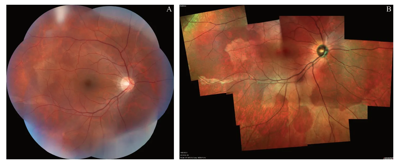
图1.病例1患者右眼底彩照及炫彩图A:彩照显示中周部视网膜呈斑片状且颜色加深;B:炫彩照显示相应改变区和正常视网膜有明显分界线Figure 1.Color photograph and multicolor image of the right eye of case 1 patient.A:Color photograph shows the dark lesion without pressure lesions on the midperipheral retina.B:Multicolor image shows sharply demarcated borders between the dark lesion and the normal retina.
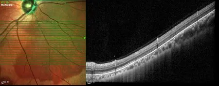
图2.病例1患者炫彩OCT可见正常椭圆体带高反射信号过渡到暗红色区域椭圆体带低反射信号Figure 2.Multicolor OCT image of case 1 patient.The OCT image shows an abrupt transition of the normal ellipsoid zone from reflectivity to hyporeflectivity photoreceptors within the lesion.
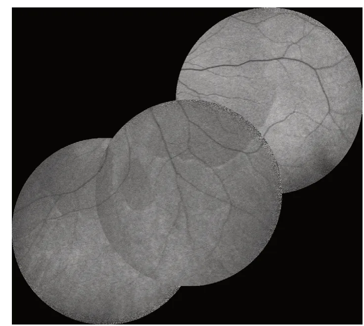
图3.病例1 患者自发荧光图显示病变区低自发荧光Figure 3.Fundus autofluorescence of case 1 patient shows hypoautofluorescence of the lesion.
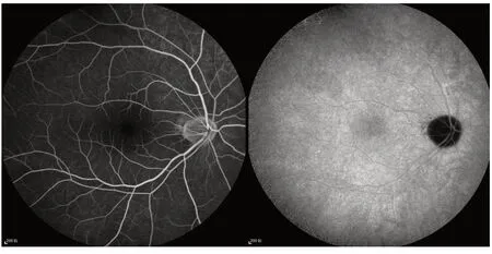
图4.病例1患者FFA和ICGA显示晚期未见明显异常荧光Figure 4.FFA and ICGA of case 1 patient show no obvious abnormal fluorescence in the late stage.
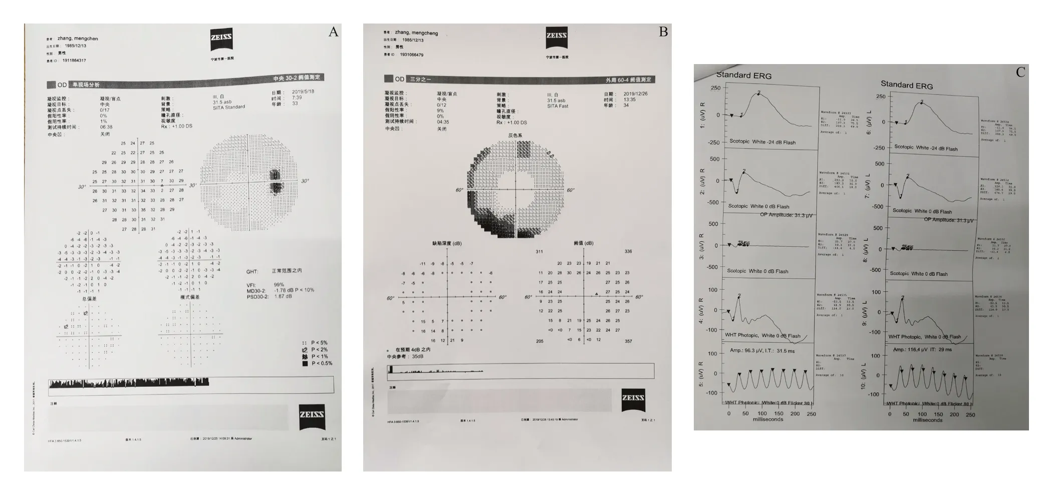
图5.病例1患者右眼视野及双眼视网膜电图检查A:右眼30-2视野图,提示右眼生理盲点扩大;B:右眼60-4视野图,提示右眼鼻下方周边暗点(对应颞上方原视网膜裂孔);C:双眼ERG图,右眼与正常左眼无明显改变Figure 5.Visual field examination of the right eye and electroretinogram of both eyes of case 1 patient.A:30-2 visual field examination shows enlargement of the blindspot.B:60-4 visual field examination shows superonasal scotoma (corresponding to the primary retinal hole in the superotemporal retina).C:ERG shows no significant change between the right eye and the normal left eye.ERG,electroretinogram.
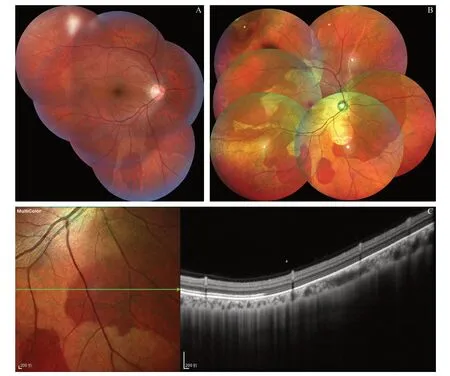
图6.病例1患者半年后眼底检查图A:眼底彩照;B:眼底炫彩照;C:眼底炫彩OCTFigure 6.Fundus examination of case 1 patient after six months.A:Color photograph.B:Multicolor image.C:Multicolor OCT.Results shows no changes over 6 months.
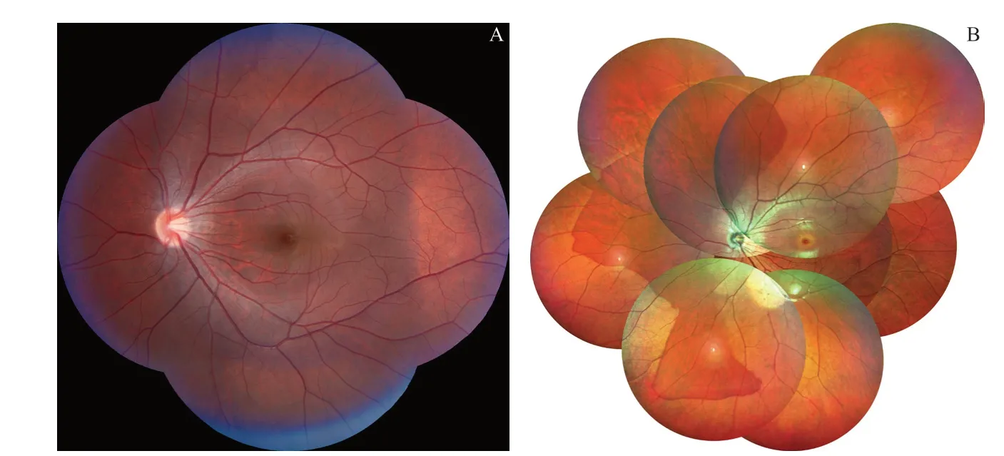
图7.病例2左眼底彩照及炫彩图A:彩照显示中周部视网膜呈斑片状且颜色加深;B:炫彩照显示相应改变区和正常视网膜有明显分界线Figure 7.Color photograph and multicolor image of the left eye of case 2 patient.A:Color photograph shows the dark lesion without pressure lesions in the midperipheral retina;B:Multicolor image shows sharply demarcated borders between the dark lesion and the normal retina.

图8.病例2炫彩OCT可见暗红色区域椭圆体带低反射信号过渡到正常椭圆体带信号Figure 8.Multicolor OCT of case 2 patient.The OCT shows hyporeflectivity of the ellipsoid zone of photoreceptors within the lesion that abruptly transitions to normal reflectivity in the surrounding normal retina.

图9.病例2非压即黑炫彩成像图A:标准炫彩像;B:红外反射成像;C:绿反射成像;D:蓝反射成像Figure 9.The dark lesion without pressure lesions on the multicolor image of case 2 patient.A:Standard multicolor image.B:Infrared reflectance image.C:Green reflectance image.D:Blue reflectance image.
猜你喜欢
小猕猴智力画刊(2024年5期)2024-06-10 04:23:03
大自然探索(2023年5期)2023-06-19 08:08:53
老友(2021年1期)2021-02-21 08:17:19
阅读与作文(小学高年级版)(2019年2期)2019-03-27 00:51:22
四川冶金(2018年1期)2018-09-25 02:39:22
初中生天地(2016年17期)2016-06-29 02:15:19
初中生学习·低(2016年11期)2016-05-30 04:50:12
作文新天地(初中版)(2016年3期)2016-03-18 09:12:04
作文新天地(2016年15期)2016-02-11 07:27:09
中国氯碱(2014年12期)2014-02-28 01:05:12