
A Case of Digital Superficial Angiomyxoma
2020-12-22 14:02:14GangLILianshengZHONG
Gang LI ,Liansheng ZHONG
1 Department of Dermatology,Suining People’s Hospital,Xuzhou,Jiangsu 221200,China.
2 Department of Dermatology,Children’s Hospital of Fudan University Xiamen Branch,Xiamen,Fujian 361006,China.
ABSTRACT Superficial angiomyxoma (SA) is a rare superficial benign tumor of the skin that is characterized by slow growth.It is painless,non-invasive,and prone to relapse.It commonly occurs on the head,neck,trunk,and limbs and in the genital region.Herein,we describe a case of a 33-year-old man who presented with a soft tender tumor measuring 1.5 × 2.0 cm on the lateral aspect of the left second finger.The histopathology of the tumor indicated that it was a lobulated tumor rich in myxoid stroma and parenchymal blood vessels in the dermis,with spindle-to-stellate tumor cells in higher-power view and no mitotic or atypical figures.Immunohistochemical studies revealed that the tumor cells were positive for CD34 and negative for S100.Based on these clinical and histological findings,the patient was diagnosed with SA.
KEY WORDS Superficial angiomyxoma; Digital; Myxoid tumors
INTRODUCTION
Superficial angiomyxoma (SA) is a rare superficial benign tumor of the skin characterized by slow growth; it is also painless,non-invasive,and prone to relapse.It is usually seen in the head,neck,trunk,and limbs,and it can also occur in the genital region[1].Herein,we describe a case of SA on the digit and review the literature about this condition.To our knowledge,this is the eighth case of SA occurring in the digital region,which might thus make this area another common site for SA.
CASE PRESENTATION
A 33-year-old man presented with a red tumor on the left second finger for 14 months.The mass grew slowly and became mildly tender 2 months ago.There were no other lesions on the skin,and no recent trauma of the affected fingernail was reported.On examination,there was a soft tender tumor measuring 1.5 × 2.0 cm on the lateral aspect of the left second finger,with some scales on the surface of the lesion (Fig.1A and 1B).Radiographic examination of the digit demonstrated soft tissue swelling without destruction of the distal phalanx(Fig.1F).Histological studies revealed a lobulated tumor rich in myxoid stroma and parenchymal blood vessels in the dermis,with spindle-to-stellate tumor cells in higherpower view,but there were no mitotic or atypical figures(Fig.1C and 1D).Immunohistochemical studies revealed that the tumor cells were positive for CD34 (Fig.1E)and negative for S100 (Fig.1C).Thus,a diagnosis of SA was established.The lesion was excised under local anesthesia,with no evidence of recurrence at the 1-year follow-up.
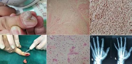
Fig.1 Physical examination revealed a soft tender tumor measuring 1.5 × 2.0 cm on the lateral aspect of the left second finger,with some scales on the surface of the lesion (A and B)
DISCUSSION
SA can occur at any age but peaks in the third to fourth decade of life.Most cases present as solitary cutaneous nodules,papules,or polypoid lesions,usually less than 5 cm in diameter.Histological findings include infiltration of the dermis,and often the subcutis,by a poorly circumscribed tumor with a multilobular growth pattern composed of spindle-shaped to stellate cells set in a copious amount of myxoid stroma.These cells generally show no nuclear atypia or hyperchromasia.Small parenchymal blood vessels are prominent.Immunohistochemically,the tumor cells are usually positive for vimentin and CD34 and negative for S-100,pan-keratin,and smooth-muscle actin.
SA can arise from anywhere in the superficial tissues,but mostly,it has a predilection for the head,neck,trunk,limbs,and genital region.To our knowledge,there have been at least 6 published reports with 7 cases of digital SA.Falidas et al.described the fourth case of digital SA and reviewed the other three cases in 2011[2].The fifth digital SA,which occurred on the index finger,was reported in 2007[3].Bhat et al.reported a case of SA on the thumb mimicking a malignant bone tumor in 2014[4].In 2014,Kura reported the seventh SA on the fourth toenail[5],which was the eighth case of digital SA.Our findings and review of the literature demonstrate that the digital region might be another common site for SA,and this condition should therefore be included in the differential diagnoses of digital tumors,especially myxoid tumors.
ETHICS DECLARATIONS
Ethics Approval and Consent to Participate
N/A as it is a case report.
Consent for Publication
All the authors have consented for the publication.
Competing Interests
The authors declare that they have no competing interests.The authors state that the views expressed in the submitted article are their own and not the official position of the institution or funder.
 Chinese Journal of Plastic and Reconstructive Surgery2020年3期
- Chinese Journal of Plastic and Reconstructive Surgery的其它文章
- Status Quo and Future Development of Female Genital Cosmetic Surgery (Intimate Surgery)
- The First Case of Free Radial Forearm Skin Flap:A 40-Year Follow-Up Study
- Meteorological Influence on Tissue Expander-Related Major Infection
- Surgical Management for Diabetic Foot Ulcer:A Bibliometric Study
- Programmed 6-Step Approach of Improved Liposuction-Curettage for Axillary Bromhidrosis
- Foreword from Professor Lee L.Q.Pu