
乳腺癌术后辅助化疗对循环肿瘤细胞影响的观察研究
2020-11-19 04:26孙晓莹马飞田朋飞李晓爽朱爱华王津京
中外医学研究 2020年26期
关键词:乳腺癌
孙晓莹 马飞 田朋飞 李晓爽 朱爱华 王津京
【摘要】 目的:本研究旨在观察乳腺癌术后辅助化疗后外周血循环肿瘤细胞阳性率及含量的变化,评价术后辅助化疗的有效性。方法:选取2015年11月-2016年8月本院收治的10例拟行术后辅助化疗的乳腺癌患者,分别于辅助化疗前后采集外周静脉血3~5 ml,通过免疫磁珠法及免疫荧光显色法检测样品中循环肿瘤细胞,对比分析化疗前后CTCs的检出率、含量及变化趋势。结果:术后辅助化疗前后CTCs的检出率分别为80%(8/10)、60%(6/10),平均CTCs含量分别为2.1(1.5,3.25)ml和0.7(0.0,1.0)ml,辅助化疗后外周静脉血中CTCs的含量较化疗前有减少趋势,差异有统计学意义(P=0.026)。结论:乳腺癌术后辅助化疗可减少患者外周血CTCs的含量。乳腺癌患者术后辅助化疗前后外周血CTCs含量检测结果对预测、评估近期治疗疗效重要指导价值。
【关键词】 乳腺癌 循环肿瘤细胞 辅助化疗
doi:10.14033/j.cnki.cfmr.2020.26.005 文献标识码 A 文章编号 1674-6805(2020)26-00-04
The Value of Adjuvant Chemotherapy for Postoperative Breast Cancer on Circulating Tumor Cells/SUN Xiaoying, MA Fei, TIAN Pengfei, LI Xiaoshuang, ZHU Aihua, WANG Jinjing. //Chinese and Foreign Medical Research, 2020, 18(26): -17
[Abstract] Objective: To analyse the changes in the content of circulating tumor cells in peripheral blood after adjuvant chemotherapy for postoperative breast cancer and to evaluate the effectiveness of chemotherapy. Method: Ten cases of postoperative breast cancer patients accepted adjuvant chemotherapy was collected from November 2015 to August 2016 in our hospital. Peripheral venous blood was taken before and after adjuvant chemotherapy. Contents of circulating tumor cells in the sample was detected by immunofluorescent staining. The detection rate and change trend of CTCs before and after chemotherapy were compared and analyzed. Result: Rates of CTCs detection before and after adjuvant chemotherapy were 80% (8/10) and 60% (6/10), and the average numbers of CTCs were 2.1 (1.5, 3.25) ml and 0.7 (0.0, 1.0) ml respectively. The contents of CTCs detected in peripheral venous blood after adjuvant chemotherapy decreased compared with that before chemotherapy, and the difference was statistically significant (P=0.026). Conclusion: Adjuvant chemotherapy can reduce the number of CTCs in peripheral blood of patients with postoperative breast cancer. Meanwhile content numbers of peripheral blood CTCs before and after adjuvant chemotherapy in breast cancer patients are valuable in predicting and evaluating adjuvant therapeutic efficacy.
[Key words] Breast cancer Circulating tumor cells Adjuvant chemotherapy
First-authors address: Beijing Chaoyang District Huanxing Cancer Hospital, Beijing 100021, China
乳腺癌是發病率最高的女性恶性肿瘤,随着手术、化疗、放疗等治疗手段的综合应用,其治愈率有了很大的提高,但仍有20%~30%乳腺癌会出现复发和转移,最终导致患者死亡[1-2]。肿瘤细胞可直接或经淋巴途径间接进入血液系统,进而在远处组织器官定植形成转移灶,这部分来自肿瘤原发灶或转移灶播散进入血液的肿瘤细胞被称为循环肿瘤细胞(circulating tumor cells,CTCs)。CTCs的出现被认为可能是肿瘤远处转移的一种标志,与乳腺癌临床预后有密切关系[3-6]。而且,CTCs的检测作为液体活检的一种重要方式,具有创伤小、可连续检测等优势,促进了相关领域的快速发展。目前诸多关于CTCs的研究更着重于癌症的早诊早筛、评估预后、早期发现肿瘤转移及指导晚期肿瘤个体化治疗等。但对于早期手术及辅助治疗的乳腺癌患者,缺乏评价辅助治疗效果的肿瘤标记物。本研究通过对乳腺癌患者术后辅助化疗前后血液中循环肿瘤细胞含量的检测,探讨CTCs动态变化与辅助化疗效果的关系及对乳腺癌患者的临床意义。
1 资料与方法
1.1 一般资料
选择2015年11月-2016年8月中国医学科学院肿瘤医院桓兴病区肿瘤内科收治的乳腺癌患者10例,均已接受手术治疗,拟行术后辅助化疗。纳入标准:(1)首次行手术切除治疗,经组织病理学检查确诊为浸润性乳腺癌;(2)临床病理资料完整;(3)未接受任何辅助治疗;(4)HER2基因均为阴性。排除标准:(1)已发生远处转移;(2)妊娠期;(3)合并其他系统恶性肿瘤;(4)合并严重心肺疾病;(5)依从性差,不能按方案辅助化疗。患者年龄33~52岁,平均41.1岁。本研究经医院医学伦理委员会审批通过,所有患者均对本研究知情并签署知情同意书。
1.2 方法
本研究患者接受表柔比星+环磷酰胺,序贯紫杉醇的密集化疗方案(1例多西他赛+卡铂化疗方案化疗)。术后肿瘤组织雌激素受体(ER)、孕激素受体(PR)及Ki-67的表达情况采用免疫组化SP法检测;人表皮生长因子受体2(HER2)采用免疫组织化学法(IHC)及原位荧光杂交技术(FISH)检测。根据美国临床肿瘤学会和美国病理学家学会公布的乳腺癌指南评估ER/PR/HER2表达状况。按照美国癌症联合会(American Joint Commission on Cancer,AJCC)2018年第8版乳腺癌TNM分期标准进行分期。
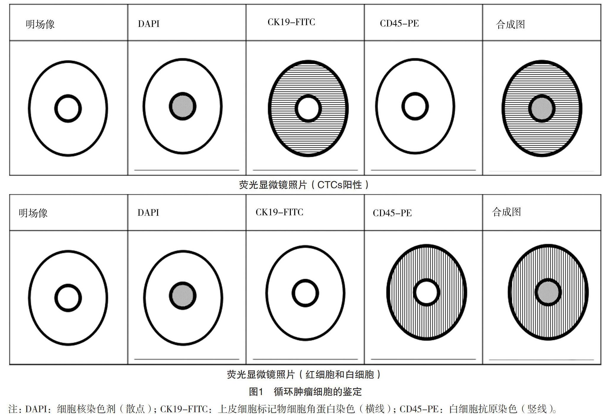
使用EDTA抗凝采血管分别于辅助化疗前后采集患者3~5 ml外周血,留取2 ml有效血液样本。本检测针对外周血液中循环肿瘤细胞(CTC)测试流程如下:将特异性识别CTC表面的上皮细胞黏附分子(EpCAM)的多肽修饰的磁性纳米颗粒与血液样品混合,利用磁力捕获血液中的CTC,用4%的多聚甲醛室温下固定细胞,然后通过免疫荧光染色方法对捕获的细胞进行鉴定。免疫荧光染色为三色标记法,分别为细胞核染色剂(DAPI)、上皮细胞标记物细胞角蛋白(CK)的抗体,以及白细胞抗原(CD45)的抗体。
DAPI可透过细胞膜对DNA染色,它在嵌入双链DNA后发出蓝色荧光;异硫氰酸荧光素(FITC)标记的CK抗体与上皮来源的肿瘤细胞表面的CK抗原结合,发出525 nm波长的绿色荧光;藻红蛋白(PE)标记的人CD45抗体与血液中白细胞表面的CD45抗原结合,发出578 nm波长的红色荧光。于多肽修饰的磁性纳米颗粒富集到的所有细胞进行三色染色,其中DAPI+/CK+/CD45-并且符合细胞形态的细胞指认为循环肿瘤细胞,见图1。
1.3 观察指标
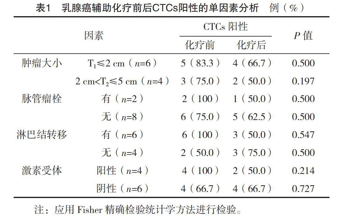
统计辅助化疗前后两组患者CTCs的阳性率(以不同CTCs组相应CTCs≥1个为阳性)及各组患者辅助化疗前后CTCs(DAPI+/CK+/CD45-)阳性值,来表示外周血肿瘤细胞含量。通过检测患者外周血CTCs,分别比较化疗前后CTCs阳性率与肿瘤大小、脉管瘤栓、淋巴结转移情况、激素受体的差异,分析CTCs阳性与临床病理特征的相关性。
1.4 统计学处理
應用SPSS 25.0统计学软件对数据进行处理。应用偏度和峰度检验对化疗前后外周血CTCs含量进行正态分布检验,在α=0.05的检验水平下,本研究数据不符合正态分布,以M(P25,P75)表示,采用非参数Wilcoxon秩和检验;计数资料以率(%)表示,组间比较采用字2检验,在适当的情况下采用Fisher精确检验进行研究。P<0.05为差异有统计学意义。
2 结果
2.1 术后辅助化疗前后CTCs阳性的单因素分析
乳腺癌患者辅助化疗前后外周血CTCs与其病理特征差异无统计学意义(P>0.05),乳腺癌辅助化疗前后外周血CTCs含量与病理特征无明确相关性,见表1。
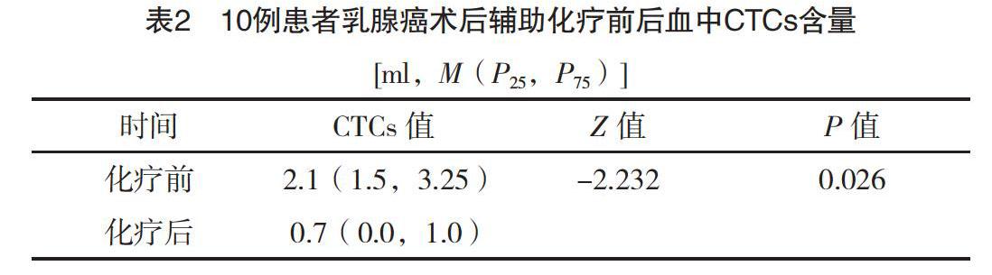
2.2 辅助化疗前后患者CTCs含量比较
近些年有关CTCs研究纳入的病例数从十几例到几百例不等[7-9]。本研究辅助化疗前循环肿瘤细胞阳性率(CTCs≥1个)为80%(8/10),平均循环肿瘤细胞含量为2.1(1.5,3.25)ml;辅助化疗后循环肿瘤细胞阳性率为60%(6/10),平均循环肿瘤细胞含量为0.7(0.0,1.0)ml,明显低于化疗前,差异有统计学意义(P<0.05),见表2。
3 讨论
乳腺癌是一种全身性疾病,30%~40%的初治乳腺癌患者骨髓、静脉血和常规病理检查区域淋巴结存在着微转移,提示微转移是导致乳腺癌患者术后复发、转移的重要原因[10]。经手术切除后的乳腺癌患者血液中仍可检测到循环肿瘤细胞,可能手术会增加肿瘤细胞脱落进入血液的风险[9,11-12],本研究中80%患者手术切除肿瘤后辅助化疗前检测血液循环中检测到CTCs,辅助化疗后CTCs的检出率降低至60%。比较患者术后辅助化疗前后血中CTCs数目,差异有统计学意义(P=0.026)。
血中循环肿瘤细胞一般是指进入机体循环血液中的实体肿瘤细胞,能够真实反映肿瘤负荷和具有发展为转移灶的潜力。CTCs在确定诊断、治疗决策和监测治疗效果中有很重要的作用。研究表明,CTCs的存在对于乳腺癌远处转移、生存都是一个独立的预后指标[13-15]。CTCs实时检测已被广泛用于评价乳腺癌的新辅助化疗、放疗、维持化疗等治疗手段的有效性,除乳腺癌外,其应用价值同样在结直肠癌、前列腺癌及非小细胞肺癌等肿瘤中被证实[16-18]。本研究中观察到术后辅助化疗可减少外周血CTCs检出率和含量,提示了化疗的有效性。
与此同时,有研究认为CTCs作为对化疗反应的预测,其表型也常随药物治疗、疾病进程等变化[19-20]。CTCs是一类异质性很强的肿瘤细胞,其中一些为肿瘤干细胞(cancer stem cells,CSCs),另一些被认为是上皮间变细胞(epithelial-mesenchymal transition,EMT),但大多数肿瘤细胞不具有上述特征,相关研究认为CSCs和EMT状态的肿瘤细胞对化疗具有抵抗性,是肿瘤复发及转移的原因[21-22]。因此,CTCs的检测需要根据肿瘤细胞生物特性进行分类,从而更精准高效的预测、评估肿瘤治疗疗效及预后。
綜上所述,本研究结果显示通过术后辅助化疗,患者外周血CTCs检出率和单位体积的含量减少,初步提示CTCs状态的变化与化疗效果有一定的关系。限于本研究纳入研究对象少,因此CTCs在术后辅助化疗效果评估中的作用价值仍需大样本、多中心临床研究进一步验证。作为一种新的实时便捷、创伤小的检测手段,CTCs检测具有很大的潜力来改变目前癌症检测的现状。然而,CTCs的临床应用还处于起步阶段,存在着诸多问题亟待解决。通过对循环肿瘤细胞生物特性进一步的分类研究和完善CTCs检测方法,最终使肿瘤患者获益。
参考文献
[1] DeSantis C E,Ma J,Gaudet M M,et al.Breast cancer statistics[J].CA Cancer J Clin,2019,69(6):438-451.
[2] Wang P P,Liu S H,Chen C T,et al.Circulating tumor cells as a new predictive and prognostic factor in patients with small cell lung cancer[J].J Cancer,2020,11(8):2113-2122.
[3]马齐襄,朱晓丹,胡凯文,等.肿瘤转移的种子与土壤学说新认识[J].肿瘤防治研究,2015,42(10):1049-1053.
[4] Maman S,Edry-Botzer L,Sagi-Assif O,et al.The metastatic microenvironment:lung-derived factors control the viability of neuroblastoma lung metastasis[J].Int J Cancer,2013,133(10):2296-2306.
[5] Wang C H, Chang C J,Yeh K Y,et al.The Prognostic Value of HER2-Positive Circulating Tumor Cells in Breast Cancer Patients:A Systematic Review and Meta-Analysis[J].Clin Breast Cancer,2017,17(5):341-349.
[6] Lv Q, Gong L,Zhang T,et al.Prognostic value of circulating tumor cells in metastatic breast cancer: a systemic review and meta-analysis[J].Clin Transl Oncol,2016,18(3):322-330.
[7] Monterisi S,Castello A,Toschi L,et al.Preliminary data on circulating tumor cells in metastatic NSCLC patients candidate to immunotherapy[J].Am J Nucl Med Mol Imaging,2019,9(6):282-295.
[8] Brown J C,Rhim A D,Manning S L,et al.Effects of exercise on circulating tumor cells among patients with resected stage Ⅰ-Ⅲ colon cancer[J].Plos One,2018,13(10):e0204875.
[9] Hall C,Valad L,Lucci A.Circulating Tumor Cells in Breast Cancer Patients[J].Crit Rev Oncog,2016,21(1):125-139.
[10] Zhang Y, Lv Y, Niu Y, et al. Role of Circulating Tumor Cell (CTC) Monitoring in Evaluating Prognosis of Triple-Negative Breast Cancer Patients in China[J].Med Sci Monit,2017,23(23):3071-3079.
[11] Li S,Yan W, Yang X,et al.Less micrometastatic risk related to circulating tumor cells after endoscopic breast cancer surgery compared to open surgery[J].BMC Cancer,2019,19(1):1070.
[12] Lozar T, Gersak K, Cemazar M,et al.The biology and clinical potential of circulating tumor cells[J].Radiol Oncol,2019,53(2):131-147.
[13] Park H S, Han H J,Lee S,et al.Detection of Circulating Tumor Cells in Breast Cancer Patients Using Cytokeratin-19 Real-Time RT-PCR[J].Yonsei Med J,2017,58(1):19-26.
[14] Wang C H,Chang C J,Yeh K Y,et al.The Prognostic Value of HER2-Positive Circulating Tumor Cells in Breast Cancer Patients: A Systematic Review and Meta-Analysis[J].Clin Breast Cancer,2017,17(5):341–349.
[15] Wu Z X,Liu Z,Jiang H L,et al.Circulating tumor cells predict survival benefit from chemotherapy in patients with lung cancer[J].Oncotarget,2016,7(41):67586-67596.
[16] Obayashi K,Akatsuka J,Endo Y,et al.Initial detection of circulating tumor cells from metastatic prostate cancer patients with a novel small device[J].Prostate Int,2019,7(4):131-138.
[17] Wang D,Yang Y,Jin L,et al.Prognostic models based on postoperative circulating tumor cells can predict poor tumor recurrence-free survival in patients with stage Ⅱ-Ⅲ colorectal cancer[J].J Cancer,2019,10(19):4552-4563.
[18] Yamauchi Y,Safi S,Blattner C,et al.Circulating and Tumor Myeloid-derived Suppressor Cells in Resectable Non-Small Cell Lung Cancer[J].Am J Respir Crit Care Med,2018,198(6):777-787.
[19] Kraan J,Sleijfer S,Foekens J A,et al.Clinical value of circulating endothelial cell detection in oncology[J].Drug Discov Today,2012,17(13):710-717.
[20] Danova M,Comolli G,Manzoni M,et al.Flow cytometric analysis of circulating endothelial cells and endothelial progenitors for clinical purposes in oncology: A critical evaluation[J].Mol Clin Oncol,2016,4(6):909-917.
[21] Cheng Y H,Chen Y C,Lin E,et al.Hydro-Seq enables contamination-free high-throughput single-cell RNA-sequencing for circulating tumor cells[J].Nat Commun,2019,10(1):2163.
[22] Lin E,Rivera-Báez L,Fouladdel S,et al.High-Throughput Microfluidic Labyrinth for the Label-free Isolation of Circulating Tumor Cells[J].Cell Syst,2017,5(3):295-304.
(收稿日期:2020-06-08) (本文編辑:何玉勤)
猜你喜欢
中国典型病例大全(2022年7期)2022-04-22
中国药学药品知识仓库(2022年1期)2022-03-23
恋爱婚姻家庭·养生版(2021年3期)2021-05-08
婚育与健康(2019年11期)2019-12-09
祝您健康(2018年5期)2018-05-16
祝您健康·文摘版(2018年1期)2018-05-14
女士(2017年11期)2017-12-12
健康博览(2017年8期)2017-12-05
妇女生活(2017年2期)2017-02-15
恋爱婚姻家庭·青春(2016年8期)2016-08-23
