Ependymal cells and multiple sclerosis: proposing a relationship
Multiple sclerosis (MS) currently affects ~2.5 million people worldwide. MS is typically diagnosed in young adults and is usually not fatal, meaning people live long lives with MS. Affected individuals usually suffer from progressive physical and/or cognitive disability,often including fatigue (89.6%), depression (53.9%), memory loss(49.0%), motor or sensory dysfunction (76.4%, 70.4%) and urinary incontinence (50.8%). This disability weighs on patients, loved ones and caretakers, and costs the economy billions of dollars each year.
MS is a chronic, progressive, demyelinating neurodegenerative disease driven by an aberrant immune system that affects the brain and spinal cord. Neuronal loss and brain atrophy, apparent even during early disease, is thought to be the major mechanism of irreversible physical and cognitive disability in MS. There remains a lack of effective treatments targeting these processes, largely due to an absence of understanding what drives neurodegeneration.What causes neurodegeneration in MS? The process of widespread progressive neurodegeneration requires more than the formation of acute demyelinating lesions driven by an autoimmune attack. Others have proposed that age, iron accumulation, chronic microglial activation, mitochondrial dysfunction and glutamate excitotoxicity(all well-characterized mechanisms that lead to neuronal damage)could be contributing factors leading to neuronal cell death in MS.Recently, it has also been suggested that there could be a direct autoimmune attack against neurons.
But let us take a step back for a moment, and understand the topographic distribution of pathology in MS. Focal lesions of active demyelination can be visualized on MRI during an MS relapse and appear throughout the spinal cord and/or brain as localized enhancing lesions that are usually associated with blood vessels.Interestingly, one other common zone of demyelination is near the periventricular region located in deep grey and white matter (Haider et al., 2014). These lesions usually directly abut the ventricles and typically follow their perimeter adjacent to the cerebrospinal fluid(CSF). In the most advanced stages of MS, striking patterns of superficial cortical lesions become widespread, where the perimeter of cortical grey matter lining the CSF-rich subarachnoid space is affected (Haider et al., 2014).
Regions of lesion formation do not have a bias for white matter or grey matter, rather their biases lie in their proximity to the bloodbrain barrier (BBB) or blood-cerebral spinal fluid barrier (BCSFB),and over time, adjacent to the CSF-rich subarachnoid space. Given that the mode of immune cell entry into the central nervous system(CNS) is via the BBB and BCSFB, these regions of lesion formation come as no surprise. It is thought that initiating mechanisms involves the infiltration of myelin-reactive T lymphocytes. At these regions of inflammation, glial cell and neurons are affected. Nearby oligodendrocytes and neurons become damaged and die. Microglia and astrocytes also become reactive contributing to disease processes. Microglia contribute to oxidative stress via oxidative burst,which is considered a major mode of pathology in MS. Astrocytes,meanwhile, assume distinctive hypertrophic morphologies, and span from active sites of demyelination to normal appearing brain matter, and likely play an early and active role in MS. Other cells,such as oligodendrocyte progenitor cells and neural stem cells,are queued to contribute to regenerative events. Oligodendrocyte progenitor cells are recruited to sites of demyelination, where they differentiate into remyelinating oligodendrocytes via a number of mechanisms. Neural stem cells possess a remarkable ability to specialize into neurons or glial cells, and have been considered targets for MS therapy. Brain endothelial cells lose efficacy as tight junction barrier cells, creating an avenue for entry of unwanted and neurotoxic substances into the CNS. Pericytes have also been implicated in MS pathology, with phenotypic changes corresponding to disease severity.Ependymal cells in MS:Ependymal cells are a type of glial cell that reside within the CNS whose role in MS is critically understudied.They are simple ciliated epithelial cells with radial glial origin, that line the entire ventricular surface of the CNS and the central canal of the spinal cord (Del Bigio, 2010). As such, ependymal cells are the most predominant cell type associated with CSF. Importantly,they are the only cell type that lie between the CSF and deep white and grey matter periventricular lesions in MS. Ependymal cells provide both an immunological barrier and a partial barrier that regulates the bidirectional transport of molecules between the ventricular CSF and interstitial fluid. Ependymal cells appear to play an integral role in clearance of toxic metabolites, nutrient sensing,and metabolic regulation within the brain. They contain a primary cilium that senses circulating molecules within the CSF and tufts of motile cilia that maintain laminar flow of CSF at the ventricle surface. Ependymal cells are also equipped with gap junctions, adherens junctions, and specialized transporters that enable selective transport of molecules (Del Bigio, 2010).
Researchers have provided evidence that suggests that ependymal cells are sensitive to inflammation and become pathological in MS (Nathoo et al., 2016; Lisanti et al., 2005; Schubert et al., 2019).Using MRI technology, Lisanti et al. (2005) discovered a unique pattern associated with the ependyma of MS patients, which they termed “the ependymal ‘Dot-Dash’ sign”. On fluid-attenuated inversion recovery images, they described a “Dot” as a round hyperintense irregularity of the ependymal undersurface with a diameter larger than the thickness of a “Dash” adjacent to it. The ‘Dot-Dash’sign is specific and sensitive to the detection of MS, especially in younger patients (Age < 50; specificity 71.9%, sensitivity 95.7%)(Lisanti et al., 2005). Our own work has suggested that these cells are particularly vulnerable to cell death; adult ependymal cells are not capable of surviving beyond several hours in vitro, even under supportive conditions (Shah et al., 2018). In cases of neurodegenerative diseases, including Alzheimer’s disease and MS, our preliminary work suggests that these cells are vulnerable to abnormal morphology in pathological brains. We found that large portions of the ependymal layer were lost (beyond normal aging); and many of the ependymal cells that remained no longer projected processes into the parenchyma (unpublished observations).
As mentioned, ependymal cells are involved in CSF circulation and these cells appear to become pathological in MS (Lisanti et al.,2005). Schubert et al. (2019) tracked CSF flow in individuals with MS, and found that the rate of CSF circulation in patients with MS was significantly decreased in comparison to healthy individuals.CSF is a major avenue for moving parenchymal byproducts, including cellular wastes that gather in the interstitial fluid, in preparation for excretion. CSF flows from the ventricular cavities, along the subarachnoid space and out of the brain and into draining lymph nodes. This flow occurs at high rates with the average person circulating CSF 2-3 times per 24 hours. Similar to MS, aging results in changes to the ependyma (Todd et al., 2018). But, unlike MS,ependymal cell dysfunction has been relatively well studied in aging. In aging, the ependymal layer thins significantly as astrocytes simultaneously form junctions with resident ependymal cells.Moreover, there are pronounced reductions in the density of motile cilia on the apical surface of ependymal cells and an accumulation of lipid. As a consequence, metabolic waste derived from the brain likely accumulates in the aged brain. Most importantly, ependymal cell damage and periventricular anomalies have been correlated with neurocognitive decline (Todd et al., 2018). Whether ependymal cell dysfunction causes cognitive decline that is observed in MS patients is yet to be studied (Chiaravalloti and DeLuca, 2008).In ependymal cell-associated gene knock-out studies, experimental animals often present with brain atrophy, neuroinflammation,periventricular demyelination and ventricular enlargement, which are features of MS pathology (Liu et al., 2014; Juurlink, 2015).Whether ependymal cell dysfunction contributes to any of these pathological features observed in MS patients has not been studied.
In MS, the early dominance of periventricular lesions, which abut the CSF, coupled with the eventual accumulation of lesions adjacent to the CSF in the subarachnoid space, makes it highly likely that the presence and progressive accumulation of CSF-associated factors contributes to disease. Given ependymal cells are the major cell type that interact with CSF, and are the major barrier cell type that lies between the CSF and periventricular MS lesions, it is highly probable that this cell is affected by CSF-associated factors in MS.There are several inflammatory cytokines and cells, as well as debris, that circulate in the CSF of MS patients; including proinflammatory factors such as, interferon-gamma (INFγ), Cxcl12, tumor necrosis factor, interleukin-2, and interleukin-22 (Magliozzi et al.,2018). Whether the accumulation of CSF-associated factors in MS is driven by ependymal cell dysfunction is unknown, and whether ependymal cell dysfunction directly contributes to MS brain pathogenesis and MS symptoms, or is a secondary consequence, remains unclear.
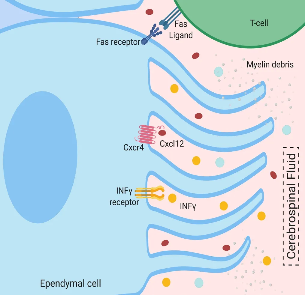
There are likely many mechanisms that drive ependymal cell dysfunction and death in pathological states (Figure 1). In particular,ependymal cells are sensitive to certain cytokines, including Cxcl12 and INFγ (Shah et al., 2018). Interestingly, INFγ has already been shown to be associated with aging and cognitive decline, associated with MS progression. Myelin fragments that enter the CSF of MS patients, likely also cause damage to ependymal cells (Laabich et al.,1991). Additionally, T-helper cells in MS likely interact directly with ependymal cells via Fas-FasL binding, and may also contribute to ependymal cell dysfunction or death (Shah et al., 2018).
Conclusion:If damage to ependymal cells, either directly or indirectly, cause dysfunction at any point within the intricate circulatory system of the brain, it is possible that the removal of cellular waste is inadequate and may cause an accumulation of toxic, damaging factors. Even though there is evidence that ependymal cells are vulnerable to dysfunction in disease, the study of ependymal cell biology in the context of MS and inflammation is critically understudied. Deciphering the precise role(s) of ependymal cells in MS will hopefully reveal new therapeutic targets to effectively treat individuals with this disease.

Figure 1 Proposed mechanisms of molecular interactions between CSF-associated factors and ependymal cells in MS.
Dale Hatrock#, Nina Caporicci-Dinucci#, Jo Anne Stratton*The Montreal Neurological Institute, McGill University, Montreal,Canada (Hatrock D, Caporicci-Dinucci N, Stratton JA)Integrated Program in Neuroscience, McGill University, Montreal,Canada (Hatrock D, Caporicci-Dinucci N)#Both authors contributed equally to this work.
*Correspondence to:Jo Anne Stratton, PhD, jo.stratton@mcgill.ca.
orcid:0000-0002-1205-1353 (Jo Anne Stratton)
Received:July 14, 2019
Accepted:August 3, 2019
doi:10.4103/1673-5374.265551
Copyright license agreement:The Copyright License Agreement has been signed by all authors before publication.
Plagiarism check:Checked twice by iThenticate.
Peer review:Externally peer reviewed.
Open access statement:This is an open access journal, and articles are distributed under the terms of the Creative Commons Attribution-NonCommercial-ShareAlike 4.0 License, which allows others to remix, tweak, and build upon the work non-commercially, as long as appropriate credit is given and the new creations are licensed under the identical terms.
Open peer reviewer:Tetsuya Akaishi, Tohoku University, Japan.
Additional file:Open peer review report 1.
- 中国神经再生研究(英文版)的其它文章
- Ethanol extract from Gynostemma pentaphyllum ameliorates dopaminergic neuronal cell death in transgenic mice expressing mutant A53T human alpha-synuclein
- Peripheral nerve injury induced changes in the spinal cord and strategies to counteract/enhance the changes to promote nerve regeneration
- Genetic targeting of astrocytes to combat neurodegenerative disease
- Pathological significance of tRNA-derived small RNAs in neurological disorders
- Applications of advanced signal processing and machine learning in the neonatal hypoxic-ischemic electroencephalography
- Protective effect of hydrogen sulfide on oxidative stress-induced neurodegenerative diseases

