Comprehensive treatment of rare multiple endocrine neoplasia type 1:A case report
Chen-Hui Ma,Huai-Bin Guo,Xin-Yan Pan,Wan-Xing Zhang,Department of Hepatobiliary,Hebei General Hospital,Shijiazhuang 050017,Hebei Province,China
Chen-Hui Ma,Graduate School of North China University of Science and Technology,Tangshan 050051,Hebei Province,China
Abstract
Key words:Multiple endocrine neoplasia type 1;Thymic carcinoid;Neuroendocrine tumor;Case report;Pancreas;Tumor
INTRODUCTION
Multiple endocrine neoplasia type 1 (MEN1),a rare hereditary disorder caused by mutations in the tumor suppressor geneMEN1,refers to two or more simultaneous or sequential occurrences of parathyroid adenomas,anterior pituitary adenomas,or entero-pancreatic neuroendocrine tumors (NETs) in an individual[1].Hormonesecreting and hormone-nonsecreting tumors may occur among some 30 tissues in MEN1[2].The most common clinical manifestations of MEN1 include parathyroid (>90%) tumors,followed by pancreatic endocrine tumors (30%-70%) and pituitary tumors (30%-40%)[3].MEN1 patients also present with other tumors,including adrenocortical tumor,carcinoids,pheochromocytoma,bronchopulmonary NET,thymic NET,gastric NET,lipomas,angiofibromas,collagenomas,and meningiomas[1,3].Herein,we report a 40-year-old male patient with MEN1 who first manifested as thymic carcinoid,then primary hyperparathyroidism and prolactinoma 8 years later,followed by a pancreatic NET grade 2 and insulinomas with intrahepatic metastases a decade later.
CASE PRESENTATION
Chief complaints
A 40-year-old male patient experiencing symptoms of hunger,such as fatigue and excessive sweating,for 2 years was admitted to the neurosurgery department at our hospital in August of 2018.The symptoms were relieved with food.
History of present illness
The patient underwent a thymectomy because of thymic carcinoid 10 years ago (July 2008).However,there were intermittent episodes of hypoglycemia symptoms in the following years.The minimum blood glucose level was 2.0 mmol/L.Hypoglycemialike symptoms can be relieved with glucose infusions.Computed tomography (CT)and magnetic resonance imaging (MRI) findings of the head and neck showed signs of hyperparathyroidism and prolactinoma 2 years prior;thus,the patient underwent a prolactinoma resection (April 2016).Although the surgery was successful,the symptoms of dizziness and hypoglycemia remained unresolved.Therefore,he was readmitted to our department for further treatment and suspected MEN1.
Personal and family history
Family history showed that his sister suffered from prolactinoma and underwent gamma knife radiosurgery.His parents,five siblings (sisters),and two offspring displayed the typical triad of amenorrhea,galactorrhea,and infertility.
Physical examination upon admission
None.
Laboratory examinations
Laboratory examination findings are summarized in Table1.The indexes for thyroidstimulating hormone,β-collagen-specific sequence,total type I collagen aminoterminal extension peptide,fasting insulin,and insulin release were abnormally elevated.
Imaging examinations
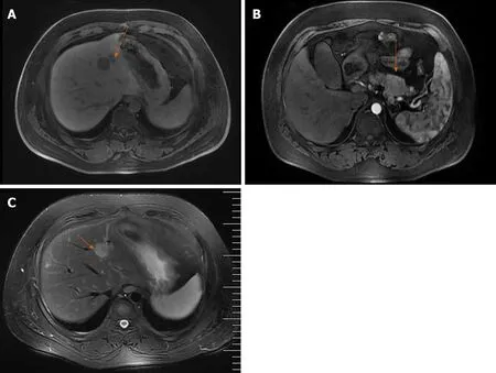
Parathyroid two-phase CT revealed a strong signal in the upper portion of the left lobes and posterior portion of the right lobes of the thyroid,in addition to the parathyroid glands (Figure1A).Pancreatic perfusion CT imaging showed irregular soft tissue densities of the pancreatic body,which was closely associated with the adjacent stomach wall,with a small reduction in blood volume (BV) and a slight increase in flow extraction product.There was increased blood flow,BV,mean transit time,and flow extraction product in nodules posterior to the pancreatic body;increased blood flow and decreased BV in nodules of the pancreatic head;and multiple circular low or slightly low-density shadows in liver parenchyma (Figure1B).MRI of the liver and pancreas revealed multiple abnormal signals in liver segment II,segment VIII,and the junction area of liver segments II and IV as well as occupied lesions in the tail of the pancreatic body (Figure2A).Enhanced MRI revealed occupied lesions in the body and tail of the pancreas (Figure2B).Enhanced MRI revealed that liver segment IV was occupied,suggesting the possibility of angiomyolipoma.In addition,there were small cysts in segments II and VIII of the liver and occupied lesions in the tail of the pancreatic body (Figure2C).
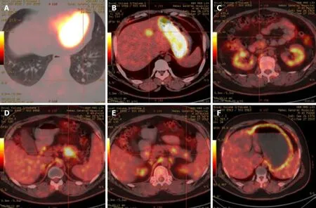
Positron emission tomography (PET)/CT imaging (Figure3) further showed strong18F-flurodeoxyglucose uptake in the tail of the pancreatic body and segment IV of the liver,indicating the presence of insulinomas with intrahepatic metastases.There were several circular nodules visible in the right and left lungs,showing partially increased signals in them.After undergoing complete surgical resection of the thymoma,ringshaped hypermetabolism became visible around the aortic root,which may be a result of fat intake.
FINAL DIAGNOSIS
A multiple disciplinary team (MDT) consisting of an endocrinologist,oncologist,pathologist,radiologist,thoracic surgeon,and hepatobiliary surgeon was incorporated to manage this patient.MEN1 with thymic carcinoid,thymoma,parathyroid and insulinomas with intrahepatic metastases was initially diagnosed.
TREATMENT
Since there were no obvious surgical contraindications,the MDT recommended a radical excision of the lesions to alleviate hypoglycemia symptoms and improve the quality of life for the patient.The patient underwent a resection of the tail of the pancreatic body,enucleation of the pancreatic head mass,and ultrasound-guided radiofrequency ablation for liver cancer.During the operation,a nodule with a diameter of about 0.4 cm was observed in the diaphragmatic surface of the right liver lobe.A mass of about 0.5 cm × 0.5 cm was palpable in the right posterior liver lobe.In addition,a tumor of about 2 cm × 2 cm in the falciform ligament about 4 cm from the hepatic margin,a mass of 1.5 cm × 1.0 cm on the pancreatic head,a mass of 5 cm × 3 cm × 3 cm on the pancreatic body,and a mass of 1.5 cm × 1 cm on the pancreatic tail were found.The intraoperative ultrasonography findings from the liver scan showed the right lobe nodules as cysts,and ultrasound-guided segment IV liver biopsy and radiofrequency ablation were performed.

Figure1 Computed tomography examination.A:Parathyroid two-phase computed tomography revealed a strong signal in the upper part of the left lobes and posterior part of the right lobes of the thyroid;B:Pancreatic perfusion computed tomography imaging revealed irregular soft tissue densities of the pancreas body,which were localized around the stomach wall.
After surgery,all diabetes factors with abnormal serum levels were returned to normal levels (Table1).The pathological findings are shown in Figure4.The pancreatic tissues showed CKpan (+),synaptophysin (+),chromogranin A (+),partial CD56 (+),p53 (-),partial PGP9.5 (+),partial SSTR2 (+),CD10 (-),partial vimentin (+),a mitotic count of 6/10 high power fields,and Ki-67 index of about 8%-20%,which confirmed the diagnosis of the pancreas NET grade 2.The liver tissues showed CKpan (+),synaptophysin (+),chromogranin A (+),partial CD56 (+),p53 (-),PGP9.5(+),a mitotic count of 4/10 high power fields,and Ki-67 index of about 10%-20%,which confirmed the diagnosis of liver NET grade 2.Moreover,there were multiple tiny nodules around the pancreatic tissues,indicating that surgical resection could not completely remove the lesions.NET grade 2 was diagnosed according to the World Health Organization 2010 classifications for gastrointestinal and pancreatic NETs[4].Furthermore,whole-exome sequencing revealed a verified pathogenic mutation c.378G>A (p.Trp126*) in theMEN1gene (reference sequence NM_130799.2).Therefore,the final diagnosis of MEN1 was confirmed.
OUTCOME AND FOLLOW-UP
The patient was free of hunger symptoms.During the follow-up visit at 6 mo,singlephoton emission CT/CT revealed multiple small nodules in the lungs.After comparing the single-photon emission CT/CT scans with the PET/CT from 4 mo prior,these nodules were suspected of being metastatic lesions.However,the nodules were too small to undergo needle biopsy,so the patient was placed under observation without additional treatment.At the last follow-up in January of 2020,the patient appeared healthy without any signs of disease reoccurrence.
DISCUSSION

Figure2 resonance imaging (MRI) examination revealed multiple abnormal signals in the liver segment II,segment VIII,and the junction area of liver segment II and IV as well as occupied lesions in the tail of the pancreatic body;B:Enhanced MRI revealed occupied lesions in the body and tail of the pancreas;C:Enhanced MRI revealed that the liver segment IV was occupied,suggesting the possibility of angiomyolipoma.There were also small cysts in segments II and VIII of the liver and occupied lesions in the tail of the pancreatic body.
MEN1 is characterized by primary hyperparathyroidism caused by a parathyroid tumor and involves the pancreas,anterior pituitary,duodenum,and adrenal gland.The annual incidence rate of MEN1 is about 1 in 30000[2].Approximately 25% of patients with thymic carcinoids are also diagnosed with MEN1[5,6],and in patients with MEN1,the incidence of thymic carcinoids is between 2% and 8%,with the disease most commonly occurring in patients 38 to 49 years of age[7,8].The patient in this study suffered from tumors of multiple organs,including the thymus,parathyroid,pituitary,pancreas,and liver.He first presented with thymic carcinoid,then primary hyperparathyroidism and prolactinoma,and later pancreatic NET with intrahepatic metastases.Moreover,the interval between thymic carcinoid and pancreatic NET was 10 years.Since the diagnosis of the thymic carcinoid,the patient had been alive for more than 11 years.MEN1-insulinoma accounts for about 10% of pancreatic NET cases.The average age of onset is less than 40 years old,oftentimes less than 20 years old,while non-MEN1 insulinoma patients are more than 40 years old[9].The symptoms of hypoglycemia often appear on an empty stomach or after physical activity,which can be relieved with food.The 72 h starvation test is the most reliable diagnostic method to demonstrate that the hypoglycemia is caused by high insulin[3].Ultrasound,CT,MRI,and PET/CT are often used for preoperative localization of lesions,and intraoperative ultrasound can improve the success rate of surgery[3].Endoscopic ultrasound (EUS) can be used to differentiate MEN1-related pancreas NET from sporadic pancreas NET[10,11].Moreover,preoperative biopsy through EUS/fine needle aspiration is useful for determining the differentiation status of pancreas NET[12-14].A well-differentiated grade 1/2 tumor is one of the primary surgical indications for pancreas NET management[15].However,the preoperative diagnosis of pancreas NET is relatively clear based on the patient's medical history,insulin level,Whipple’s triad,CT,MRI,and PET/CT findings.Hence,EUS was not performed,which is a limitation of the present case.
Generally,patients with MEN1 should be managed by a MDT consisting of relevant specialists from endocrinology,radiology,oncology,pathology,and surgery[3].For this disease,intraoperative monitoring of the insulin release index is useful for determining whether the tumor is successfully removed.Chemotherapy is recommended for patients with metastatic insulinomas and may include streptomycin,5-fluorouracil,doxorubicin,or hepatic artery embolization[16-18].The patient in this study suffered from tumors of multiple organs,including the pancreas,parathyroid,and pituitary.The progression of MEN1 was successfully controlled by MDT in the current study.
CONCLUSION
In conclusion,we presented a case of MEN1 that appeared sequentially as thymic carcinoid,primary hyperparathyroidism and prolactinoma,and pancreatic NET with intrahepatic metastases.Through the consultation of MDT for complete preoperative diagnosis and postoperative treatment and management,the patient was successfully treated in our clinic.If a patient is diagnosed with primary hyperparathyroidism with pituitary tumors or gastrointestinal NET,and the family history shows similar symptoms,MEN1 should be considered,and genetic testing is recommended.We hope that this study will add new insight into the diagnosis and treatment of patients with MEN1.

Figure3 Positron emission tomography/computed tomography examination.A,B,D and E:Hypermetabolic lesions in the lungs (A),intrahepatic segment IV(B),and the body (D) and tail (E) of the pancreas;C:No metabolic round nodules in the anterior pancreas;F:Diffusely increased metabolism in the stomach wall.

Figure4 Pathological examination.Hematoxylin &eosin staining showed (magnification,40 ×;100 ×) multiple nodules next to the pancreatic tumor,in the pancreatic tissue around the pancreatic body,and next to the pancreatic tail.Immunohistochemistry showed (magnification,40 ×;100 ×) CKpan (+),synaptophysin(+),chromogranin A (+),partial CD56 (+),p53 (-),partial PGP9.5 (+),partial SSTR2 (+),CD10 (-),partial vimentin (+) in pancreas tissues,and CKpan (+),Syn (+),CgA (+),partial CD56 (+),p53 (-),and PGP9.5 (+) in liver tissues.Bar,100 μm.Syn:Synaptophysin;CgA:Chromogranin A.
 World Journal of Clinical Cases2020年12期
- World Journal of Clinical Cases的其它文章
- Assessment of diaphragmatic function by ultrasonography:Current approach and perspectives
- Computer navigation-assisted minimally invasive percutaneous screw placement for pelvic fractures
- Research on diagnosis-related group grouping of inpatient medical expenditure in colorectal cancer patients based on a decision tree model
- Evaluation of internal and shell stiffness in the differential diagnosis of breast non-mass lesions by shear wave elastography
- Real-time three-dimensional echocardiography predicts cardiotoxicity induced by postoperative chemotherapy in breast cancer patients
- Lenvatinib for large hepatocellular carcinomas with portal trunk invasion:Two case reports
