Magnetic resonance imaging features of minimal-fat angiomyolipoma and causes of preoperative misdiagnosis
Xiao-Long Li,Qi-Cong Du,Wei Wang,Li-Wei Shao,Ying-Wei Wang,Department of Radiology,First Medical Center of Chinese People’s Liberation Army General Hospital,Beijing 100853,China
Li-Xin Shi,Department of Urology Surgery,First Medical Center of Chinese People’s Liberation Army General Hospital,Beijing 100853,China
Abstract
Key words:Minimal-fat angiomyolipoma;Magnetic resonance imaging;Misdiagnosis;Preoperative diagnosis
INTRODUCTION
Angiomyolipoma (AML) is the most common benign tumor of the kidney and is composed of mature adipose tissue,smooth muscle,and varying amounts of vessels.The typical imaging manifestations of AML make it easy to confirm by magnetic resonance imaging (MRI)[1-5].However,there are certain AMLs that lack a sufficient amount of fat (a fat content less than 25%) or do not contain adipose tissue,which cannot be distinguished by the naked eye on MRI.These AMLs are called minimal-fat angiomyolipoma (mf-AML).Because mf-AMLs are lack of typical AML imaging characteristic,they are often misdiagnosed as renal cell carcinoma before surgery,thus causing worthless surgical removal of the carcinoma[6,7]and bringing unnecessary burden to the patients.Therefore,this study retrospectively analyzed ten cases of mf-AML confirmed by surgical pathology in combination with the features of MRI manifestations to improve the accuracy of preoperative imaging diagnosis and pathological diagnosis.
MATERIALS AND METHODS
Clinical material
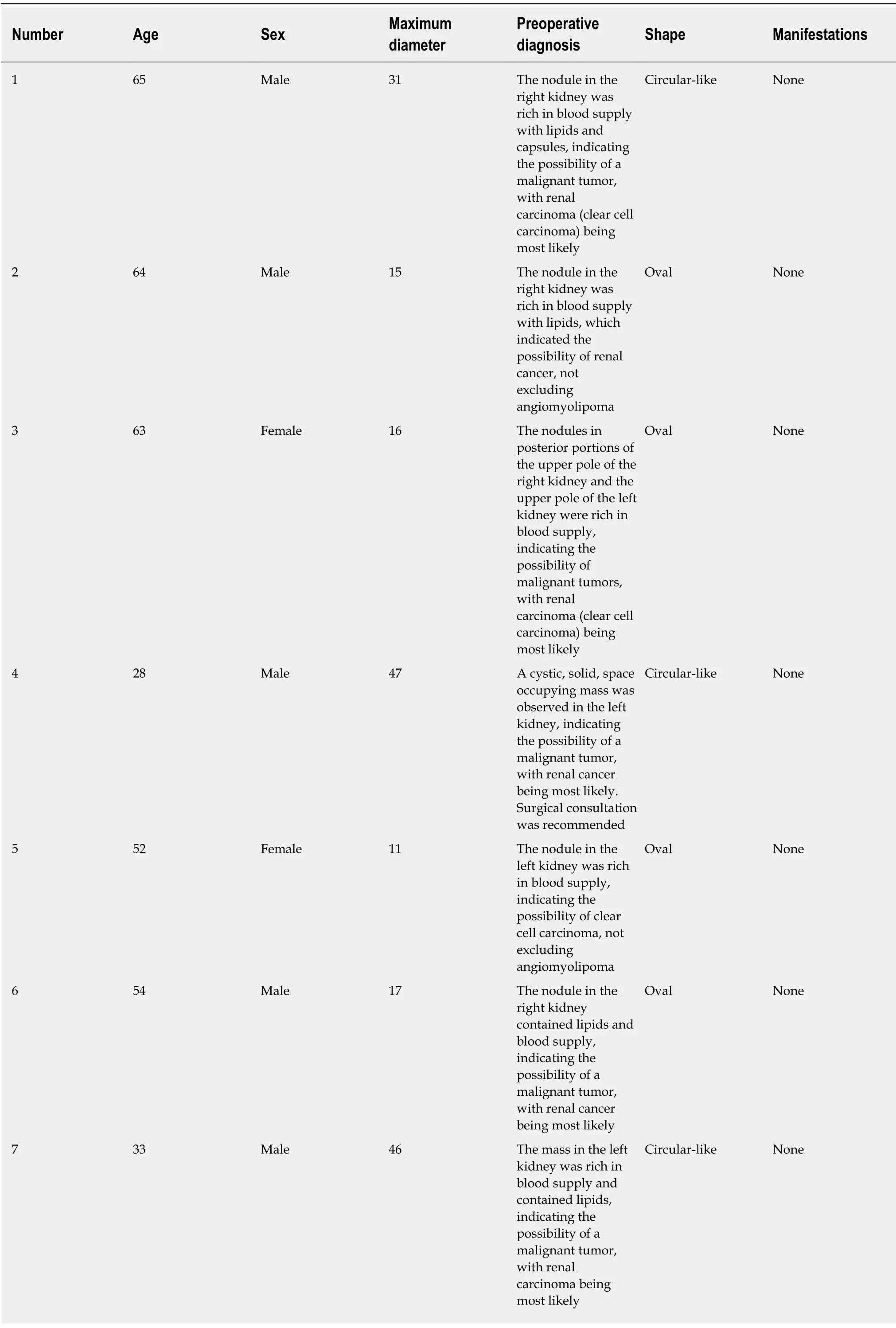
The study selected ten patients [five males and five females,with an average age of 50 years (28-65)] with mf-AML confirmed by surgical pathology in the First Medical Center of the People’s Liberation Army General Hospital between January 2018 and December 2018.Among the ten patients,eight were found to have space-occupying lesions on physical examination,and two had urinary retention or lower abdominal pain as the chief complaint.One patient had left renal clear cell carcinoma and right renal angiomyolipoma,one had hyperplasia of the left adrenal gland,and one had left multiple renal angiomyolipoma (Table1).Clinical data did not indicate tuberous sclerosis or Von Hippel-Lindau syndrome.Among the ten cases,eight were diagnosed with renal cell carcinoma by preoperative MRI,while the diagnosis could not be confirmed in the other two (renal cell carcinoma was likely to be diagnosed by MRI,but AML could not be completely excluded).
Examination method
All patients were examined with a GE 3.0 T scanner.The basic scanning methods involved fast spin-echo (FSE,) T2-weighted imaging (T2WI) and T2WI without fat suppression,including transverse and coronal positions,and T1WI inphase/opposed-phase examination.The parameters were:Time of repeatation:2000-6000 ms;time of echo:80-104 ms;matrix:320 × 224;layer thickness:5-6 mm;spacing:1 mm;and field of view (FOV):36 cm × 36 cm-40 cm × 40 cm.Thebvalue of the diffusion weighted imaging (DWI) sequence was set at 0 and 800-1000 s/mm2.A 3D liver acquisition with volume acceleration (LAVA) sequence was used in the multitemporal dynamic enhancement scan to perform a prescan of fat suppression on T1WI.The contrast agent Gd-diethylenetriamine pentaacetic acid was used in the enhanced scan at a dose of 0.1 mmol/kg (body weight),and the injection was performed with a high-pressure syringe at a rate of 1.5 mL/s.Cortico-medullary scanning was performed 18-21 s after contrast agent injection,while nephrographic phase and excretory phase scanning was performed at 45-60 s and 5-6 min after contrast agent injection,respectively.
Image data analysis

Table1 General patient information

8 61 Female 21 The nodule in the left kidney was rich in blood supply,indicating the possibility of a malignant tumor,with renal cancer being most likely;a fat embolism had formed in the intrahepatic segment of the inferior vena cava and right renal vein Circular-like Superior vena cava and right renal vein fat embolus 9 48 Female 13 The nodule in the right kidney was rich in blood supply and contained lipids,indicating the possibility of a malignant tumor,with renal cancer(clear cell carcinoma) being most likely Oval None 10 34 Female 36 The mass in the left kidney was rich in blood supply,indicating the possibility of a malignant tumor,with renal cancer(clear cell carcinoma) being most likely Oval None
The images were analyzed to observe the lesion morphology (circular-like/oval),size,signal characteristics (T2WI/T1WI/DWI/lipid/hemorrhage/cystic changes),surrounding capsule,and tumor enhancement characteristics on MRI images.Among them,tumor enhancement was confirmed based on the renal cortex signals as the reference standard:Nephrographic phase lesions whose signal intensity was hypointense to renal cortex were defined as mild or insignificant enhancements,while lesions whose signal intensity was isointense to renal cortex were defined as moderate enhancements,and lesions whose signal intensity was hyperintense to renal cortex were defined as significant enhancements[8-10].
RESULTS
In this study,nine lesions showed different degrees of exophytic growth.Four cases presented as round-like masses and six as oval masses.Lipid signals were observed in seven cases with different degrees of opposed-phase signal decrease.Lipid composition was not observed in three cases.An identifiable capsule was observed in nine cases but not in one case.The diameter of the tumors ranged from 11 to 47 mm,with a diameter of less than 20 mm in five tumors and less than 40 mm in eight tumors.There were eight cases of solid masses,including one case of a cystic solid mass and one case of a cystic lesion due to massive hemorrhage.
Eight cases of tumors had a homogeneous and slightly low T1W1 signal,and one had a homogeneous isointense T1 signal.One case had a heterogeneous,short T1 signal due to mass bleeding,while the other nine had no bleeding.Eight cases had a homogeneous,low T2 signal or slightly low T2 signal,one had a heterogeneous,low signal due to small-scale cystic changes,and one had a heterogeneous,short T2 signal due to mass bleeding.Six cases had DWI isointense or slightly high signals,and four had a high signal.Seven cases of tumors showed significant heterogeneous enhancement,and three showed moderate enhancement.In the excretory phase,eight cases had tumor washout,and two showed continuous enhancement.Intratumoral vasculature was not observed in eight cases but was visible in the other two (Figure1).

Figure1 Intratumoral vasculature was not observed in eight cases but was visible in the other two.A:An oval,homogeneous,and short T2 signal was found in the right kidney,showing a surrounding capsule.The lesion in the left kidney was pathologically confirmed as renal clear cell carcinoma;B and C:A mass in the renal parenchyma of the left kidney showed decreased signal intensity on opposed-phase MRI,indicating lipid composition;D:An oval,short T2 signal was found in the left kidney,showing a surrounding capsule;E and F:The round-like tumor in the left kidney showed heterogeneous and significant enhancement in cortico-medullary phase and decreased signal intensity(washout) of tumor in the excretory phase.
DISCUSSION
AML is a benign soft tissue tumor that is susceptible to the influence of adjacent normal kidney tissues.When adjacent normal renal parenchyma or renal mass is squeezed by the membrane,AML will present with typical morphological features,such as angular interface sign,mushrooming,and bubble-over sign[11-15].However,nine cases in this study,instead of presenting with a typical morphology such as angular interface sign,presented with round-like or oval shapes,indicating the possibility of mf-AML in a solid texture due to the lack of fat and leading to roundlike,oval,or ellipse morphological features.In particular,among the six cases of oval or ellipse lesions,the long diameter of the lesions was parallel to the renal medulla,suggesting that the tumors had different degrees of compression.Therefore,this study suggested that round-like or oval lesions could be regarded as the morphological characteristics of AML,which are more common in mf-AML.An oval or ellipse lesion shape has great significance for the diagnosis of mf-AML.A circularlike lesion shape can be considered an atypical morphological feature of mf-AML.
In this study,eight cases of tumors were less than 40 mm in diameter,and the masses were primarily characterized by uniform T1WI and T2WI signals on MRI,a feature of solid masses.Meanwhile,hemorrhage cystic changes were seldom found,indicating little secondary progression of mf-AML.However,a larger sample size is needed to confirm these observations.Regarding enhancement characteristics,in this study there were seven cases of tumors with significant and heterogeneous enhancement,and eight patients had tumor washout in the excretory phase,indicating that mf-AML is rich in blood supply,thus allowing for the enhancement seen on MRI.
The formation of capsule results from ischemia and necrosis caused by the growing tumor pressing on the surrounding normal renal parenchyma,which eventually leads to the formation of fibrous tissue[16-18].An identifiable capsule is a typical MRI sign of renal cell carcinoma.In this study,nine cases of tumors had capsules,which explained the high rate of preoperative misdiagnosis.This is probably because mf-AML has a lower level of fat,leading to a slightly harder tumor texture and causing compression on the surrounding normal renal parenchyma to form a capsule.Therefore,the identifiable capsule can also be considered an atypical MRI manifestation of mf-AML.However,a larger sample size is needed to confirm whether the capsule thickness,capsule integrity,and enhancement of the mf-AML pseudo capsule are provided with diagnostic significance.
Lipid change,also known as fatty change or steatosis,is defined pathologically as excessive triglyceride accumulation in the cytoplasm of nonfat cells such as liver cells,cardiomyocytes,and renal tubular epithelium and skeletal muscle cells[19-21].The presence of a small amount of lipid components can be identified on MRI when the opposed-phase signal is reduced.In this study,seven cases were found with small lipid signals in the mass,indicating that lipids could be present in mf-AML.The presence of in-phase and opposed-phase lipid signals is usually a clear and direct sign for the diagnosis of renal cell carcinoma,especially clear cell carcinoma[22-24].Therefore,the presence of lipid components detected by in-phase and opposed-phase in renal tumors could indicate the possibility of mf-AML based on morphological characteristics.
Based on the above analysis,mf-AML can present with oval or round-like morphological features,particularly oval or ellipse features,which are of great significance for diagnosis.Capsules,lipid composition,and washout are atypical manifestations of mf-AML.The presence of these atypical MRI manifestations should be combined with the morphological characteristics for the preoperative diagnosis of mf-AML.
ARTICLE HIGHLIGHTS
Research background
Minimal-fat angiomyolipoma (mf-AML) is often misdiagnosed as renal cell carcinoma before operation,which leads to unnecessary operation.Improving the rate of preoperative diagnosis is helpful to reduce unnecessary surgical treatment.
Research motivation
The magnetic resonance imaging (MRI) features of mf-AML are different from those of typical AML.Summarizing and analyzing the imaging features of mf-AML are helpful to improve the understanding of the disease and avoid unnecessary surgery.
Research objectives
To summarize the MRI features of mf-AML in order to improve the rate of preoperative diagnosis of mf-AML.
Research methods
The MRI features of mf-AML confirmed by operation and pathology were retrospectively analyzed,including morphological features,lipids,capsule,washout and so on.These lesions were diagnosed as renal cell carcinoma before operation or could not be diagnosed clearly.
Research results
A retrospective analysis of the results of ten cases of AML revealed a circular-like mass in 4/10(40%) patients,an oval mass in 6/10 (60%),a mass with a capsule in 9/10 (90%),and a mass with a lipid component in 7/10 (70%).But it still needs studies with a larger sample size to prove it.
Research conclusions
An oval morphological characteristic is strong evidence for the diagnosis of mf-AML,while a capsule and lipids are atypical manifestations of mf-AML.
Research perspectives
The imaging features of mf-AML are not typical,morphological features are very important for the diagnosis of renal tumors,and lipids and capsules can also be MRI findings of mf-AML.Some imaging features of mf-AML overlap with renal cell carcinoma,so it is necessary to comprehensively analyze its imaging features to improve the rate of preoperative diagnosis.
 World Journal of Clinical Cases2020年12期
- World Journal of Clinical Cases的其它文章
- Elevated serum growth differentiation factor 15 in multiple system atrophy patients:A case control study
- Clinical outcomes of sacral neuromodulation in non-neurogenic,non-obstructive dysuria:A 5-year retrospective,multicentre study in China
- Recovery from prolonged disorders of consciousness:A dual-center prospective cohort study in China
- Gene testing for osteonecrosis of the femoral head in systemic lupus erythematosus using targeted next-generation sequencing:A pilot study
- Epidemiological and clinical characteristics of COVID-19 patients in Hengyang,Hunan Province,China
- Demyelinating polyneuropathy and lymphoplasmacytic lymphoma coexisting in 36-year-old man:A case report
