
Biopharmaceutical Identification of Adventitious Buds and Root Bark of Aralia elata Seem.
2020-06-30 01:57:16FeiQIShizhaoXU
Medicinal Plant 2020年3期
Fei QI, Shizhao XU
Dalian Municipal Central Hospital, Dalian 116033, China
Abstract [Objectives] This article aimed to systematically study the biopharmaceutical characteristics of root bark and adventitious buds (tender stems and young leaves) of Aralia elata Seem. and to distinguish its root bark from that of Acanthopanax gracilistylus, in order to avoid confusion in clinical medication and guide root segment cutting technology of A. elata Seem. [Methods] Samples were obtained by field collection, and using trait and microstructure identification techniques, systematic biopharmaceutical identification of samples was carried out. [Results] The trait and microscopic characteristics of the root bark and adventitious buds (tender stems and young leaves) of A. elata Seem. were clarified. [Conclusions] The traits, cross-sections and powder of root bark, tender stems and young leaves of A. elata Seem. all have unique identification characteristics. The secondary phloem of A. elata Seem. is particularly developed, and it provides adventitious buds with sufficient nutrients for their meristem.
Key words Aralia elata Seem., Pharmacognostics, Identification
1 Introduction
AraliaelataSeem. (Araliaceae:Aralia) is a perennial deciduous tree or shrub, also known as Longya Congmu and Cilongya. It is mainly distributed in Liaoning, Jilin and Heilongjiang[1].A.elataSeem. is a medicinal and edible plant, and is often used as medicine with its root bark, with effects of invigorating qi, calming the nerves, dispelling wind and eliminating dampness. The germinated top bud (adventitious bud) ofA.elataSeem. is a natural green food rich in nutrients, and it is called "the king of wild vegetables". It is expensive, and has earned foreign exchange through exports[2]. In recent years, due to deforestation, the wild resources ofA.elataSeem. have almost been exhausted. At present, root segment cutting technique is used for its breeding. This article systematically studies the root bark and adventitious buds ofA.elataSeem. to provide a basis for the establishment of quality standards forA.elataSeem. and theoretically guide the root cutting, thereby improving the survival rate and expand the resources ofA.elataSeem.
2 Materials
The samples were collected at the seedling base of Danangou Village, Balidianzi Town, Huanren County, Benxi City (124°49′-124°52′ E, 41°16′-41°20′ N, 500-600 m a.s.l.). Annual seedlings ofA.elataSeem., with a seedling age of about 6 months, were investigated.
3 Methods and results
3.1ComparisonofrootbarkbetweenA.elataSeem.andAcanthopanaxgracilistylus
3.1.1Trait characteristics. (i)A.elataSeem. Rolled, about 0.3 cm thick, surface cork coming off as long sheets, outer surface yellow-brown, horizontal rings and horizontal long lenticels visible; inner surface with longitudinal wrinkles; slightly airy, slightly bitter, and tingling. Fracture surface mostly lamellar, powdery, gray, yellowish white under ultraviolet light. (ii)A.gracilistylusIrregularly rolled, about 0.2 cm thick. Outer surface gray-brown, with slightly distorted vertical wrinkles and horizontal lenticel-like scars; inner surface light yellow or grayish yellow, with fine vertical lines. Light, brittle, easy to break, uneven section, off-white. Slightly fragrant and slightly spicy and bitter[3].
3.1.2Microscopic characteristics of cross section. (i)A.elataSeem. Periderm phellem composed of 8-20 rows of flat cells, closely arranged; cortex about 1/2 of the cross section, multiple resin ducts arranged in a ring with a diameter of 15-30 μm; cracks and decadent tissues common; parenchyma cells in cortex containing a large number of starch granules; clustered crystals of calcium oxalate more common. Phloem cells closely arranged, secondary phloem well developed, multiple resin ducts arranged in lageniform rings in the phloem, with a larger diameter of about 25-50 μm, with drip-shaped tan secretions. Cambium cells approximately rectangular, connected into loops. Intact adventitious buds occasionally visible, composed of young leaves and growth cones, developed from the phloem. (ii)A.gracilistylusPhellem layer composed of numerous rows of cells. Interior narrow, a few secretory canals scattered. Phloem broad with fissures on the outside, each ray composed of 1-5 rows of cells; many secretory canals, with 4-11 secretory cells around. Parenchyma cells containing clustered crystals of calcium oxalate and fine starch granules[3](Fig.1).
3.1.3Microscopic characteristics of root bark powder. (i)A.elataSeem. Light brown, phellem cells approximately rectangular or polygonal, slightly brown, wall lignified. Fragments of resin ducts easily visible, with yellow-brown secretions. Fiber long shuttle-shaped, blunt at both ends, thick in wall, with a diameter of 10-20 μm. Many clustered crystals of calcium oxalate with a diameter of 18-60 μm. Many starch granules, spherical or oblong, umbilical point herringbone, dot-shaped, star-shaped,etc., compound granule composed of 2-6 granules. (ii)A.gracilistylusOff-white, clustered crystal of calcium oxalate 8-64 μm in diameter, sometimes crystal-containing cells connected and clustered crystals arranged in rows. Phellem cells rectangular or polygonal, and thin in wall; wall of phellem cells in old root bark sometimes unevenly thickened with a few pits. Fragments of secretory canals more common[3], containing colorless or pale yellow secretions. Many starch granules, polygonal or spheroidal, and complex granule composed of 2-10 granules.
3.2IdentificationofadventitiousbudsandtenderstemsofA.elataSeem.
3.2.1Trait characteristics. Un-spread young leaves with tender stem, adventitious buds 5-20 cm in total length (the best harvesting time); young leaves were bipinnate or tripinnate compound leaves; petioles of young leaves were plump and thick, 5-10 cm long, 0.2-0.3 cm in diameter, hairy or glabrous, and stipules and petiole base were gamogenic, bract-like amplexicaul; stems were 0.2-0.5 cm long, with residual dark brown bud scales[4].
The buds ofA.elataSeem. were divided into two types. In male plant, the leaves were purple, the petioles and veins all had sharp spines, the bases of the leaf axis and pinna rachis usually had short spines, and the short spines were purple; in female plants, the leaves were green (often chosen for artificial cultivation), and the bases of the leaf axis and pinnate rachis usually had no spines; the pinna rachises had 7-11 leaflets, the leaflets were opposite, there were erect spines in the center of the connection with the total petiole, and there were one pair of leaflets at base; the leaflets were papery or membranous, ovate to oval-ovate, 2-5 cm long, 1.5-3.0 cm wide, apex acuminate, base round to heart-shaped, thin wedge-shaped, glabrous or pubescent and finely bristled on both sides of veins, sparsely serrated at edges, sometimes finely serrated, thin and wavy, 6-8 pairs of lateral veins, obvious on both sides, reticulate veins not obvious.
3.2.2Microscopic characteristics of cross-section of tender stems. The epidermal cells were approximately square, and 1-2 rows were closely arranged. The surface was densely covered with non-glandular hairs, and most of them were multi-row non-glandular hairs, and a few were multicellular non-glandular hairs. The number of intact cells was 3-7. The collenchyma was composed of 5-7 layers. The cortical cells were nearly round and thin-walled, and contained brownish-yellow secretions. A total of 10-20 cortical cells were closely arranged. The resin ducts were arranged in a ring in the cortical parenchyma cells. The vascular bundles were bi-tough. The phloem was semilunar. The xylem was separated or connected. The vessels had become lignified, and their size was different, 16-71 μm in diameter. The ray cells were round and had 2-6 rows. The parenchyma cells in the pith were nearly round, with multiple resin ducts distributed in 6-8 rings (Fig.2).
3.2.3Microscopic characteristics of powder. The powder was gray-green or tan; the epidermal cells of tender stems were parallelogram or trapezoidal, and the vertical peripheral wall was straight and thickened like beads. The mesophyll cells were pale brownish yellow, and oblong or polygonal. The vessels were mainly scalariform and reticulate, and spiral vessels were visible, with a diameter of 9-35 μm. The crystals of calcium oxalate were different in size, and square or almost square. Multi-row non-glandular hairs were numerous, 50-200 μm, 20 μm in diameter (Fig.3).

Fig.1 Cross section of root bark of Aralia elata Seem. (10×40)
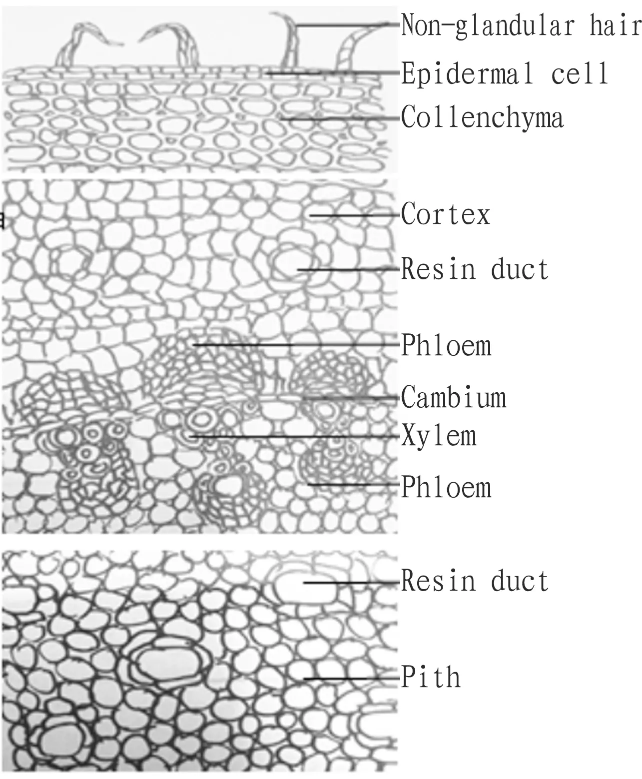
Fig.2 Cross section of tender stem of Aralia elata Seem. (10×40)
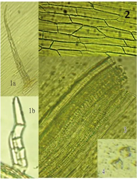
Note: 1a. multiple-row non-glandular hair; 1b. multiple-cell non-glandular hair; 2. epidermal cell; 3. vessel; 4. crystal of calcium oxalate.
Fig.3 Micrograph of powder of adventitious bud of Aralia elata Seem.
4 Conclusions and discussion
4.1IdentificationofrootbarkbetweenA.elataSeem.andA.gracilistylusThe root bark ofA.elataSeem. and the root bark ofA.gracilistylusare easily confused. Although both of them are used as medicine, the efficacy is different. It is of practical significance to compare and analyze the trait and microscopic characteristics of the two. The main distinguishing characteristics are as the follows. (i) In appearance, the root bark ofA.elataSeem. is relatively thick. The cork peels off in long sheets. The fracture surface is mostly lamellar, powdery and yellowish white under ultraviolet light. The root bark ofA.elataSeem. tastes tingling. The root bark ofA.gracilistylusis thin and tastes slightly spicy and bitter. (ii) In cross section ofA.elataSeem., the cortex resin ducts are arranged in a ring, and the phloem has multiple resin ducts arranged in 6-8 rings, which is its typical distinguishing feature. (iii) In terms of microstructure of powder, the root bark powder ofA.elataSeem. is brown, and that ofA.gracilistylusis off-white and darker in color. Both of them contain clustered crystals of calcium oxalate, but the clustered crystals inA.gracilistylusare arranged in rows.
4.2IdentificationofadventitiousbudsandtenderstemsofA.elataSeem.A.elataSeem. carries un-spread young leaves with tender stems. The leaves of male plants are purple, and their surface is densely covered with purple spines. The leaves of female plants are green, and spines are rare. The resin ducts in the cortical parenchyma cells are arranged in a ring. The vascular bundles are bi-tough. The phloem is semilunar. The parenchyma cells in the pith are round, and the resin ducts are arranged in 6-8 rings. The epidermal cells of the tender stem are parallelograms or trapezoids. The vessels are mainly scalariform and reticulate. Multi-row non-glandular hairs are numerous.
4.3ConclusionsIn this paper, a comparative study was conducted for the root bark ofA.elataSeem. andA.gracilistylus. The root cortex ofA.elataSeem. contains one ring of resin ducts, and the phloem contains 8 rings of resin ducts, which are its exclusive microscopic features. Its tender stems have many resin ducts. The rich secretion contained in the resin ducts is the material basis of the special flavor of tender stems ofA.elataSeem. The vascular bundles of tender stems are bi-tough vascular bundles. The secondary phloem is well-developed and contains abundant organic matters, which provide adventitious buds with sufficient nutrients for their meristem.

- Medicinal Plant的其它文章
- Optimization of Quality Control Method and Ethanol Extraction Process of Psoralen and Bergapten in Ficus pandurata
- Optimization of Processing Technology for Roasted Licorice with Water by Orthogonal Method
- Study on the Quality Standard of Shema Mixture
- Optimization of Extraction Process and Antioxidant Activity of Polysaccharide in Embelia parviflora Wall. by RSM
- Systematic Review and Meta-analysis of Efficacy of Banxia Xiexin Decoction for Treatment of Bile Reflux Gastritis
- Effects of Dachengqi Decoctions Made from Raw Rhubarb and Vinegar-processed Rhubarb on Levels of Endotoxin, NO and TNF-α in Serum