短花针茅荒漠草原单条土壤线虫分子鉴定方法研究
王嘉宝 孟元 史明易 刘美琪 贾美清 张国刚


摘 要:土壤線虫在土壤中具有重要的生态作用,目前大多通过形态学鉴定方法研究土壤线虫,但该方法有很多弊端。为了更好地在分子水平上研究土壤线虫群落及其多样性,对内蒙古短花针茅荒漠草原土壤线虫进行了单条线虫的分子鉴定,通过比较4种不同的DNA提取方法(试剂盒法、冻融法、切割法、切割冻融法),确定了土壤线虫最佳的DNA提取方法为切割冻融法。同时,选出10对土壤线虫的通用引物经过重复试验,从10对引物中筛选出3对引物(分别为线虫rDNA的18S区引物G18SU/R18Tyl1、28S区引物D2A/D3B、ITS区引物TW81/AB28)能达到较高的成功率,其中适用于内蒙古短花针茅荒漠草原土壤线虫的最佳通用引物是:D2A/D3B(5‘-ACAAGTACCGTGAGGGAAAGTTG-3;5‘-TGCGAAGGAACCAGCTACTA-3)。本研究通过明确短花针茅荒漠草原土壤线虫最佳DNA提取方法和最佳通用引物,为进一步通过高通量测序技术在分子水平上进行土壤线虫研究奠定基础。
关键词:内蒙古;短花针茅荒漠草原;土壤线虫;分子鉴定方法
中图分类号:S154.5;S154.38+6 文献标识码:A DOI 编码:10.3969/j.issn.1006-6500.2020.02.017
Molecular Identification of Soil Nematodes in Stipa breviflora Desert Steppe
WANG Jiabao1, MENG Yuan1, SHI Mingyi3, LIU Meiqi1, JIA Meiqing2, ZHANG Guogang1
(1.Life Science College, Tianjin Normal University, Tianjin 300387, China; 2.Tianjin Key Laboratory of Water Resources and Environment, Tianjin Normal University, Tianjin 300387, China; 3.School of Geographic and Environmental Sciences, Tianjin Normal University, Tianjin 300387, China)
Abstract:Soil nematodes play an important ecological role in soil. At present, the study of soil nematodes mostly uses morphological methods, but this method has many disadvantages. In order to better study the community characteristics and diversity of soil nematodes at the molecular level, the researchers carried out molecular identification of single nematodes on soil nematodes in the desert steppe of Inner Mongolia.We compared a total of four different DNA extraction methods: kit, freeze-thaw, cutting and cutting freeze-thaw, and determined that the best DNA extraction method for soil nematodes is the cutting freeze-thaw method.At the same time, the researchers selected a total of 10 pairs of universal PCR primers for soil nematodes.After repeated verification experiments, 3 pairs of optimal primers were selected from the 10 pairs of primers (18S region primers G18SU / R18Tyl1 and 28S region primer D2A of the rDNA of the nematode, and D3B, ITS region primer TW81 / AB28 respectively).These 3 pairs of PCR primers can achieve a high success rate of amplification, and the best universal primers for soil nematodes in the Stipa breviflora desert steppe of Inner Mongolia are: D2A / D3B(5'-ACAAGTACCGTGAGGGAAAGTTG-3'; 5'-TGCGAAGGAACCAGCTACTA-3'). In this study, we identified the best DNA extraction method and the best universal primer for soil nematodes in the Stipa breviflora desert steppe, laying a foundation for the further study of soil nematodes at the molecular level by using high-throughput sequencing technology.
Key words: Inner Mongolia; Stipa breviflora desert steppe; soil nematode; molecular identification method
土壤线虫是土壤动物的重要组成部分,其广泛存在于各种土壤中,如草原、森林等[1-3]。土壤线虫不仅能够促进养分循环,改变土壤理化性质,还能影响地上和地下生态系统[4-6]。其具有种类丰富、繁殖周期较短、对环境变化反应敏感等特点,因此经常被用作指示生物来反映土壤受污染的程度[7],是土壤指示生物中的典型代表[8]。在土壤线虫的鉴定方面,目前使用最多的是依据其形态学特征的传统鉴定方法,不过这种方法存在许多弊端,如鉴定结果不准确、鉴定速度慢等,同时也受到了经济和时间的成本限制[9]。随着分子生物技术的产生与发展,越来越多的学者开始采用分子方法鉴定线虫[10-11]。
在线虫分子鉴定方面,ITS区对于线虫属下种的区分与鉴定有较好的应用[12-14],如可应用于伞滑线虫属、茎线虫属的鉴定等[15]。然而ITS序列并不能区分所有线虫属下物种,核糖体DNA(rDNA)中除了ITS序列,还有18S rDNA基因和28S rDNA基因中的D2A/D3B区在线虫的分子鉴定中也具有非常重要的作用[16]。我们在形态学分类的基础上,通过比较4种不同的DNA提取方法和筛选线虫rDNA的ITS区、18S区、28S区通用引物,确定了短花针茅荒漠草原土壤线虫的最佳DNA提取方法和最佳通用引物,为进一步在分子水平上研究土壤线虫奠定基础。
1 材料和方法
1.1 研究区概况
样地位于内蒙古自治区乌兰察布市四子王旗农牧科学院长期定位试验站(N41°47′17″,E111°53′46″),海拔高度为1 456 m,所属气候为温带大陆性气候,平均气温为3.4 ℃。植被类型为典型荒漠草原,植物组成匮乏,建群种为短花针茅(Stip abreviflora),优势种为冷蒿(Artemisia frigida Willd)和无芒隐子草(Cleistogenes songorica)。土壤的主要类型为棕钙土。
1.2 土壤样品采集和线虫形态学鉴定
在实验区内选择3块典型的短花针茅荒漠草原样地,在每块样地内分别用土壤环刀采集0~5 cm、5~10 cm、10~20 cm、20~30 cm 4层土壤样品,3次重复,为了减少土壤线虫空间分布差异所造成的影响,每次重复对样地内进行多点取样,每个取样点地上植被类型必须为典型短花针茅荒漠草原群落,取样后将土壤装入密封袋中,置于-20 ℃冰箱中保存备用。
收集線虫的方法为改良的贝尔曼漏斗法,在漏斗中的铁丝网上放入纱布包裹好的土壤,漏斗上端设置有60 W的白炽灯,下端装有带止水夹的乳胶管,漏斗中加入蒸馏水浸没土壤,20 ℃室温条件下分离,期间不断加入蒸馏水,经过24 h后,将液体收集到离心管中,65 ℃水浴加热30 min。将收集好的土壤线虫置于培养皿中,于倒置显微镜及体式显微镜下,依据《中国土壤动物检索图鉴》、《植物线虫分类学》观察鉴定。通过形态学鉴定方法共鉴定出土壤线虫共计24个属,并以此为试验材料进行分子鉴定方法研究。
1.3 土壤线虫的分子鉴定方法
1.3.1 DNA提取方法 在形态学鉴定的基础上,将形态学鉴定出来的土壤线虫进行分类。由于土壤线虫的体壁较厚,为其DNA的提取增加了难度。因此,如何高效地提取土壤线虫DNA便成为实现分子鉴定的关键。本项研究为了获得最佳DNA提取浓度,共设置4种DNA提取方法[17],方法如下。
(1)试剂盒法。采用柱式抽提试剂盒直接提取线虫DNA。
(2)冻融法。挑取传统形态学鉴定分类的单条线虫,用灭菌去离子水将线虫清洗干净,将清洗干净的线虫放入200 μL PCR管中,PCR管中加入8 μL去离子水,放入离心机瞬时离心5 s,取出PCR管后放入液氮中1 min,迅速转移到80 ℃水浴锅中2 min,重复3次,最后加入蛋白酶裂解液2 μL,56 ℃保温1 h,95 ℃加热10 min。
(3) 切割法。挑取传统形态学鉴定分类的单条线虫,用灭菌去离子水将线虫清洗干净,放入含8 μL灭菌去离子水的200 μL PCR管中,放入离心机瞬时离心5 s,取出后将PCR管外壁擦拭干净,在显微镜下用小型解剖针将PCR管内的线虫切开,最后加入蛋白酶裂解液2 μL,56 ℃保温1 h,95 ℃加热10 min。
(4)切割冻融法。挑取传统形态学鉴定分类的单条线虫,用灭菌去离子水将线虫清洗干净,放入含8 μL灭菌去离子水的200 μL PCR管中,放入离心机瞬时离心5 s,取出后将PCR管外壁擦拭干净,在显微镜下用小型解剖针将PCR管内的线虫切开,放入离心机瞬时离心5 s,取出后将PCR管放入液氮中1 min,迅速转移到80 ℃水浴锅中2 min,重复3次,最后加入蛋白酶裂解液2 μL,56 ℃保温1 h,95 ℃加热10 min。
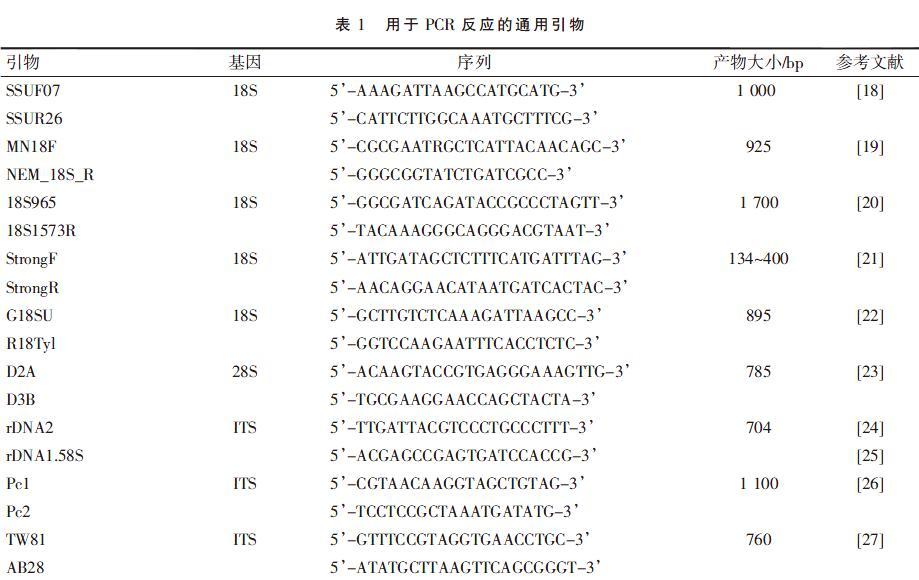
1.3.2 引物筛选 经查阅文献[18-27],本研究利用线虫核糖体DNA的ITS区、18S区、28S区具有种内高度保守、种间变化大且受环境变化影响较小的特点,在线虫rDNA的ITS区、18S区和28S区的高保守区筛选出10对土壤线虫通用引物,用于确定内蒙古荒漠草原土壤线虫分子方法鉴定的最佳引物。10对通用引物序列如表1所示。
由于引物不同,选择相应的PCR反应体系及反应条件也不同,用以下体系及反应条件为核心反应体系和模板,根据不同引物在此基础上进行修改。
PCR扩增体系:模板DNA10 μL,10×PCR Buffer (Mg2+) 5 μL,2 μL dNTP (2.5 mmol·L-1),1 μL引物F (10μmol·L-1),1μL引物R(10μmol·L-1);rTaq DNA 聚合酶(5U·L-1)0.4 μL;模板DNA 10 μL;无菌ddH2O 30.6 μL。
PCR反應条件:95 ℃预变性3 min;95 ℃变性40 s;55 ℃退火40 s;72 ℃ 延伸 1 min,40个循环,72 ℃保温10 min。
应用琼脂糖凝胶电泳检测PCR产物,检测合格后送样测序。
通过NCBI数据库中的BLAST工具对测序结果进行比对分析。
2 结果与分析
2.1 短花针茅荒漠草原土壤线虫DNA提取的最佳方法
用4种不同的方法对形态学鉴定得到的24个属的线虫进行了DNA提取,以研究柱式抽提试剂盒、冻融法、切割法、切割冻融法对土壤线虫DNA的提取效果,采用D2A/D3B作为通用引物进行PCR扩增,由于该引物只能扩增出24个属中的22个,所以只能应用这对引物来扩增22个属的DNA,来研究最佳的DNA提取方法,研究结果如下。
由图1可见,柱式抽提试剂盒提取DNA的效率很低,对于大多数土壤线虫属都不能提取,用柱式抽提试剂盒只能提取出丽突属、拟丽突属线虫DNA,且提取浓度和成功率都很低。对PCR产物进行测序发现,由柱式抽提试剂盒提取DNA的PCR产物测序结果会有杂带,测序成功率低。
由图2可知,冻融法提取DNA的效率有所提升,能提取出丽突属、拟丽突属、鹿角唇属、滑刃属、真滑刃属线虫的DNA,提取浓度有所升高。但是成功率依然很低,而且冻融法提取的DNA测序结果仍有杂带,测序成功率不高。
由如图3可见,切割法提取的DNA的效率升高,能提取出20个属的DNA,且提取的DNA浓度很高,测序结果仍有杂带,但测序成功率有所升高。
应用切割冻融法提取22个属的rDNA,扩增产物电泳检测结果如图4所示。
conditions[J]. Ecological indicators,2016,60:310-316.
[9]KERFAHI D,PARK J, TRIPATHI B M, et al. Molecular methods reveal controls on nematode community structure, and unexpectedly high nematode diversity, in Svalbard High Arctic Tundra[J]. Polar biology,2016,40(4):765-776.
[10]DEVRAN Z, POLAT I, MISTANOGLU I, et al. A novel multiplex PCR tool for simultaneous detection of three root-knot nematodes[J]. Australasian plant pathology,2018,47(4):389-392.
[11]BRAUN-KIEWNICK A, KIEWNICK S. Real-time PCR, a great tool for fast identification, sensitive detection and quantification of important plant-parasitic nematodes[J]. European journal of plant pathology,2018,152(2):271-283.
[12]SUBBOTIN S A, VIERSTRAETE A, LEY P D, et al. Phylogenetic relationships within the cyst-forming nematodes (Nematoda, Heteroderidae) based on analysis of sequences from the ITS regions of ribosomal DNA[J]. Molecular phylogenetics & evolution,2001,21(1):1-16.
[13]ZHUO K, WANG H H, YE W, et al. Cryphodera sinensis n. sp. (Nematoda: Heteroderidae), a non-cyst-forming parasitic nematode from the root of ramie Boehmeria nivea in China[J]. Journal of helminthology,2014,88(4):468-480.
[14]HE Y, SUBBOTIN S A, RUBTSOVA T V, et al. A molecular phylogenetic approach to Longidoridae (Nematoda: Dorylaimida)[J]. Nematology,2005,7(1):111-124.
[15]SUBBOTIN S A, STURHAN D, CHIZHOV V N, et al. Phylogenetic analysis of Tylenchida Thorne, 1949 as inferred from D2 and D3 expansion fragments of the 28S rRNA gene sequences[J]. Nematology,2006,8(3):455-474.
[16]WAITE I S, O'DONNELL A G, HARRISON A, et al. Design and evaluation of nematode 18S rDNA primers for PCR and denaturing gradient gel electrophoresis(DGGE) of soil community DNA[J]. Soil biology & biochemistry,2003,35(9):1165-1173.
[17]容万韬,王金成,刘鹏,等.单条线虫DNA提取方法研究[J].华北农学报,2014,29(2):127-132
[18]FLOYD R, ABEBE E, PAPERT A, et al. Molecular barcodes for soil nematode identification[J]. Molecular ecology,
2002,11(4):839-50.
[19]BHADURY P, AUSTEN M C, BILTON D T, et al. Development and evaluation of a DNA-barcoding approach for the rapid identification of nematodes[J]. Marine ecology progress,
2006,320(1):1-9.
[20]MULLIN P G, HARRIS T S, POWERS T O. Phylogenetic relationships of Nygolaimina and Dorylaimina(Nematoda: Dorylaimida) inferred from small subunit ribosomal DNA sequences[J]. Nematology,2005,7(1):59-79.
[21]LABES E M, WIJAYANTI N, DEPLAZES P, et al. Genetic characterization of Strongyloides spp. from captive, semi-captive and wild Bornean orangutans (Pongo pygmaeus) in Central and East Kalimantan, Borneo, Indonesia[J]. Parasitology, 2011, 138(11):1417-1422.
[22]CHIZHOV V N, CHUMAKOVA O A, SUBBOTIN S A. Morphological and molecular characterization of foliar nematodes of the genus Aphelenchoides: A. fragariae and A. ritzemabosi (Nematoda: Aphelenchoididae) from the Main Botanical Garden of the Russian Academy of Sciences, Moscow[J]. Russian journal of nematology,2006,14(2):179-184.
[23]KANZAKI N, GREEFF J M, W?魻HR M, et al. Molecular and morphological observations on Parasitodiplogaster sycophilon Poinar, 1979 (Nematoda: Diplogastrina) associated with Ficus burkei in Africa[J]. Nematology,2014,16(4):453-462.
[24]VRAIN T, WAKARCHUK D, LAPLANTELEVESQUE A, et al. Intraspecific rDNA restriction fragment length polymorphism in the Xiphinema americanum group[J]. Fundamental & applied nematology,1992,15(6):563-573.
[25]CHERRY T, SZALANSKI A L, TODD T C, et al. The internal transcribed spacer region of Belonolaimus (Nemata: Belonolaimidae)[J]. Journal of nematology,1997,29(1):23-29.
[26]余永廷,薛召东,曾粮斌,等.1种苎麻根腐病线虫的鉴定[J].西北农林科技大学学报(自然科学版),2011,39(7):105-109.
[27]SUBBOTIN S A, MADANI M, KRALL E, et al. Molecular diagnostics, taxonomy, and phylogeny of the stem nematode ditylenchus dipsaci species complex based on the sequences of the internal transcribed spacer-rDNA[J]. Phytopathology,2005,
95(11):1308-1315.
[28]王江岭, 张建成, 顾建锋.单条线虫DNA提取方法[J].植物检疫,2011(2):38-41.
[29]杜宇,邓宗汉,丁元明,刘冰洋,李旻,雷屈文,林萌.四种短体线虫的形态和分子生物学鉴定[J/OL].植物病理学报:1-22[2019-12-04].https://doi.org/10.13926/j.cnki.apps.000410.
[30]刘海璐,王暄,李红梅,等. 我国黄淮麦区10个短体线虫样品种类的分子鉴定[J].中國农业科学,2018,51(15):78-92.
[31]GEISEN S, SNOEK L B, TEN HOOVEN F C, et al. Integrating quantitative morphological and qualitative molecular methods to analyze soil nematode community responses to plant range expansion[J]. Methods in ecology and evolution,2018,9(6):1366-1378.
[32]张晓珂,梁文举,李琪.我国土壤线虫生态学研究进展和展望[J].生物多样性,2018,26(10):34-47.
[33]薛蓓.基于高通量测序分析西藏北部高寒草甸不同深度土壤线虫群落分布特征[J].生态学报,2019,39(11):4088-4095.
[34]PEHAM T, STEINER F M, SCHLICK-STEINER B C, et al. Are we ready to detect nematode diversity by next generation sequencing[J]. Ecology and evolution,2017,7(12):4147-4151.

