
Paraplegia from spinal intramedullary tuberculosis: A case report
2020-04-07 02:13:14LiMeiQuDiWuLiangGuoJinLuYu
World Journal of Clinical Cases 2020年24期
Li-Mei Qu, Di Wu, Liang Guo, Jin-Lu Yu
Li-Mei Qu, Di Wu, Liang Guo, Department of Pathology, The First Hospital of Jilin University,Changchun 130021, Jilin Province, China
Jin-Lu Yu, Department of Neurosurgery, The First Hospital of Jilin University, Changchun 130021, Jilin Province, China
Abstract BACKGROUND Tuberculosis (TB) mostly attacks the lungs, and extrapulmonary TB involving the central nervous system is uncommon; among these cases, spinal intramedullary TB is even more rare. The clinical manifestations of spinal intramedullary TB are similar to those of intramedullary spinal cord tumors. Therefore, it is necessary to make a careful differential diagnosis of spinal intramedullary lesions to achieve the appropriate treatment and favorable prognosis. We report a rare case of a young male patient with paraplegia due to spinal intramedullary TB, which is uncommon and regrettable.CASE SUMMARY A 23-year-old male presented with fever accompanied by nausea and vomiting lasting for 2 mo and was then diagnosed with tubercular meningitis. After anti-TB treatment, his symptoms were significantly improved. However, 2 mo after the diagnosis of tubercular meningitis, the patient felt numbness below the costal arch level, which lasted for 1 wk, and he paid no attention to this symptom. What followed was paraplegia and urine/fecal incontinence. Magnetic resonance imaging of the thoracic spine showed a ring-enhanced intramedullary cord lesion at T8-T9. Lesion exploration showed enlargement of the spinal cord at T8-T9, and the lesion could be observed by incision. The lesion was adhered to the peripheral tissue and was grayish-white and tough with a poor blood supply and a diameter of approximately 0.8 cm. The lesion was resected completely. The results of pathological examination by both hematoxylin-eosin staining and acid-fast bacilli staining confirmed TB, accompanied by acute and chronic suppurative inflammation and granulation tissue formation. The patient was instructed to continue anti-TB treatment after the operation, but he did not follow the medical advice. Follow-up continued for ten years, the patient had persistent paraplegia,the numbness disappeared and urine/fecal sensation recovered.CONCLUSION Although TB is a kind of benign disease, some cases progress rapidly. Moreover,spinal intramedullary TB may seriously endanger quality of life and still needs timely diagnosis and proper treatment.
Key Words: Tuberculosis; Intramedullary; Extrapulmonary; Central nervous system;Paraplegia; Case report
INTRODUCTION
According to global data, India has the highest incidence of tuberculosis (TB), which is mainly pulmonary TB[1]. While the incidence of cases of pulmonary TB has shown a downward trend in China, these are not uncommon[2,3]. TB of the central nervous system accounts for approximately 1% of all cases of TB, and 50% of these cases involve the spine[4]. Intramedullary involvement is rare in extrapulmonary TB, and less than 50 cases of this disease have been reported in the literature. The clinical manifestations of spinal intramedullary TB are similar to those of intramedullary spinal cord tumors with diverse manifestations[5]. Although spinal intramedullary TB is a kind of benign disease, timely diagnosis and proper treatment are still needed to achieve a better prognosis. We retrospectively collected the only case of spinal intramedullary TB confirmed by pathological examination at the First Hospital of Jilin University from 2010 to 2020, reported as follows.
CASE PRESENTATION
Chief complaints
The patient was a 23-year-old male who presented with fever accompanied by nausea and vomiting lasting for 2 mo. He had paraplegia and urine/fecal incontinence lasting for 7 d.
History of present illness
Two months ago, the patient was diagnosed with tubercular meningitis, and his symptoms improved obviously after anti-TB treatment. Seven days ago, he felt numbness below the costal arch level followed by paraplegia and urine/fecal incontinence.
History of past illness
The patient had no significant past medical history or surgical history and did not take any medications.
Personal and family history
His family members denied a history of TB.
Physical examination
The temperature was 37.4 °C, the heart rate was 80 beats per minute, the respiratory rate was 16 breaths per minute and the blood pressure was 126/97 mmHg. The patient had clear consciousness and clear and fluent speech. His sensations of pain, warmth and deep stimuli were absent below the costal arch level. The muscular tension and tendon reflexes of the lower limbs were absent with grade 0 muscle strength. The bilateral Babinski sign was negative.
Laboratory examinations
The laboratory examination findings were all within normal limits, including the complete blood count, routine urine test results and liver function test results. The results of hepatic serology were also negative. The fasting blood glucose level increased to 6.43 mmol/L (3.9-6.1 mmol/L). Neither lung digital radiography nor thoracic vertebrae digital radiography showed involvement on radiography.
Imaging examinations
Magnetic resonance imaging (MRI) of the thoracic spine showed a ring-enhanced intramedullary cord lesion at T8-T9 (Figure 1).
MULTIDISCIPLINARY EXPERT CONSULTATION
There is a possibility of tumor but need to exclude nontumor disease.
FINAL DIAGNOSIS
Spinal intramedullary TB with acute and chronic suppurative inflammation and granulation tissue formation.
TREATMENT
Surgical resection was performed.
OUTCOME AND FOLLOW-UP
After the operation, the patient stopped anti-TB treatment. Follow-up continued for ten years. The patient had persistent paraplegia, the numbness disappeared and urine/fecal sensation recovered without new TB lesions.
DISCUSSION
TB can involve the central nervous system, accounting for approximately 10% of all cases, and it can occur due to latent infection reactivation, transmission from organ donors or new nosocomial infections. Most cases of central nervous system TB are intracranial TB, while spinal intramedullary TB is even rarer with a ratio between the two at approximately 42:1[6]. The pathways by whichMycobacterium tuberculosisreach the spinal cord include hematogenous dissemination and cerebrospinal fluid. While the probability of spinal intramedullary TB in the thoracic, cervical and lumbar segments decreases successively, intramedullary TB can also occur near the end of the spinal cord and in the medullary cone[7]. The spinal intramedullary TB focus in this case was located in the intramedullary cord at T8-T9.
In some cases of spinal intramedullary TB, there are clinical manifestations of pulmonary or systemic TB, and recent history of tubercular meningitis is not uncommon. In this patient, the disease involved the spinal cord after the onset of tubercular meningitis for 2 mo. There have been a few case reports describing intramedullary suppurative and subdural abscesses[8]. The postoperative pathological examination in this case revealed many neutrophils in the lesion as well as granulation tissue formation, indicating the trend of suppurative inflammation and abscess formation.
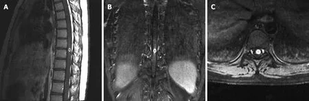
Figure 1 Magnetic resonance imaging examination before the operation showing a ring-enhanced intramedullary cord lesion at T8-T9. A:Sagittal image; B: Coronal image; C: Axial image.
Patients with spinal intramedullary TB often present with signs of spinal cord compression, such as progressive lower limb weakness, paresthesia, quadriplegia,paraplegia and bladder and bowel dysfunction. However, as reported in the literature,almost all of these symptoms resolved in patients who underwent surgical resection of the lesion. The urine/defecation function recovered to normal, and the muscle strength of the lower limbers recovered to grade 4. Unfortunately, the paraplegia in our reported 23-year-old male patient failed to recover after surgical resection of the spinal intramedullary lesion. His urine/fecal sensation recovered, but he could still not excrete on his own. Furthermore, he remained in a wheelchair for 10 years after the operation. Cases of such poor postoperative conditions have rarely been reported. This case us unusual because the pathological examination showed acute suppurative inflammation in the lesion, which had already formed granulation tissue. Whether the above pathological changes induced the failure to recover from the paraplegia needs to be further discussed.
The diagnosis of spinal intramedullary TB requires the combination of clinical,imaging and pathological examinations. Special attention should be paid to this disease in patients with medical history of pulmonary TB or systemic TB, especially those with immunodeficiency or a history of organ transplantation. In terms of imaging, MRI is valuable for diagnosis, while pathological examination is the most accurate diagnostic method. The criteria for the diagnosis of TB under microscopy are caseous necrosis, epithelioid nests and Langhans giant cells (Figure 2). But in different cases or stages, there will be different manifestations, and the proportion of these three changes will vary greatly. Some cases mainly show caseous necrosis, while other cases show proliferative lesions dominated by epithelioid cell masses. TB is often accompanied by acute and chronic inflammation as well, such as the acute infection and granulation tissue formation reported in this paper.
The treatment of spinal intramedullary TB mainly consists of anti-TB drugs. In addition, it has been reported that steroids in combination with anti-TB drugs have achieved positive clinical results[9]. Abnormal reactions to anti-TB drugs, such as the enlargement of existing tuberculomas or emergence of new tuberculomas as the treatment begins, have been reported in a small number of cases of spinal intramedullary TB. This is believed to be the result of increased inflammation caused by the anti-TB drugs relieving cell-mediated immunosuppression[10]. The course of disease in our case may be in accordance with this theory. Surgical indications include paralysis due to spinal compression or increasing spinal intramedullary lesions despite anti-TB drug treatment or pathological findings in patients with an unclear diagnosis indicating the need for lesion excision.
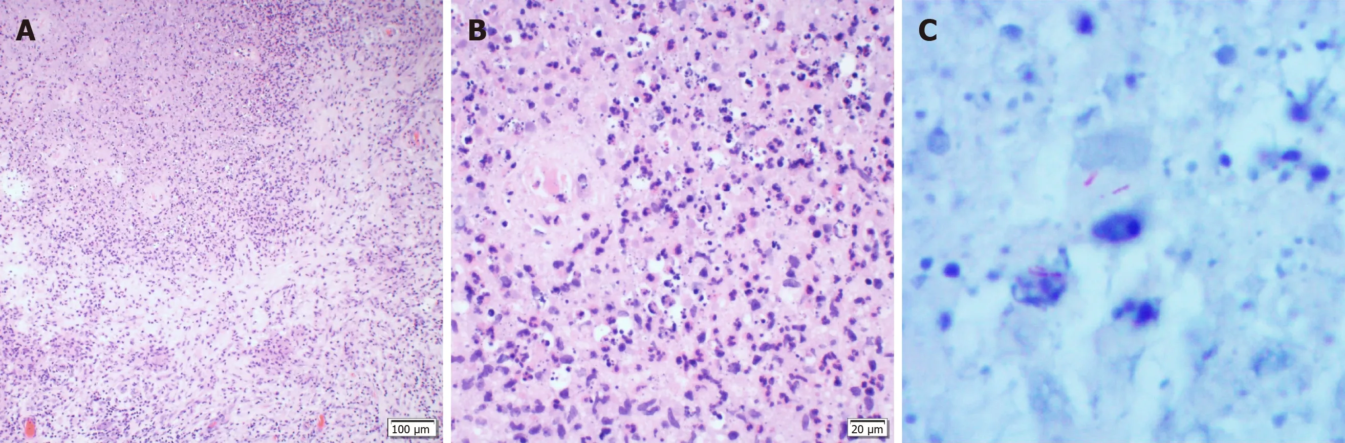
Figure 2 Pathological examination. Under the microscope, caseous necrosis was observed in the center of the focus, surrounded by a few epithelioid cells and incomplete multinucleated giant cells. Meanwhile, more neutrophils, lymphocytes and plasma cells were observed forming granulation tissue. A: Hematoxylin-eosin staining at 100 ×; B: Hematoxylin-eosin staining at 400 ×; C: Acid-fast bacilli staining revealed a small amount of Mycobacterium tuberculosis in the necrotic tissue.
CONCLUSION
Spinal intramedullary TB is extremely rare among cases of the central nervous system TB, and in these cases, the clinical manifestations are similar to subacute myelopathy.The diagnosis of spinal intramedullary TB is based on clinical features, MRI findings and pathological and microbiological examination. Most patients have a good prognosis after surgical resection of the lesion. However, patients whose pathological examinations show acute suppurative inflammation in the lesion, which has already formed granulation tissue, may have poor prognosis, such as the case we reported.
 World Journal of Clinical Cases2020年24期
- World Journal of Clinical Cases的其它文章
- Primary duodenal tuberculosis misdiagnosed as tumor by imaging examination: A case report
- Successful endovascular treatment with long-term antibiotic therapy for infectious pseudoaneurysm due to Klebsiella pneumoniae: A case report
- Idiopathic adulthood ductopenia with elevated transaminase only: A case report
- Takotsubo cardiomyopathy associated with bronchoscopic operation: A case report
- Extracorporeal shock wave therapy treatment of painful hematoma in the calf: A case report
- Rare case of drain-site hernia after laparoscopic surgery and a novel strategy of prevention: A case report