Biosensors for monitoring mitochondrial Ca2+ signaling
, ,
(Kansas City University of Medicine and Biosciences,Kansas Missouri 64106,USA)
[Abstract] The universality of Ca2+ as a mediator of different biological processes heavily relies on the stringent control in the spatiotemporal profile of this ion.Hence,the investigation of Ca2+ signaling in the context of cell,tissue,and intact animals entails the development of tools for imaging and quantification of Ca2+ dynamics.Compared to Ca2+ sensitive chemical dyes,the Ca2+ biosensors possess irreplaceable advantages,such as self-renewed production inside the cell,specific targeting to subcellular compartments,low toxicity,availability of a large spectrum of Ca2+ affinity,etc.They are gradually becoming popular tools for organelle-specific,long-term,and deep-tissue Ca2+ imaging.In this review,we covered the major categories of the genetically encoded Ca2+ indicators (GECIs) currently available and highlight GECIs targeted to mitochondria,especially those that have already been applied in excitable tissues,such as skeletal muscle,cardiac muscle and neurons.The drawbacks of GECIs and the perspectives of their future applications were also briefly discussed.
[Key words] Ca2+ imaging; organelle targeting; mitochondria
Ca2+is an extremely versatile signal involved in the regulation of numerous biochemical,biomechanical and bioelectric processes,such as gene transcription,cell proliferation,apoptosis,differentiation,migration,adhesion,contraction,mechanotransduction,exocytosis,metabolism,membrane hyperpolarization,transcellular ion transport,egg fertilization,axis induction in developing embryos,etc[1-5].Focusing on the excitable tissue specifically,in skeletal and cardiac muscle cells,elevated cytoplasmic free Ca2+promotes contraction and ATP production[1].In neurons,Ca2+influx at synaptic endings triggers exocytosis of neurotransmitters,while the influx at the cell body and dendrites modulates cyclic AMP production and gene transcription[1].Additionally,glutamate-induced Ca2+signaling at synaptic endings contribute to long-term potentiation (LTP) and long-term depression (LTD),which affects learning and memory[1].During development,Ca2+-dependent signaling pathways are associated with neuronal migration,axon and dendrite growth and regeneration,as well as synaptic plasticity[6].Such great universality of Ca2+signaling heavily relies on the stringent regulation in the spatiotemporal properties of the Ca2+transients,including the amplitude,frequency and subcellular distribution[1].Thus the study of Ca2+signaling in the context of live cell,tissue or intact organism entails the development of Ca2+sensors with different Ca2+binding affinities,kinetics,and targeted towards different subcellular compartments.
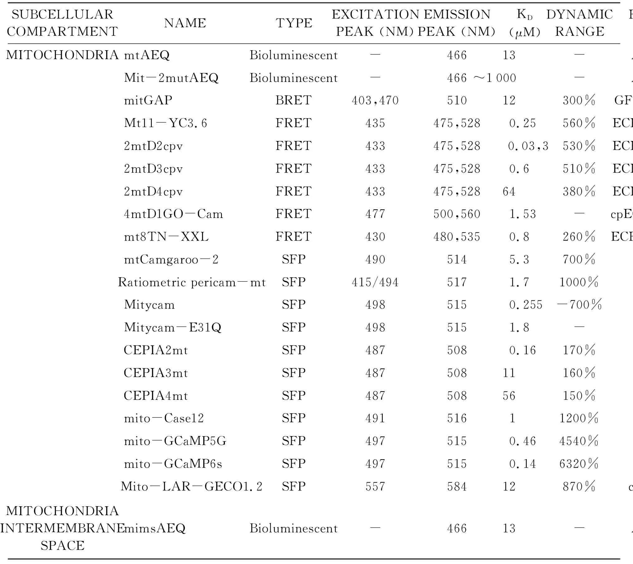
The current tools available for monitoring spatiotemporal Ca2+dynamics at subcellular levels include Ca2+sensitive fluorescent chemical dyes and genetically encoded Ca2+indicators (GECIs).The major drawback of chemical dyes is that they have to be introduced into the cell either structurally through linking with acetoxymethyl ester (AM) or physically through patch clamp (whole cell configuration),which greatly restricts its application in deep tissue and could introduce artifacts caused by diffusion,meanwhile the byproduct of AM hydrolysis could be toxic.Furthermore,targeting of these dyes to subcellular compartments are usually not specific[7-8].The appearance of GECIs helps solve some of the issues.They are directly produced inside the cell so the diffusion has reached steady state long before the imaging starts.They could be fused with peptide sequences for specific subcellular compartments easily through genetic editing.They are continuously produced by the cell,which enables prolonged imaging for days or even weeks.They have lower cellular toxicity.Additionally,a large spectrum of Ca2+affinities can be acquired through cite-specific mutations[7,9].In this review we cover the major categories of GECIs developed in recent years,with focus on those targeted to mitochondria and already applied in excitable tissues such as skeletal muscle,cardiac muscle and neurons (Table 1).
Tab1SummaryofmitochondriatargetedGECIs

SUBCELLULAR COMPARTMENTNAMETYPEEXCITATIONPEAK (NM)EMISSIONPEAK (NM)KD (μM)DYNAMIC RANGEFLUOROPHORETARGETING SEQUENCEREFMITOCHONDRIAmtAEQBioluminescent-46613-AequorinCOX VIII[37]Mit-2mutAEQBioluminescent-466~1000 -AequorinCOX VIII[39]mitGAPBRET403,47051012300%GFP,AequorinCOX VIII[38]Mt11-YC3.6FRET435475,5280.25560%ECFP,cpVenusCOX IV[42]2mtD2cpvFRET433475,528 0.03,3530%ECFP,cpVenusCOX VIII[17]2mtD3cpvFRET433475,5280.6510%ECFP,cpVenusCOX VIII[46]2mtD4cpvFRET433475,52864380%ECFP,cpVenusCOX VIII[47]4mtD1GO-CamFRET477500,5601.53-cpEGFP,mKOkCOX VIII[18]mt8TN-XXLFRET430480,5350.8260%ECFP,cpCitrineCOX VIII[47]mtCamgaroo-2SFP4905145.3700%cpEYFPCOX VIII[30] Ratiometric pericam-mtSFP415/4945171.71000%cpEYFPCOX IV[31]MitycamSFP4985150.255-700%cpEYFPCOX VIII[48]Mitycam-E31QSFP4985151.8-cpEYFPCOX VIII[49]CEPIA2mtSFP4875080.16170%cpEGFPCOX VIII[26]CEPIA3mtSFP48750811160%cpEGFPCOX VIII[26]CEPIA4mtSFP48750856150%cpEGFPCOX VIII[26]mito-Case12SFP49151611200%cpEGFPCOX VIII[50]mito-GCaMP5GSFP4975150.464540%cpEGFPCOX VIII[52]mito-GCaMP6sSFP4975150.146320%cpEGFPCOX VIII[52]Mito-LAR-GECO1.2SFP55758412870%cpmAppleCOX VIII[33]MITOCHONDRIA INTERMEMBRANE SPACEmimsAEQBioluminescent-46613-AequorinN626 of glycerol phosphate dehydrogenase[40]
- Data not available.
1 Major categories of GECIs
1.1 Bioluminescent and BRET GECIs The development of GECI can be traced back to 1962,when the Ca2+sensitive bioluminescent protein aequorin was discovered[10].The modified aequorins,combined with the use of different coelenterazine derivatives,could be used to measure Ca2+in the micromolar,or even millimolar range(such as the aequorin with Asp119Ala and Asn28Leu double mutation)for long periods of time[11].Additionally,they exhibit high signal-to-noise ratio and low pH sensitivity.Their interference with endogenous Ca2+buffering proteins is negligible[12].All these features make aequorin derivatives suitable for organelles with high Ca2+levels and non-neutral pH.However,aequorin has very low quantum yield.The bioluminescent signals are not conveniently compatible with modern time microscopy and hard to calibrate between cells/organelles with heterogeneous Ca2+levels.The cells also need to be pretreated with coelenterazine,which could potentially introduce spatial variations due to diffusion and consumption.Additionally,aequorin must be regenerated after emission upon Ca2+binding,which takes more than 10 minutes[9,12-13].
The bioluminescence signals of aequorin is so dim that the exposure time is usually more than several tens of seconds,which makes it impossible to monitor fast Ca2+transients in real time.To overcome this issue,the bioluminescence resonance energy transfer(BRET)probes have been developed by fusing Ca2+sensitive bioluminescent protein with a fluorescent protein[13].Similar to fluorescence resonance energy transfer (FRET),BRET probes take advantage of the differential efficiency of resonance energy transfer from the donor to the receptor moiety at the Ca2+bound and Ca2+unbound state,except that the donor is now a chemiluminescent protein.The exemplary probes include GAP(GFP-Aequorin Protein),BRAC (RLuc8-Venus)and Nano-lantern(split RLuc8-Venus)[9].For BRAC and Nano-lantern,calmodulin(CaM) and its physiological binding partner M13 were adopted to regulate the efficiency of resonance energy transfer in response to Ca2+levels[13-14].
1.2 FRET and SFP GECIs On the other hand,GECIs purely based on fluorescent proteins have been developed,including FRET-based sensors and single fluorescent protein (SFP) sensors.Most of the FRET-based sensors incorporate CaM-M13 as the Ca2+sensors to adjust the proximity of the donor and receptor fluorophores,including FIP-CBsmand cameleon derivatives such as D1-D4,D1cpv-D4cpv,D1GO-Cam,YC2-4 series,YC-Nano[15-20].One drawback of CaM-M13 based sensors is that the cell endogenous CaM could compete for the M13 binding.In D2cpv-D4cpv the CaM-M13 interface has been reengineered based on computational design to reduce nonspecific interactions with wild type CaM[17].One alternative approach is to use troponin-C to replace CaM.This type of FRET sensor includes Twitch and TN-XXL[21-22].The other alternative approach is to use the kringle domain of apolipoprotein A as the Ca2+sensing unit.The conformation change of this domain requires the activities of Ca2+dependent chaperones in endoplasmic reticulum (ER)[23].FRET-based sensors are ratiometric,which overcomes problems like the heterogeneity of expression levels between different cells,photobleaching,cell movement or focal plane shift during imaging[9,12],thus provides quantitative evaluation of intracellular Ca2+dynamics.Meanwhile the blue donor (such as CFP),yellow receptor (such as Venus or Citrine) pair generate the highest ratio change upon Ca2+binding,hence this fluorophore pair is the most common ones seen in current ratiometric Ca2+sensors[9].
The SFP sensors are mostly non-ratiometric (with some exceptions),but they have superiorities in other aspects.For example,a large spectrum of excitation/emission wavelength is available for these sensors.Blue/green ones include GEM-GECO,GEM-CEPIA,G-CEPIA,GCaMP,Case,CatchER,NTnC,Pericam (excitation/emission peak blue shifted).Yellow/orange ones include Camgaroo-2,O-GECO1.Red ones include R-GECO,CAR-GECO,LAR-GECO,REX-GECO (excitation peak blue-shifted),R-CEPIA,jRGECO,RCaMP,jRCaMP,K-GECO[24-36].The development of Ca2+sensors with different excitation spectra facilitate users to simultaneously monitor the dynamics of Ca2+and other molecules,or Ca2+dynamics in different subcellular compartments[9].The sensors with red-shifted excitation spectra generate lower background fluorescence,has lower phototoxicity and better penetration in deep tissue[32].Meanwhile these sensors also enable simultaneous Ca2+imaging and optogenetic manipulation of channel activities[9].However,some red-shifted Ca2+sensors (especially those derived from R-GECO) have blue-light-activated photoswitching behavior,which could complicate result interpretation when used together with channelrhodopsin-2 (ChR2) or other blue light activated optogenetic tools[35-36].
2 Monitor mitochondrial Ca2+ dynamics with GECIs
2.1 Application of bioluminescent and BRET GECIs The functional role of Mitochondria Ca2+uptake reported previously includes regulation of ATP production,activation of cell apoptosis and buffering cytosolic Ca2+increases[12].In 1992 the first organelle-targeted GECI was developed by fusing the mitochondrial pre-sequence of cytochrome c oxidase (COX) subunit VIII with aequorin (mtAEQ)[37].The application of this GECI revealed that agonist-induced elevations of cytosolic Ca2+evoke rapid and transient increases of mitochondrial Ca2+,which could be prevented by pretreatment with a mitochondrial uncoupler FCCP[37].Later on similar experiments were also done for the aequorin derived BRET sensor mitGAP[38].As the accumulation of Ca2+in mitochondria rapidly occurs in a few seconds,the low Ca2+affinity aequorin mutant (mit-2mutAEQ) help demonstrate the mitochondrial Ca2+elevation reaches and maintains at a steady state of 2~3 mM without phosphate and 0.5~1 mM with phosphate,respectively[39].Aequorin was also fused with fragment of glycerol phosphate dehydrogenase to target the mitochondrial inner membrane space (mims)[40].mimsAEQ revealed that active inositol 1,4,5-triphosphate (IP3)-gated channels of the ER establish high Ca2+microdomains between ER and mitochondria,which could trigger the opening of the low Ca2+affinity mitochondrial Ca2+uniporter (MCU) channel[40].
2.2 Application of FRET and SFP GECIs For FRET based Ca2+sensors,cameleon derivatives such as YC2,YC3.6,D1cpv-D4cpv,D1GO-Cam,TN-XXL have been targeted towards mitochondrial matrix in different systems[16-17,41-47].YC3.6 is suitable for mitochondrial Ca2+imaging due to the relatively large dynamic range (560%) and relatively high pH stability (cpVenus has a pKaof 6.0)[19].In skeletal muscle fibers,simultaneous observation of mitochondrial Ca2+dynamics (using mt11-YC3.6,which is YC3.6 fused with 11 tandem repeats of COX IV) and cytosolic Ca2+transients revealed a coupling between mitochondrial Ca2+uptake and voltage induced Ca2+release into the cytosol[42].Interestingly,in G93A mice (amyotrophic lateral sclerosis model) the defective mitochondria had a notable decrease of Ca2+uptake compared to the health ones[43].D2cpv is sensitive to a wide range of Ca2+concentrations and hence has been proposed to use in mitochondria.However,its biphasic Ca2+response curve may put challenges to quantification[17].D3cpv has been applied in measuring plant mitochondrial Ca2+dynamics but seems to be less sensitive than YC3.6,potentially due to its larger Kd(600 nM vs 250 nM of YC3.6)[46].D4cpv has a very large Kd(64 μM) and hence is more suitable for monitoring high level Ca2+[17].For example,mitochondria targeted D4cpv has been applied in Drosophila motor neuron terminals to measure the maximum mitochondrial Ca2+level,while TN-XXL (Kd= 800 nM) was used for measuring minimum mitochondrial Ca2+level[47].A positive correlation between membrane hyperpolarization and mitochondrial Ca2+level has been observed in this study[47].Mitochondria targeted D1GO-Cam (4mtD1GO-Cam) also has a relatively large kd(1.53 μM).The red-shifted excitation wavelength of its donor fluorophore (cp173-mEGFP) enables it to be used together with fura-2 for simultaneous observation of mitochondrial and cytosolic Ca2+dynamics.Meanwhile 4mt D1GO-Cam showed less off-targeting effect than 4mtD3cpv[18].
SFP sensors that have been applied for monitoring mitochondrial Ca2+dynamics include Camgaroo-2,pericams,CEPIAs,Case12,GCaMPs,and LAR-GECO1.2[26,30-31,33,48-52].Camgaroo-2 has a large Kd(5.3 μM) suitable for detecting variations at high Ca2+concentrations.It is also relatively photostable and resistant to pH changes (The fluorophore Citrine in Camgaroo-2 has a pKaof 5.7) compared to Ca2+sensors based on first generation YFP (pKa of 6.9-7.1)[30].Mitochondria targeted Camgraoo-2 showed response to histamine and ionomycin treatment,but has a much smaller dynamic range than those in cytosol[30].Ratiometric pericam is one of the few examples of ratiometric SFP sensors,as the excitation wavelength changes in a Ca2+dependent manner.Its dynamic range is larger than most cameleon type GECIs[31].Application of mitochondria targeted ratiometric pericam in Hela cells confirmed systematic increase of mitochondrial Ca2+after perfusion of histamine.Interestingly,transient Ca2+uptake by some mitochondria was also observed during the falling phase after the administration of histamine[31].Another pericam family member:inverse pericam has also been fused with COX VIII (termed Mitycam) and used for mitochondrial Ca2+imaging in cardiomyocytes[48].Because inverse pericam has a Kdof about 200 nM,additional mutations were introduced to produce low Ca2+affinity Mitycam[49].CEPIAs are derived from the GECO sensors by introducing mutations in CaM to bring down the Ca2+affinity and hence are more suitable for high Ca2+cellular compartments[26].Application of mitochondria targeted CEPIAs in Hela cells help reveal the heterogeneity of mitochondrial Ca2+response to agonist induced ER Ca2+release,despite of the uniform Ca2+elevation in cytoplasm[25].Mitochondria targeted Case12 has been applied to reveal correlations between mitochondrial Ca2+and motility in hippocampal neuronal axons[50].GCaMPs are very popular Ca2+biosensors these days due to their extremely large dynamic range and great photostability[24].They have also been adopted for imaging mitochondrial Ca2+dynamics in neurons and astrocytes[51-52].However,GCaMPs are known to be sensitive to pH changes,which may complicate the result interpretation when used in mitochondria[53].The red-shifted SFP sensor LAR-GECO1.2 has a large Kdof 12 μM[33].It has been targeted to mitochondria in dorsal root ganglion and hippocampal neurons from mice (primary culture) to demonstrate that KCl induced depolarization evoked coincident and rapidly reversible increases in mitochondrial and cytosolic Ca2+(monitored by fura-2)[33].
3 Prospect of combinatory use of GECIs,other sensors and photoactivatable tools
3.1 Simultaneously monitoring Ca2+dynamics in multiple cellular compartments One of the major advantages of GECIs is the ease of introducing targeting sequences against different subcellular compartments,including cytosol,plasma membrane,nucleus,ER/SR,mitochondria,Golgi apparatus,endosome,secretory granules,lysosome and even primary cilia,although the targeting efficiency for some of these compartments have large rooms to improve[54].Since Ca2+channels,pumps and binding proteins exist in many of these compartments,the major and minor downstream effects of Ca2+signaling can be distinguished better when the Ca2+dynamics in multiple compartments could be monitored simultaneously.This would also help clarify the order of events along the signal cascade,and unveil the presence of potential positive/negative feedback loops.
3.2 Simultaneously imaging the dynamics of Ca2+and other molecules The physiological and pathological function of Ca2+signaling relies on its crosstalk with other signaling pathways and molecular activities.For example,the denervation of muscle from motor neuron causes muscle atrophy,which is accompanied by increased production of reactive oxygen species (ROS) in the mitochondria,elevated Ca2+content in both mitochondria and cytosol[42,55].Simultaneous observation of mitochondrial ROS,Ca2+as well as cytoplasmic Ca2+would help clarify the cause-and-effect and feedback relationships between them.
3.3 Combining Ca2+imaging with optogenetic tools Since the discovery of channelrhodopsin[56],more and more photo-regulatable channels and binding proteins,including Ca2+specific ones,have been developed,such as PACR,LOVS1K,OptoSTIM1,and Opto-CRAC[57-60].These tools could artificially induce cytosolic Ca2+transients with customizable altitude and time span.Most optogenetic tools currently available are activated by blue light,making them compatible with red-shifted,organelle-targeted GECIs to investigate the impact of cytosolic Ca2+transients on organelle Ca2+profile.The photoswitching behavior of some red-shifted GECIs need to be solved before this combinatory application takes place.On the other hand,attempts to specifically and efficiently targeting these tools to different organelles would greatly facilitate the investigation of inter-organelle relationships in Ca2+regulation.
GECIs have been proven to be useful tools for imaging Ca2+dynamics in cells,tissue culture and intact animals due to their unique advantages that are irreplaceable by Ca2+sensitive chemical dyes.Continuous efforts have been made to optimize the properties of GECIs including dynamic range,kinetics,Kd,pH stability,photostability,excitation wavelength variation,targeting towards different cellular compartment etc,which would further broaden the application of GECIs.

