农药残留的表面增强拉曼光谱快速检测技术研究现状与展望
张文强,李 容,许文涛
农药残留的表面增强拉曼光谱快速检测技术研究现状与展望
张文强1,李 容1,许文涛2
(1. 中国农业大学工学院,北京 100083; 2. 中国农业大学食品科学与营养工程学院,北京 100083)
果蔬农药残留危害人类健康,施药后,农药分布于其表皮和内部组织,果蔬表面农药绝对残留量低、不均匀,直接光谱检测表征难,而表面增强拉曼散射(surface-enhanced Raman scattering, SERS)技术具有分子级检测精度,可以有效扩增信号,在实现微量物质检测方面优势明显。为此,论文综述了国内外表面增强拉曼散射技术的研究现状,特别是详细介绍了通过设计合理的表面增强拉曼基底结构,实现农药残留信号增强的主要技术手段和表面增强拉曼光谱信号分析方法。在此基础上,指出农药残留的表面增强拉曼检测技术研究中的前沿热点问题,探讨并展望了表面增强拉曼技术在农药残留快速检测方面的发展趋势。基于表面增强拉曼的农药高灵敏度、快速检测表征技术,将在农药违禁使用和农药残留超标监管中有广阔应用前景。
农药;光谱分析;无损检测;残留;表面增强拉曼散射光谱
0 引 言
中国作为农药生产和施用大国,已有农药制剂1 000多种,农药用量达到337万t/a,防治面积达3 亿 hm2以上[1];但农药只有20%~30%的利用率,不适时、不对症和过量用药,带来了农药残留毒性、病虫抗(耐)药性上升、环境污染等一系列问题[2]。农药在施用到果蔬表面后,不同种类的农药迁徙特性不同,在表皮、果浆、果肉的分布与迁徙规律也不尽相同,已有的农药残留检测方法,需要对果蔬进行破碎后,采用化学萃取等前处理手段富集农药后再进行表征[3],操作复杂,不能适应现场快速、实时检测的需求。光谱检测法尽管具有对农药快速、无损的检测特征,且研究表明,利用红外、拉曼光谱等手段可以实现多种农药的一致检测。但分布于果蔬表皮表面的农药残留占果蔬整体农药残留比例较低,光谱信号弱,需要采用大型高灵敏设备进行探测,不能满足现场快检的需求。近来研究发现,采用表面增强拉曼的方法,不仅可以极大提升农药的特征拉曼光谱信号强度,还突破了液体中拉曼光谱信号弱、荧光干扰的问题,有可能实现无损条件下的现场微量甚至分子级农药的快速检测。采用商品级纳米金/银球与农药溶液混合的方法进行增强检测,由于团聚、氧化、组分不稳定的原因,难以使分子有效附着在增强基底表面,信号增强效果有限,定量分析表征难。为此,国内外表面增强拉曼光谱领域的研究者通过研制多种功能材料的纳米表面增强拉曼基底,优化获得各种农药最佳的特征拉曼信号增强单元或组合结构,力图实现微量农药快速检测并建立低浓度农药特征拉曼信号的表征模型。此外,为实现基于表面增强拉曼散射(surface-enhanced Raman scattering, SERS)技术的农药残留现场快速检测,对农药拉曼光谱进行分析必不可少。拉曼光谱分析技术是以拉曼效应为基础建立起来的分子结构表征技术,其信号来源于分子的振动和转动[4]。拉曼光谱分析的研究方向一般分为定性分析(不同的物质具有不同的特征光谱)、结构分析(对光谱谱带的分析)及定量分析(根据物质对光谱的吸光度计算所得)。由于不同农药具有不同的拉曼特征峰,因此对被检测物的分析主要运用“特征峰位移”进行定性、定量判定。农药的实际使用过程中多是几种农药混合使用,有必要研究快速、准确判定混合农药残留的特征拉曼光谱计算模型。本文从表面增强活性基底的制备、农药残留表面增强拉曼光谱分析两方面综述了农药残留SERS检测技术的研究现状,展望农药残留的SERS检测方法发展趋势,以期为SERS技术在指导农药施用、农产品生产收获、控制食品安全等方面提供参考。
1 农药残留表面增强拉曼光谱研究
1.1 表面增强基底研究进展
1928年Raman和Krishman首次在液体的散射光中发现了拉曼散色现象[5],对CCl4的检测限一般只能达到10-3g/L,而当激发光波长落在分子的电子跃迁吸收波长区间时,拉曼散射强度就会提高103~104倍[6]。1974年Fleischmann等[7]发现吡啶分子吸附在粗糙金银表面时,拉曼信号强度提高显著,同时信号强度随着电极所加电位的变化而变化。直至1977年,Jeanmaire等[8]与Albrecht等[9]经过系统的试验研究和理论计算后,将这种与金、银、铜等粗糙表面相关的增强效应称为表面增强拉曼散射效应,对应的光谱则称为表面增强拉曼光谱。SERS效应发现以来,研究者们长时间运用粗糙化的Au、Ag金属作为增强基底,其中Frens法[10]至今仍是制备纳米金粒子的经典方法,此外还有机械研磨法、金属蒸汽溶剂化法、还原含金/银化合物法、晶种生长法等。1985年张鹏翔等[11]根据Creighton描述的纳米银胶体制备法,用NaBH4还原AgNO3水溶液得到粒径约20 nm的银溶胶增强基底,将其运用于苯甲酸饱和水溶液SERS检测中,得到的拉曼信号与普通苯甲酸饱和水溶液谱图相比,其谱峰强度约提高1倍,增强因子达2.3×10⁴。Emory等[12]利用分布过滤的方法分离不同粒径的银粒子,并发现粒径在80~100 nm之间SERS信号最强。王健等[13]通过柠檬酸三钠还原HAuCl4制备了粒径分别约为16、26、40、66 nm的金粒,直接获取偶连层分子的SERS信号,发现在所研究的粒径范围内,SERS增强因子随粒径的增大而增加。金银溶胶的经典制备方法虽然比较方便,但是直接使用纳米金/银胶体作为拉曼增强基底,受纳米金属的氧化和团聚等因素的影响,便携式激光拉曼光谱仪的检测灵敏度会降低1~2个数量级,不能达到农药残留量标准检测要求[14-15]。此外采用贵金属作为增强基底材料,存在成本高、不可重复利用等缺点,制约其在现场快速检测中的应用。因此拉曼增强基底从简单纳米Au、Ag微粒,研究逐渐趋向于廉价金属纳米微粒、各向异性纳米氧化物微粒、纳米阵列结构等方向发展[16]。
1.2 农药残留表面增强拉曼基底研究
1.2.1 表面增强拉曼光谱活性基底制备研究
国内外研究表明通过优化设计、选择合适的材料、完善制造工艺参数等手段可以获得SERS性能显著提升的活性基底,在果蔬表面农药残留快速检测中已经取得一定的进展。Zhao等[17]通过化学反应将碳点(carbon dots, CDs)与Ag纳米颗粒(Ag nanoparticles, Ag NPs)结合形成Ag NPs/CDs杂化物作为新的表面增强拉曼散射(SERS)基底,可以检测浓度低至10-9mol/L的氨基苯硫酚(-aminothiophenol, PATP)。宾夕法尼亚州立大学的Feng团队[18]通过控制氮掺杂石墨烯的费米能级(fermi level, EF)偏移使电荷转移增强,从而显著扩大分子的振动拉曼模式。试验证实此基底可以实现浓度为10-11mol/L的罗丹明B、结晶紫和亚甲蓝分子的检测。Kim等[19]利用银镜为表面增强基底,采集不同pH值下的苯并咪唑类表面增强拉曼光谱,对拉曼谱峰进行了归属,其试验结果与密度泛函理论计算值吻合较好。Carrillo-Carrión等[20]制备了具有CdSe/ZnS颗粒的Ag-QD纳米结构,使其形成核-壳量子点SERS活性基底。使用微分散器与海绵状SERS活性基底结合形成精确的液相色谱检测系统,对注射农药(绿麦隆、莠去津、敌敌畏和特丁津)进行SERS检测,检测限可达0.2~0.5 ng/L,精确度在10.2%~12.5%之间。Li等[21]和Kubackova等[22]分别研究了基于改性纳米银微粒和二氧化硅包覆纳米金微粒做基底材料,对有机氯农药和对硫磷农药进行快速检测,证实改性和控制微粒排布有利于检测灵敏度提升,如图1所示。
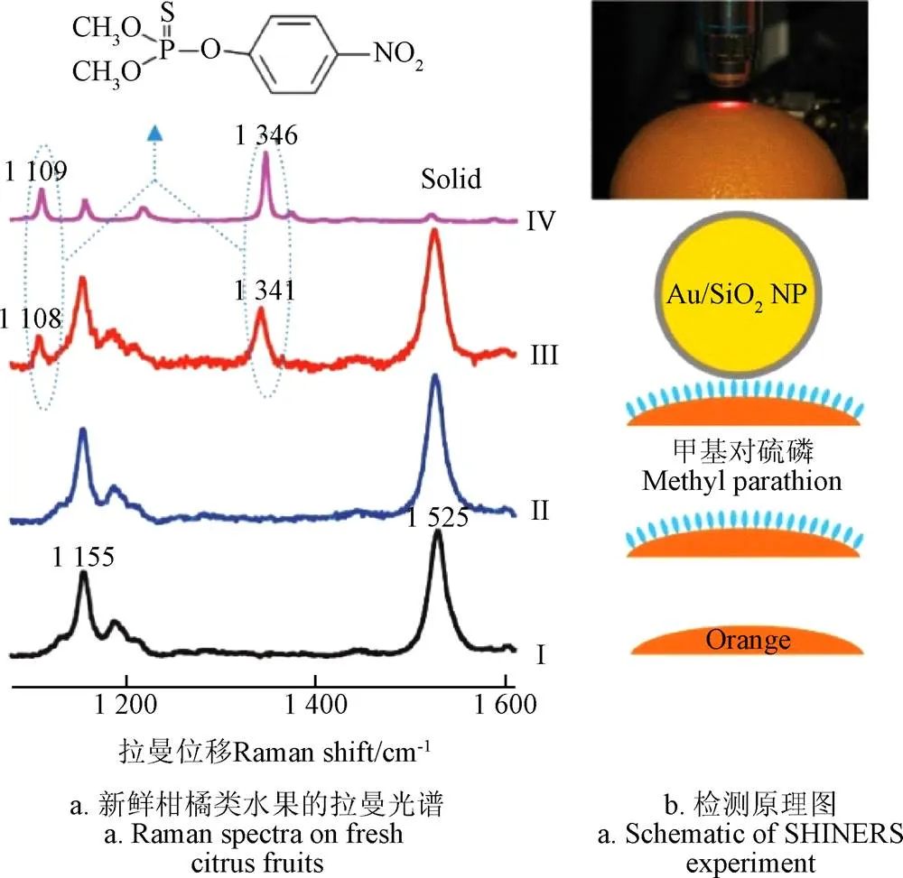
注:I新鲜柑橘的拉曼光谱曲线;II含有对硫磷的果皮拉曼光谱;III金/二氧化硅纳米颗粒修饰的柑橘表面的拉曼光谱;IV固体甲基对硫磷拉曼光谱。(激光功率为0.5 mW,采集次数为30 s)
Dai等[23]采用三步法制备了多功能TiO2/Ag纳米粒子(Ag NPs)复合基底,该基底对苹果汁中福美双的检测限低于美国环境保护局(U.S. environmental protection agency, EPA)规定的水果中最大残留限量(如图2所示)7 ppm(2.9×10⁻⁵mol/L)。一般三维纳米颗粒制备过程中都会引入十六烷基三甲基溴化铵(hexadecyl trimethyl ammonium bromide, CTMAB)等表面活性剂[24],而这些活性剂会抑制痕量样品的SERS检测,因此Xu等[25]开发了一种无须表面活性剂的方法制备出爆米花状金纳米颗粒,并借助纳米孔之间形成的“热点”,对水果样品表面毒死蜱残留进行检测,检测浓度低至1mol/L,符合国家标准。Li等[26]通过加热回流法获得由硬脂酸(stearic acid, SA)和聚乙烯吡咯烷酮(polyvinyl pyrrolidone, PVP)混合物改性合成的Ag/ZnO纳米复合材料。改性的Ag/ZnO纳米结构与普通亲水性Ag/ZnO基底相比,具有3倍的增强效果。Li等[27]制备的Ag2O@Ag核-壳型纳米结构活性增强基底,可以避免环境中杂质带来的干扰,从而获得更稳定的信号;同时超薄的内壳结构让基底灵敏度更高、均匀性和重现性更好,对R6G的检测浓度低至10-11mol/L,增强因子高达106。实际应用中,Ag2O@ Ag/PMMA混合形成的柔性SERS基底对黄瓜和苹果皮上的毒死蜱进行实时原位检测,检测浓度低至10-7mol/L。Li等[28]通过制备PS/Ag纳米颗粒作为动态SERS检测活性基底,对有机磷杀虫剂对氧磷和杀螟松进行低浓度检测,检测浓度低至10 nmol/L时仍有较好的检测效果。Wang等[29]在2D PS模板上通过溅射Ag和SiO2材料交替地制备了Ag/SiO2多层的“柱帽”形阵列。在氩气下退火加速了Ag和SiO2之间的界面扩散,一些Ag颗粒通过SiO2孔隙进入SiO2层间,根据退火温度不同,Ag纳米粒子的尺寸从2~5 nm不等,在Ag颗粒和底层Ag之间产生1~2 nm SiO2的分离,为SERS提供了大量的热点位点。
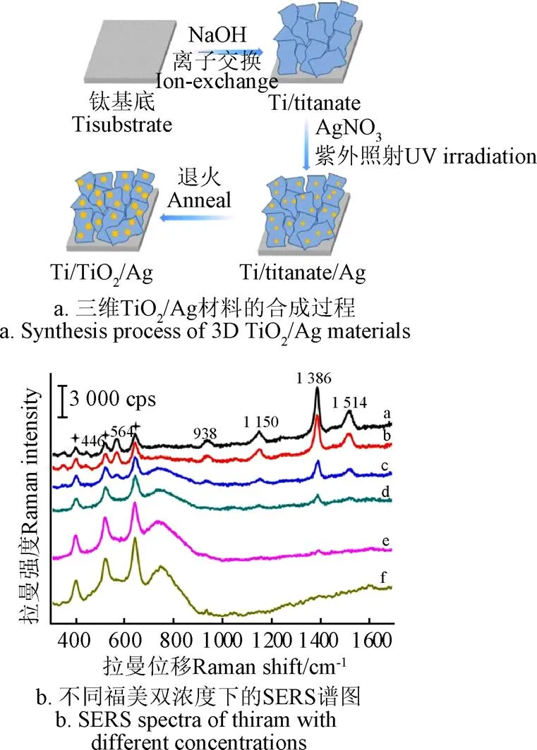
注:图b中a-f为在SERS基底上分别掺加10-3、10-4、10-5、10-6、10−7、0 mol×L-1福美双。
1.2.2 表面沉积纳米颗粒的阵列SERS基底研究
阵列SERS结构可以有效提升快速检测结果的均匀性、一致性、准确性,如Zhu等[30]构筑了具有强电磁场耦合效应的银纳米棒簇有序阵列,该阵列的表面增强拉曼光谱增强因子高达108,并具有较好的信号均匀性和重现性,能够同时检测水果中多种痕量农药,如甲基对硫磷和2-4-二氯苯氧乙酸(如图3所示)等。Kumar等[31]通过将Ag纳米棒嵌入聚二甲基硅氧烷(PDMS)聚合物中来制造新型表面增强拉曼光谱阵列基底。在机械拉伸应变条件下对这些柔性基底进行原位表面增强拉曼光谱测量。研究结果表明,柔性表面增强拉曼光谱基板可以承受高达30%的拉伸应变()值,而不会损失表面增强拉曼光谱性能。通过简单的“粘贴和剥离”方法从果皮中直接提取微量(约10-9g/cm2)的福美双(thiram)杀虫剂,证明了银纳米颗粒(Ag NRs)嵌入PDMS组成表面增强拉曼光谱基底的高灵敏检测功能。
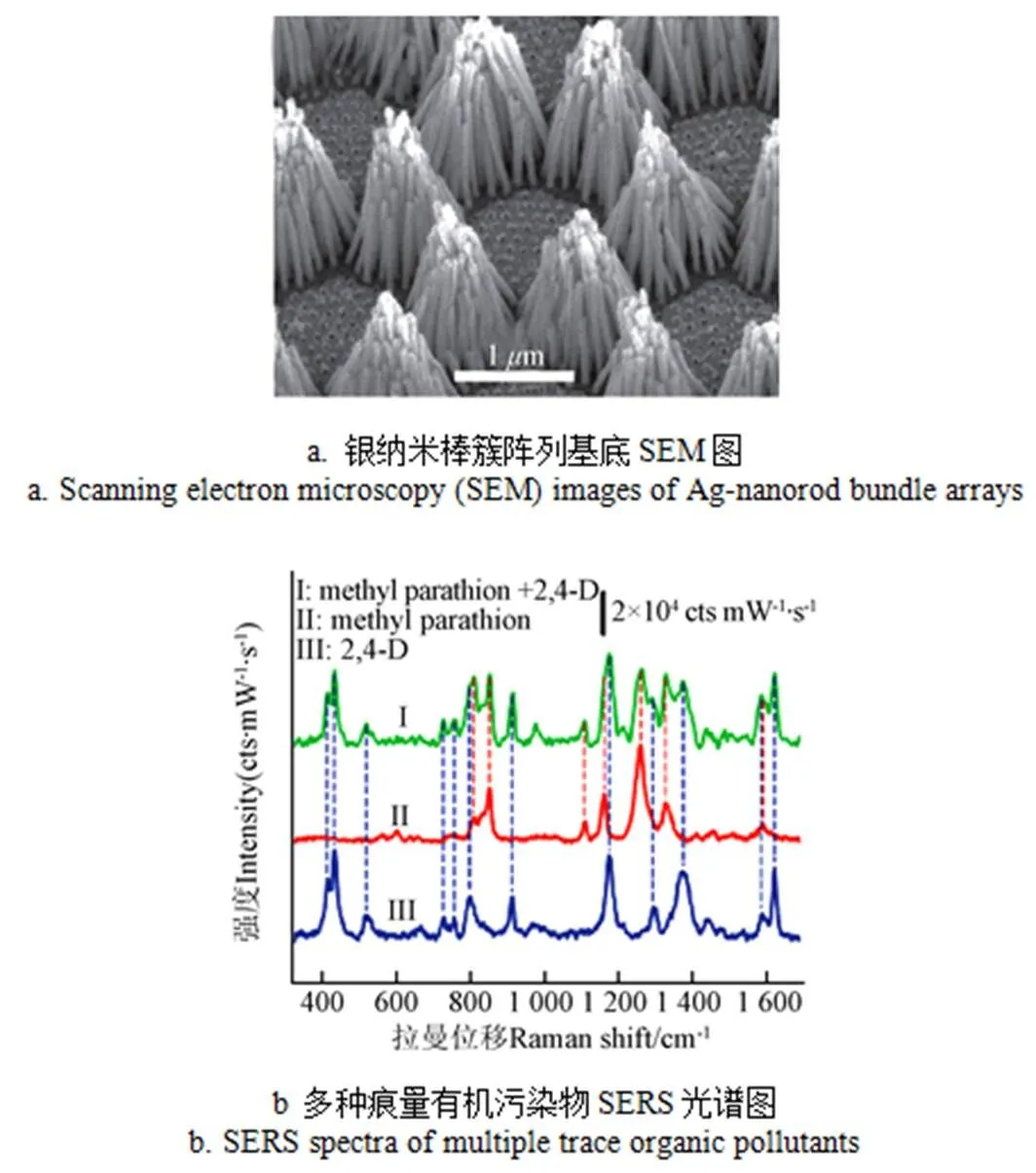
图3 痕量有机污染物表面增强拉曼检测[30]
近年来,如何低成本、快速制造纳米阵列表面增强拉曼光谱结构备受关注。Wei等[32]通过使用Au纳米颗粒/蜻蜓翼(Au NPs/DW)阵列作为SERS活性基底,通过简单的二步法在蜻蜓双翼(DW)表面上沉积组装金纳米颗粒(Au NPs)作为增强检测基底,对罗丹明6G(R6G)的检出限可达10-8mol/L,对于三维Au NPs/DW的应用,也可以定量检测西维因和甲萘威的微量样品,检出限达到10-7mol/L。Ren等[33-34]、Kwon等[35]分别利用硅藻多孔及其吸附特性和透明介质属性,在其表面自组装了银、金纳米颗粒(如图4,图5所示),检测发现拉曼增强效果好于单纯的纳米颗粒、薄膜。
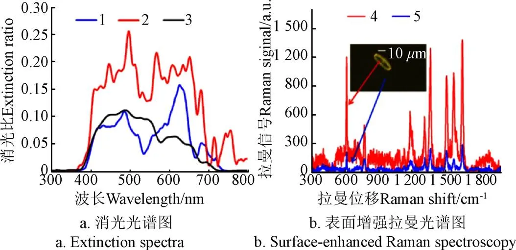
注:1纳米颗粒置于平板玻璃上;2纳米颗粒置于硅藻上;3未加修饰的硅藻;4 硅藻壳上的表面增强拉曼散射;5 玻璃基底上表面增强拉曼散射。
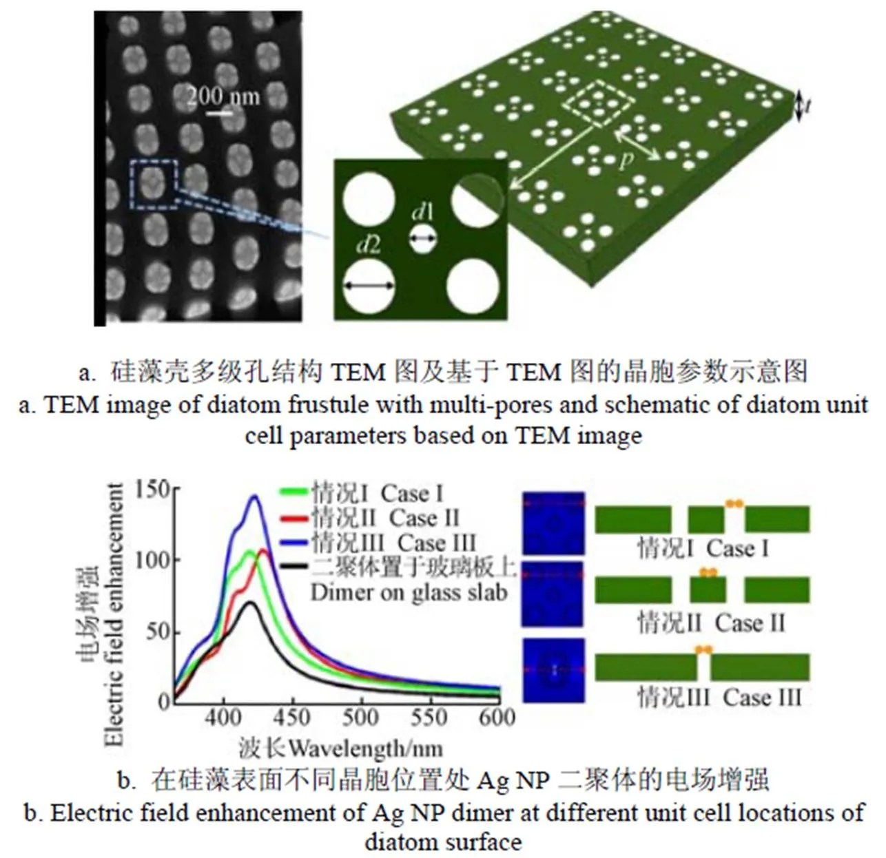
注:情况Ⅰ和情况Ⅲ, Ag NPs二聚体位于孔上方;情况ⅡAg NPs二聚体位于壳体上方;
Zhan等[36]推出了制造具有有序纳米结构且低成本的柔性SERS基底。将聚二甲基硅氧烷(polydimethylsiloxane, PDMS)滴加到自组装聚苯乙烯(polystyrene, PS)纳米颗粒(NP)模板上时,PDMS溶液在固化过程中的流动性可以更好地进入PS纳米球(NP)之间的间隙以形成更紧密的填充,使PDMS膜上形成有序纳米结构。该基底用于弯曲物体上测试分析物,增强因子约为107,重复性好,偏差小于13%。并有望扩展到灵敏传感器和执行器的未来应用中。刘绍根等[37]制备了具有柔韧性和透光性的Au NPs/PMMA表面增强拉曼基底,其对鱼表面残留孔雀石绿的原位检测下限达到 0.1mol/L。Sivashanmugan等[38]运用聚焦离子束与纳米压痕法制备出Au/Ag/Au纳米棒阵列,以罗丹明6G作为探针分子进行SERS检测,得到增强因子达2.15×108,并用该阵列基底对氯菊酯、氯氰菊酯、甲萘威和亚胺硫磷杀虫剂进行痕量检测,检测浓度可低至10-8mol/L。
此外,本课题组系统地研究了基于硅藻的功能结构制造技术,已经实现了基于硅藻的微纳米功能单体、器件、微流体芯片的制造;如利用空气液面自组装方法实现硅藻的一致定向密排[39]。以硅藻为模板,在自组装前提下,实现纳米级金微柱阵列的制造,并用结晶紫作为检测探针得到极强的拉曼增强效果(如图6a所示)[40],其增强因子达到7.64×10⁴;试验证实硅藻壳体的多级孔系结构,对蛋白分子、脂质体分子、微米级的碳素微粒、神经毒气、喹硫磷均有吸附富集作用[41-42]。对比发现,硅藻富集吸附后,利用生物分子马达对农药喹硫磷的检测限值从5g/mL下降到0.5g/mL。此外,通过硅藻的多级孔系结构及其本身带负电性质成功吸附纳米Au颗粒,以此制备的SERS基底呈单层稳定状态,试验也证明了该基底对核酸的拉曼检测有较强增强作用(如图6b所示)。结合上述国内外研究文献资料,不难发现,基于复合拉曼增强基底的低浓度、非均匀、便携快速的高灵敏检测,以及低成本制造,多种结构阵列化耦合技术等将成为未来一段时间内的研究热点。
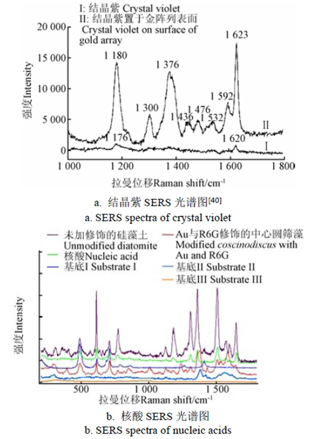
注:基底Ⅰ,Au NPs置于修饰中心圆筛藻上;基底Ⅱ,Ag NPs和R6G置于修饰中心圆筛藻上;基底Ⅲ,Au NPs和R6G置于中心圆筛藻上。
2 基于SERS的农药检测分析
农药残留分析不仅需要考虑其痕量检测拉曼光谱信噪比低、微弱信号极易被荧光信号泯灭等问题,还需考虑农药混合使用复杂体系中其它未知组成成分的干扰因素。为此,国内外开展了借助拉曼光谱检测分析方法对SERS谱图进行定性、定量分析算法的研究。拉曼振动峰的位置只与化学键的振动频率或转动频率有关,不同位置的拉曼峰代表了不同的化学键,反映了分子的结构信息,所以根据拉曼光谱可以确定分子的结构,而分子的化学键等结构信息同样表现在拉曼光谱上。因此确定各种农药的特征拉曼位移是进行农药残留表面增强拉曼光谱检测的前提条件[43]。黄双根等[44]应用密度泛函理论对3种有机磷类农药分子(乐果、敌百虫和伏杀硫磷)进行了几何结构优化和频率计算,并将试验拉曼光谱、理论计算拉曼光谱和表面增强拉曼光谱进行比较,对这3种有机磷类农药分子在400~1 800 cm-1范围内的振动频率进行了全面地归属。得出乐果明显拉曼振动峰位于494、646、764、908、1 164和1 136 cm-1处;敌百虫的位于438、618、784、1 026和1 238 cm-1;伏杀硫磷的位于650、692、755、1 109、1 238和1 283 cm-1处。赵琦等[45]采用距离匹配和判别分析的方法对苹果汁中马拉硫磷和二嗪农进行定性分析,再结合偏最小二乘法(partial least squares, PLS)对2种农药的表面增强拉曼光谱分别进行数学建模分析。结果表明表面增强拉曼散射技术对无损快速定性分析马拉硫磷和二嗪农具有较高的准确性,而定量分析二者的含量也具有较高的可行性;其中马拉硫磷定量模型相关系数为0.99,校正均方根误差为0.02,校正样本的拟合值与真实值的最大残差为0.059 mg/kg;二嗪农定量模型相关系数为0.99,校正均方根误差为0.01,校正样本的拟合值与真实值最大残差为0.03 mg/kg。李晓舟等[46]通过对苹果表面残留农药进行检测,从获得的谱图中选取了728、1 512 cm-1处的拉曼信号分别作为甲拌磷与倍硫磷定量分析特征峰,并采用内定法分别建立了甲拌磷和倍硫磷的线性回归模型。利用拉曼光谱的方法对物质检测时,每种物质特征峰的检出数量,除了与增强基底有关,还与物质的浓度密切相关,所以当检测低浓度农药时就不可能将其所有的特征峰找出并一一对应,必要研究并找到被测物的特征明显且在不同浓度下均能稳定检出的特征峰。对此邹明强等[47]对百草枯经过多样品试验得出其SERS强度与对应含量进行线性分析,明确百草枯最明显特征峰在1 665 cm-1处。王晓彬等[48]检测脐橙果肉提取液中三唑磷溶液最低检测浓度为0.5 mg/L,选用2 257 cm-1处乙腈C≡N伸缩振动峰为内标峰,以三唑磷1 409 cm-1特征峰与乙腈2 257 cm-1特征峰强度比值作为相对强度,得出其相对强度在三唑磷溶液浓度为0.5~20 mg/L范围内具有良好的线性关系,同时,建立的模型具有较好的预测效果,其预测误差在小于0.12 mg/L,预测回收率为98.1%~102.5%。He等[49]开发了一种快速(约10 min)简单的表面擦拭捕获农药的方法,利用不同浓度的噻苯达唑(thiabendazole,TBZ)建立标准校准模型,在计算得到释放因子为66.6%基础上对苹果表面噻菌灵进行回收率计算和定量检测,得到不同水平下噻菌灵回收率在59.4%~76.6%之间,擦拭-表面增强拉曼法最终计算准确度可达89.2%~115.4%。上述文献表明,对单一品种农药的拉曼光谱特征信号增强及拉曼光谱特征峰值曲线表征与计算方法可行,开展结合实际工况的复杂环境下(多种混合分子条件下)的拉曼光谱特征曲线表征研究,建立对应的标准校准模型,实现痕量农药的精准定量表征尤为重要。
3 农药残留的SERS检测技术发展趋势展望与应用价值探讨
从农药使用现状来看,经营者多只看到高产量带来的高经济收益而忽略消费者健康与生态环境保护,不合理配药、施药使作物抗性增强。因此市场监管部门需要对农药毒性划分并能给予消费者直观的判断;如何运用SERS检测技术对农药毒性进行快速区分尤为重要,其难点不在于浓度的高低而是如何快速划分并直观显示检测结果。从果蔬购买者来说,由于种植者为提高果蔬产量会在实际种植中使用杀虫剂、除草剂等农药,使得最终果蔬表面残留多种混合农药,而能否即检即测农药残留是否超标,是果蔬购买者最为关心的问题;在未知农药类型和微量无损条件下,农药残留的有效、快速低成本感知技术研究十分必要,相对其他快速检测方法来说SERS检测技术对农药的检测限虽低,但是其难点在于仅仅通过特征峰判别种类对于混合农药的检测存在困难,要解决该难点就需要针对常用农药在低浓度状态下提取每种农药特征峰并建立农药残留拉曼图谱库。考虑到在实际运用中农药残留的SERS检测分析谱图并非与所有待测农药之间存在线性关系,必要借助建立多局部线性模型[50]或者使用人工神经网络方法(artificial neural network, ANN)[51]等非线性建模手段对待测物质进行定量分析。
金属表面产生SERS增强通常被解释为表面等离子体共振(surface plasmon resonance,SPR)引起的[52]。同时,表面等离子体共振激发主要集中在纳米颗粒之间的空隙(称为热点)[53]。而SERS检测均匀性、重现性不好的主要因素就是因为制备的金属胶体稳定性差,容易聚集,大大减少SERS检测热点;使得信号强度提升小、测试结果不稳定。本文综述的文献表明,制备三维有序阵列的增强基底,由于3D纳米结构的比表面积较大、结构稳定有序,在进行SERS检测时,吸附待测分子的能力显著,获取的SERS信号稳定性和均匀性更好。同时,通过精确控制增强基底阵列纳米颗粒之间形成的间隙大小可以产生更高密度的检测“热点”,从而具有更好的重现性。所以未来农药残留检测的SERS基底应考虑农药富集浓缩结构和图形化表面增强拉曼结构在微流控芯片上的集成制造。该结构一方面能最大程度增加单位面积上的农药含量,提升信号强度;另一方面,让微量农药与“带有指纹”功能的图形化多种表面增强拉曼复合结构充分接触,提升特征信号增强效果,实现混合农药的定性定量分析。此外,通过设计制造一种具有阵列结构的柔性透明表面增强拉曼贴片,在施药后,立即将功能贴片贴敷在果蔬表皮,通过拉曼光谱仪照射贴片,可以快速、直观获取果蔬表面的农药分布和含量,为合理施药提供指导。
然而,就目前的现状来看,上述柔性透明阵列基底距离实际应用仍有一段距离。首先,从活性基底的制造工艺来看,有序阵列基底的制备工艺流程复杂,成本较高;此外,柔性基底对材料的要求高,目前集中在柔性碳材料及部分高分子聚合物上。其中柔性碳材料的制作工艺困难,而高分子聚合物本身的拉曼信号干扰又会增加光谱分析难度。因此在果蔬农药残留实际检测过程中,仍需努力突破以上瓶颈;进一步,结合各类农药的迁徙和实效模型,建立果蔬农药残留状态的精准预测模型,才能最终实现服务互联网+农业和安全消费的目标。
4 结 论
表面增强拉曼光谱技术作为一种快速检测技术,在农药痕量检测,尤其是农药残留快速筛选方面有较大的应用潜力。由于表面增强拉曼光谱信号的稳定性和重现性主要取决于于表面增强基底的纳米材料选择、阵列结构等,研制方便易用的表面增强基底并不断提升其灵敏度,建立复杂条件下农药特征拉曼光谱表征模型,将有效拓宽表面增强拉曼光谱的应用领域,为安全合理施药提供技术支持,保障食品安全。
[1] 谢昌会. 当今农药的使用现状及存在的问题[J]. 农业开发与装备,2017, (2):99.
Xie Changhui. Current status and problems of pesticide use[J]. Agricultural Development and Equipments, 2017, (2): 99. (in Chinese with English abstract)
[2] 朱赫,纪明山. 农药残留快速检测技术的最新进展[J].中国农学通报,2014,30(4):242-250.
Zhu He, Ji Mingshan. Recent advances in rapid detection technology of pesticide residue[J]. Chinese Agricultural Science Bulletin, 2014, 30(4): 242-250. (in Chinese with English abstract)
[3] Sagratini Gianni, Mañes Jordi, Giardiná Dario, et al. Determination of isopropyl thioxanthone (ITX) in fruit juices by pressurized liquid extraction and liquid chromatography-mass spectrometry[J]. Journal of Agricultural and Food Chemistry, 2006, 54(20): 7947-7952.
[4] 李琼. 微型拉曼光谱仪的拉曼光谱数据处理方法研究[D]. 重庆:重庆大学,2008.
Li Qiong, Study on Data Processing of Raman Spectrum Based on Mini-Spectroscopy[D]. Chongqing: Chongqing University, 2008. (in Chinese with English abstract)
[5] Cialla D, Maerz A, Boehme R, et al. Surface-enhanced raman spectroscopy (SERS): Progress and trends[J]. Analytical & Bioanalytical Chemistry, 2012, 403(1): 27-54.
[6] 董学锋. 拉曼光谱传递与定量分析技术研究及其工业应用[D]. 杭州:浙江大学,2013.
Dong Xuefeng. Research and Industrial Application of Raman Spectra Transfer and Quantitative Analysis[D]. Hangzhou: Zhejiang University, 2013. (in Chinese with English abstract)
[7] Fleischmann M, Hendra P J, McQuillan A J. Raman spectra of pyridine adsorbed at a silver electrode[J]. Chemical Physics Letters, 1974, 26(2): 163-166.
[8] Jeanmaire D L, Van Duyne R P. Surface Raman spectroelectrochemistry. Part I Heterocyclic aromatic and aliphatic amines adsorbed on the anodized silver electrode[J]. Electroanal. Chem, 1977, 84(1): 1-20.
[9] Albrecht M G, Creighton J A. Intense Raman spectra at a roughened silver electrode[J]. Electrochimica Acta, 1978, 23(10): 1103-1105.
[10] Frens G. Controlled nucleation for the regulation of the particle size in monodisperse gold suspensions[M]// Elementary economics, Macmillan company, 1973,241(105): 20-22.
[11] 张鹏翔,高小平,庄为平. 苯甲酸邻经基苯甲酸和对经基苯甲酸在银胶粒中的表面增强喇曼散射[J]. 物理学报,1985,34(12):1603-1612.
Zhang Pengxiang, Gao Xiaoping, Zhuang Weiping. Surface enhanced raman scattering from benzoic acid salicylic acid and P-hydroxybenzoic acid in Ag colloids[J]. Acta Physica Sinica, 1985, 34(12): 1603-1612. (in Chinese with English abstract)
[12] Emory Steven R, Nie Shuming. Screening and enrichment of metal nanoparticles with novel optical properties[J]. 1998, 102(3): 493-497.
[13] 王健,朱涛,张续,等. 表面增强拉曼散射强度与金纳米粒子粒径关系[J]. 物理化学学报,1999(5):476-480.
Wang Jian, Zhu Tao, Zhang Xu, et al. The SERS intensity vs the size of Au nanoparticles[J]. Chinese Journal of Physical Chemistry, 1999,15(5): 476-480. (in Chinese with English abstract)
[14] 刘涛. 水果表面农药残留快速检测方法及模型研究[D]. 南昌:华东交通大学,2011.
Liu Tao. Study on Rapid Determination Methods and Models of Pesticide Residues on the Surface of Fruits[D]. Nanchang: East China Jiaotong University, 2011. (in Chinese with English abstract)
[15] 唐慧容. 银溶胶表面增强拉曼光谱(SERS)定性和定量分析农药残留的方法研究[D]. 上海:华东理工大学,2012.
Tang Huirong. Study on Quantitative Analysis Methods of Pesticide Residues by Surface-Enhanced Raman Spectroscopy (SERS) Using Silver Colloid[D].Shanghai: East China University of Science an Technology, 2012. (in Chinese with English abstract)
[16] Sun Xin, Li Hao. A Review: Nanofabrication of surface-enhanced raman spectroscopy (SERS) substrates[J]. Current Nanoscience, 2016, 12(2): 175-183.
[17] Zhao Hongyue, Guo Yue, Zhu Shoujun, et al. Facile synthesis of silver nanoparticles/carbon dots for a charge transfer study and peroxidase-like catalytic monitoring by surface-enhanced Raman scattering[J]. Applied Surface Science, 2017, 410: 42-50.
[18] Feng Simin, Santos Maria Cristina Dos, Carvalho Bruno R, et al. Ultrasensitive molecular sensor using N-doped graphene through enhanced Raman scattering[J]. 2016, 2(7): e1600322.
[19] Kim Mak-Soon, Kim Min-Kyung, Lee Chul-Jae, et al. Surface-enhanced raman spectroscopy of benzimidazolic fungicides: Benzimidazole and thiabendazole[J]. Bulletin of the Korean Chemical Society, 2009, 30(12): 2930-2934.
[20] Carrillo-Carrión Carolina, Simonet Bartolomé M, Valcárcel Miguel, et al. Determination of pesticides by capillary chromatography and SERS detection using a novel Silver-Quantum dots “ sponge ” nanocomposite[J]. Journal of Chromatography A, 2012, 1225: 55-61.
[21] Li Jianfeng, Huang Yifan, Ding Yong, et al. Shell-isolated nanoparticle-enhanced Raman spectroscopy[J]. Nature, 2010, 464(7287): 392-395.
[22] Kubackova Jana, Fabriciova Gabriela, Miskovsky Pavol, et al. Sensitive Surface-enhanced raman spectroscopy (SERS) detection of organochlorine pesticides by alkyl dithiol- functionalized metal nanoparticles-Induced plasmonic hot spots[J]. Analytical Chemistry, 2014, 87(1): 663-669.
[23] Dai Haichao, Sun Yujing, Ni Pengjuan, et al. Three- dimensional TiO2supported silver nanoparticles as sensitive and UV-cleanable substrate for surface enhanced Raman scattering[J]. Sensors and Actuators B: Chemical, 2017, 242: 260-268.
[24] Pei Yuwei, Wang Zhuyuan, Zong Shenfei, et al. Highly sensitive SERS-based immunoassay with simultaneous utilization of self-assembled substrates of gold nanostars and aggregates of gold nanostars[J]. Journal of Materials Chemistry B, 2013, 1(32): 3992-3998.
[25] Xu Qin, Guo Xiaoyu, Xu Li, et al. Template-free synthesis of
SERS-active gold nanopopcorn for rapid detection of chlorpyrifos residues[J]. Sensors and Actuators B: Chemical, 2017, 241: 1008-1013.
[26] Li Zhenjiang, Zhu Kaixing, Zhao Qian, et al. The enhanced SERS effect of Ag/ZnO nanoparticles through surface hydrophobic modification[J]. Applied Surface Science, 2016, 377: 23-29.
[27] Li Chonghui, Yang Cheng, Xu Shicai, et al. Ag2O@Ag core-shell structure on PMMA as low-cost and ultra-sensitive flexible surface-enhanced Raman scattering substrate[J]. Journal of Alloys and Compounds, 2017, 695: 1677-1684.
[28] Li Pan, Dong Ronglu, Wu Yiping, et al. Polystyrene/Ag nanoparticles as dynamic surface-enhanced Raman spectroscopy substrates for sensitive detection of organ phosphorus pesticides[J]. Talanta, 2014, 127: 269-275.
[29] Wang Yaxin, Zhang Mengning, Yan Chao, et al. Pillar-cap shaped arrays of Ag/SiO2multilayers after annealing treatment as a SERS—active substrate[J]. Colloids and Surfaces A: Physicochemical and Engineering Aspects, 2016, 506 (Supplement C): 96-103.
[30] Zhu Chuhong, Meng Guowen, Zheng Peng, et al. A hierarchically ordered array of silver-nanorod bundles for surface-enhanced raman scattering detection of phenolic pollutants[J]. Advanced Materials, 2016, 28(24): 4871-4876.
[31] Kumar Samir, Goel Pratibha, Singh Jitendra P. Flexible and robust SERS active substrates for conformal rapid detection of pesticide residues from fruits[J]. Sensors and Actuators B: Chemical, 2017, 241: 577-583.
[32] Wei Yong, Zhu Yan-ying, Wang Ming-li. A facile surface-enhanced Raman spectroscopy detection of pesticide residues with Au nanoparticles/dragonfly wing arrays[J]. Optik, 2016, 127(22): 10735-10739.
[33] Ren Fanghui, Campbell Jeremy, Wang Xiangyu, et al. Enhancing surface plasmon resonances of metallic nanoparticles by diatom biosilica.[J]. Optics Express, 2013,21(13):15308-15313.
[34] Ren Fanghui, Campbell Jeremy, Hasan Dihan, et al. Surface-enhanced Raman scattering on diatom biosilica photonic crystals[J]. Bioinspired, Biointegrated, Bioengineered Photonic Devices, 2013, 8598: 85980N-85980N-8.
[35] Kwon Sun Yong, Park Sehyun, Nichols William T. Self-assembled diatom substrates with plasmonic functionality[J]. Journal of the Korean Physical Society, 2014, 64(8): 1179-1184.
[36] Zhan Haoran, Cheng Fansheng, Chen Yanqiu, et al. Transfer printing for preparing nanostructured PDMS film as flexible SERS active substrate[J]. Composites Part B: Engineering, 2016, 84: 222-227.
[37] 刘绍根,尹君,郑煜铭,等. 基于柔性SERS基底的快速原位检测环境污染物的方法[J]. 环境科学学报,2014,34(8):2157-2162.
Liu Shaogen, Yin Jun, Zheng Yuming, et al.Flexible SERS substrates-based in situ method for rapid detection of environmental pollutant[J]. Acta Scientiae Circumstantiae, 2014, 34(8): 2157-2162. (in Chinese with English abstract)
[38] Sivashanmugan Kundan, Lee Han, Syu Chiu-Hua, et al. Nanoplasmonic Au/Ag/Au nanorod arrays as SERS-active substrate for the detection of pesticides residue[J]. Journal of the Taiwan Institute of Chemical Engineers, 2017, 75: 287-291.
[39] 张文强,张锌. 厘米级盘形硅藻微粒单层密排研究[J]. 无机材料学报,2015,30(11):1208-1212.
Zhang Wenqiang, Zhang Li. Centimeter level monolayer close-packed of disk-Shaped diatomite particles[J]. Inorganic Materials Journal, 2015,30(11): 1208-1212. (in Chinese with English abstract)
[40] 潘骏峰. 表面多级结构的可调生物成形基础研究[D]. 北京:北京航空航天大学,2014.
Pan Junfeng. Basic Study on Hierarchical Surface Structure Based on Adjustable Bio-forming Technology[D]. Beijing: Beihang University, 2014. (in Chinese with English abstract)
[41] Wang Yu, Zhang Deyuan, Pan Junfeng, et al. Key factors influencing the optical detection of biomolecules by their evaporative assembly on diatom frustules[J]. Journal of Materials Science, 2012, 47(17): 6315-6325.
[42] Li Aobo, Cai Jun, Pan Junfeng, et al. Multi-layer hierarchical array fabricated with diatom frustules for highly sensitive bio-detection applications[J]. Journal of Micromechanics and Microengineering, 2014, 24(2): 25014.
[43] 孙旭东,郝勇,刘燕德. 表面增强拉曼光谱法检测农药残留的研究进展[J]. 食品安全质量检测学报,2012,3(5):421-426.
Sun Xudong, Hao Yong, Liu Yande. Progress of detection of pesticide residues by surface-enhanced Raman spectroscopy[J]. Journal of food safety and quality control, 2012, 3(5): 421-426. (in Chinese with English abstract)
[44] 黄双根,吴燕,胡建平,等. 有机磷类农药的密度泛函理论计算及拉曼光谱研究[J]. 光谱学与光谱分析,2017,37(1):135-140.
Huang Shuanggen, Wu Yan, Hu Jianping, et al. Density functional theory calculations and Raman spectroscopic studies of organophosphorus pesticides[J]. Spectroscopy and Spectral Analysis, 2017,37(1): 135-140. (in Chinese with English abstract)
[45] 赵琦,刘翠玲,孙晓荣,等. 基于SERS法的苹果中农药残留的定性及定量分析[J]. 光散射学报,2016,28(1):6-11.
Zhao Qi, Liu Cuiling, Sun Xiaorong, et al. Qualitative and quantitative analyzing on pesticide residue in apple using SERS[J]. The Journal of Light Scattering, 2016, 28(1): 6-11. (in Chinese with English abstract)
[46] 李晓舟,于壮,杨天月,等. SERS技术用于苹果表面有机磷农药残留的检测[J]. 光谱学与光谱分析,2013,33(10):2711-2714.
Li Xiaozhou, Yu Zhuang, Yang Tianyue, et al. Detection of organophosphorus pesticide residues on the surface of apples using SERS[J]. Spectroscopy and Spectral Analysis, 2013, 33(10): 2711-2714. (in Chinese with English abstract)
[47] 邹明强,齐小花,张孝芳,等. 一种用于果蔬中百草枯现场快速检测的拉曼光谱法:104597024[P]. 2015-05-06.
Zou Mingqiang, Qi Xiaohua, Zhang Xiaofang, et al. Raman spectrometry used for rapidly detecting paraquat in fruit and vegetable on site: CN104597024A[P]. 2015-05-06. (in Chinese with English abstract)
[48] 王晓彬,吴瑞梅,凌晶,等. 表面增强拉曼光谱法快速检测脐橙果肉中三唑磷农药残留[J]. 光谱学与光谱分析,2016,36(3):736-742.
Wang Xiaobin, Wu Ruimei, Ling Jing, et al. Study on the rapid detection of triazophos residues in flesh of navel orange by using surface-enhanced raman scattering[J]. Spectroscopy and Spectral Analysis, 2016,36(3): 736-742. (in Chinese with English abstract)
[49] He Lili, Chen Tuo, Labuza Theodore P. Recovery and quantitative detection of thiabendazole on apples using a surface swab capture method followed by surface-enhanced Raman spectroscopy[J]. Food Chemistry, 2014, 148: 42-46.
[50] Chang Shih-Ying, Baughman Ernie H, McIntosh Bruce C. Implementation of locally weighted regression to maintain calibrations on FT-NIR analyzers for industrial processes[J]. Applied Spectroscopy, 2001, 55(9): 1199-1206.
[51] Balabin Roman M, Lomakina Ekaterina I, Safieva Ravilya Z. Neural network (ANN) approach to biodiesel analysis: Analysis of biodiesel density, kinematic viscosity, methanol and water contents using near infrared (NIR) spectroscopy[J]. Fuel, 2011, 90(5): 2007-2015.
[52] Zhang X L, Zhang J, Fan T, et al. Three-dimensional vertically aligned CNTs coated by Ag nanoparticles for surface-enhanced Raman scattering[J]. Spectroscopy and Spectral Analysis, 2014, 34(9): 2444-2448.
[53] Mosier-Boss P A, Sorensen K C, George R D, et al. SERS substrates fabricated using ceramic filters for the detection of bacteria[J]. Spectrochimica Acta Part A Molecular and Biomolecular Spectroscopy, 2016, 153: 591-598.
Research status and prospect of rapid detection technology of pesticide residues based on surface-enhanced Raman scattering
Zhang Wenqiang1, Li Rong1, Xu Wentao2
(1.,,100083,; 2.,,100083,)
Pesticide residues in fruits or vegetables are detrimental to human health seriously. After spraying pesticides, residual pesticides existed on the surface and internal tissues of fruits or vegetables. The surface pesticide residues were few and uneven, being difficult to distinguish quantitatively losslessly and rapidly with simple spectral detection. In order to prevent acute or chronic toxicity to human health through their residues in agricultural products and foodstuffs, monitoring pesticide residues was extremely crucial to ensure that pesticides in agricultural products were in permitted levels. Therefore, it was important to choose rapid and convenient technique with various advantages, like high sensitivity and selectivity, efficiency, and easy to operate for rapid detection of pesticide residues. The surface-enhanced Raman scattering (SERS) spectroscopy is a high sensitive analytical technology in the detection of trace material, and has been widely used in the field of public security, environmental monitoring and food safety, owing to the advantages of sharp bandwidth, effective signal amplification, rich molecular information and molecular level detection accuracy. Developing an SERS substrate which could be applied in rapid efficient sampling and direct detection in on-spot assay has been one of the keys of SERS research. Hence, the research status of SERS technology was summarized in this paper. In particular, the main technical methods to realize the pesticide residues signal enhancement by designing a reasonable surface-enhanced Raman substrate, and the surface-enhanced Raman spectral signal analysis methods were described in detail. By fabricating an ordered array with so-called “hot spot” of reinforced substrates, the SERS detection signal strength was greatly enhanced compared to the original SERS detection technique in controlling excellent uniformity, reproducibility and stability. The qualitative identification method of pesticide residue by SERS was mainly based on the mathematical model of pesticide characteristic peak shift. Then partial least squares were used to build the quantitative model, and the results showed that the qualitative identification based on SERS to detect trace levels of pesticide residues had high accuracy, but the quantitative analysis still needed further efforts. By the way, the frontier hotspots in the research of SERS detection technology for pesticide residues were pointed out in this paper. According to the actual situation of pesticide residues in fruits and vegetables, much attention has been paid to the flexible SERS substrate due to its powerful properties that met the food or vegetable demand. At the same time, it was proposed that SERS detection instruments should be more miniaturized and integrated, possess multi-channel detection, and have wireless communication and higher stability and repeatability in the development of future pesticide residue detection. In addition, the development trends of SERS technology in rapid detection of pesticide residues were discussed and forecasted. The rapid detection of pesticide residues and characterization techniques with high sensitivity, pollution-free and lossless nature based on SERS technology would have broad application prospects for the supervision on pesticides using.
pesticide; spectrum analysis; non-destructive detection; residues; surface-enhanced Raman scattering (SERS) spectroscopy
10.11975/j.issn.1002-6819.2017.24.035
S126
A
1002-6819(2017)-24-0269-08
2017-07-26
2017-10-23
国家自然科学基金(51305446)
张文强,男,博士,副教授,主要从事农业机器人、智能农业装备、生物制造方面研究。Email:zhangwq@cau.edu.cn
张文强,李 容,许文涛. 农药残留的表面增强拉曼光谱快速检测技术研究现状与展望[J]. 农业工程学报,2017,33(24):269-276. doi:10.11975/j.issn.1002-6819.2017.24.035 http://www.tcsae.org
Zhang Wenqiang, Li Rong, Xu Wentao. Research status and prospect of rapid detection technology of pesticide residues based on surface-enhanced Raman scattering[J]. Transactions of the Chinese Society of Agricultural Engineering (Transactions of the CSAE), 2017, 33(24): 269-276. (in Chinese with English abstract) doi:10.11975/j.issn.1002-6819.2017.24.035 http://www.tcsae.org

